INSTITUTO DE HIGIENE E MEDICINA TROPICAL
UNIVERSIDADE NOVA DE LISBOA
C
HARACTERIZATION AND
F
UNCTIONAL
A
NALYSIS OF
C
ELL
-M
EDIATED
I
MMUNITY TO
L
EISHMANIA INFANTUM
A
NÁLISE
F
UNCIONAL DA
I
MUNIDADE
C
ELULAR
NA INFECÇÃO POR
L
EISHMANIA INFANTUM
O
LIVIAR
OOSR
ODRIGUESL
ISBONDissertation in support of Candidature for "Grau de Doutor no Ramo das Ciências
Biomédicas, Especialidade de Parasitologia" by the Universidade Nova de Lisboa, Instituto
de Higiene e Medicina Tropical.
This work was done at the Unidade de Leishmanioses, Laboratório Associado Centro de
Malária e Outras Doenças Tropicais, Instituto de Higiene e Medicina Tropical,
Universidade Nova de Lisboa, under the supervision of Professor Gabriela Santos-Gomes
from May 2004 to April 2008. The financial support by the Fundação para Ciência e
Tecnologia was granted with a PhD fellowship SFRH/BD/12250/2003 and research project
POCI/CVT/55113/2004 of the programme POCI 2010 and FSE, partially funded by the
European Union Fund FEDER.
XIII
ACKNOWLEDGMENTS
To Prof. Gabriela Santos-Gomes my supervisor, to whom I will always be grateful, for her invaluable teachings, for close guidance, for constant incentive and motivation, for strong support and recognition in good and not so good times during these past years, which undoubtly contributed to my quest in science.
To Prof. Lenea Campino, Director of the Leishmaniasis Unit for providing the work conditions for this study.
To Dr. Sandra Gomes-Pereira, Researcher at the Parasite Disease Group at the Institute for Molecular and Cell Biology and Prof. Celso Cunha, Head of the Molecular Biology Unit of IHMT, members of my tutorial committee, I convey my uptmost gratitude and sincere appreciation for their critical discussions and suggestions, providing crucial guidance during the entire course of this work.
To Dr. Marta Soares-Clemente, my sincerest indebtedness for her endless energy, critical and constructive suggestions, hard work and dedication in achieving what was set out to do. For those long and after-hour work sessions in the laboratory Treging! For all those rhetorical ‗tás feliz?!‘ moments, thank you.
XIV
To Dr. Cláudia Marques for her undeniable support and helpful lab discussions, a definite contribution to this study.
To Dr. Nuno Rolão for teaching the technical art of Real-time PCR and for his important contribution and critical and always helpful discussions related to this work.
To Prof. Virgílio E. do Rosário, the Head of the Malaria Unit, for providing good working conditions and logistical support in efficiently bringing this work to good term.
To Prof. Luís Távora Tavira, Coordinator of CMDT-LA for logistics and support for this work and its‘ participation in international meetings.
To Dr. Dinora Lopes, Head of the Animal Facility of IHMT for all the extra effort made in providing for housing conditions and well-being of the animals used in this study.
To all staff members of IHMT, in special to those of the Units of Molecular Biology, Malaria, Mycobacteria, Opportunistic Protozoa/HIV and Other Protozooses, Leptospirosis and Lyme Borreliosis, Virology, and from the Administrative Services for having directly or indirectly contributed to this work.
XV To Dr. Alexandre Salvador, Dr. Simon Monard and Dr. Maria del Cármen Algueró, the ―FACS‖ experts, for their time and interest, effort and enthusiasm in this work, and for transmitting their ―know-how‖ in flow cytometry.
To Prof. Damo Xu and Dr. Haiying Liu from the Department of Immunology and Bacteriology, Division of Infection, Immunology and Inflammation, University of Glasgow, UK for so kindly accepting me in their laboratory and having provided their expertise on Treg biology and technical training of several immunological methods for Treg analysis.
To Prof. Shizou Akira, Head of the Department of Host Defense at the Research Institute for Microbial Diseases, Research Center for Emerging Infectious Diseases, University of Osaka, Japan, for his immediate collaboration in this work and for developing the TLR-2 knock-out mice that were used in this study.
To Prof. Helena Ferronha, Head Researcher of the Department of Cell Biology at the Laboratório Nacional de Investigação Veterinária, Lisbon for demonstrated interest, incentive and collaboration in this work and for providing access to the laboratory facilities and equipment.
To Prof. Rui Appelberg, Head Researcher of the Laboratory of Microbiology and Immunology of Infection Institute for Molecular and Cell Biology, Porto for his interest and incentive in this work and for providing full access to the laboratory facilities and equipment and to Dr. M. Salomé Gomes for kindly providing access to the TLR-2 knock-out mice, bred and housed at IBMC animal facility.
XVI
To Prof. Hélder Trindade, Director of the Centro de Histocompatibilidade do Sul, Lisbon and Dr. Álice Lima, Head of the Laboratory of Flow Cytometry and Cell Sorting for their collaboration in kindly granting access to the laboratory facilities and equipment.
To Dr. Ana Paula Ferreira Martins Head of the Serviço de Anatomia Patológica, Hospital de Santa Cruz, Carnaxide and members of staff for technical support in histopatholgy.
To all past and present members that have crossed my path in the Leishmaniasis Unit and my special thanks to D. Arminda Barbosa, Sr. João Ramada and José Manuel Cristóvão for all their help and support in this work.
To my dearest friends and colleagues: Marta Soares-Clemente, Nuno Rolão, Sofia Cortes, Carina Esteves, Mónica Nunes, Rita Moura, Marco Barbosa, Cláudia Marques, Ricardo Rosa, and Maria José Capela for all your support, someway or the other, scientifically or otherwise, in this work but also most importantly never forgetting our unforgetable TGIFs, as a Unit.
To all my new Italian and Non-Italian friends who had to put up with me and my thesis, my thesis and I, grazie. To my old Friends who know who they are, I thank you.
To my family spread all over the world, to Tessa and Bizkit, to Francisco, to Amanda and above all to my Mum and Sis, to whom I don‘t have to say what is always being said.
XVII
SUMMARY
Leishmania infantum is the causative agent of zoonotic visceral leishmaniasis (ZVL), a disease
frequently characterized by specific impairment of cell-mediated immune responses and uncontrolled parasitization. Regulatory T cells (Treg) have been shown to be involved in the direct induction of immunosuppresion of effector immune response during chronic Leishmania infections. The present study aims firstly to investigate the possible involvement of Treg cells during L. infantum infection in a susceptible animal model. Results indicated that CD4+CD25+ regulatory T cells are present in L. infantum-infected BALB/c mice and exhibit phenotypic and functional characteristics of Treg. The presence of high levels of foxp3 gene expression and surface expression of E 7 integrin (CD103) suggest a predisposition for Treg retention within sites of L.
infantum infection, as is the case of the spleen and draining lymph nodes, consequently influencing
local immune response and increased susceptibility. However, no evident Th polarization despite chronic parasitism in both spleen and liver was observed during L. infantum infection in this model. Th1 and Th2 effector immune responses seemed inadequate, perhaps due to Treg expansion. Foxp3-expressing-CD4+CD25+ T cells are indeed capable of producing TGF- and may contribute to immunosuppression and better control of parasite-mediated-immunopathology during infection. Surprisingly, IL-10 producing-CD4+CD25-Foxp3- T cells were also identified as an additional source of IL-10 and represent a type 1 regulatory T (Tr1) cell subset that is being induced in vivo by L. infantum parasites. These findings suggest that distinct regulatory T cells develop in response to L. infantum and may play a possible role in promoting parasite persistence and the establishment of chronic infection in this particular experimental model of infection. Having demonstrated that Treg-mediated immunosuppression is evident in L. infantum susceptible infection model, the next step would be to verify if in a resistant experimental model of infection, immunosuppresive Treg can or cannot be modulated by the parasite. This would represent the development of a parasite strategy able to upregulate immunosuppressive cells, dampen effector immune response in their favour, and promote expansion, ultimately regulating the regulator. So to elucidate on
XVIII
involved in the direct host-parasite interactions and immune regulation, the second part of this study focuses on the role of TLR-2 on Treg function during L. infantum experimental murine infection by investigating the influence of TLR-2 on Treg kinetics, immune response, parasite-associated pathology and the outcome of L. infantum infection. To achieve this, TLR-2 deficient mice (TLR-2-/-) and their wild-type C57BL/6 mice (WT) were infected with L. infantum parasites and comparative analysis was done to see whether or not the presence or absence of TLR-2 produces any differential effect on the host parameters related to Treg dynamics and protective immunity. Defective TLR-2 signalling had a visible effect on outcome of L. infantum infection. Higher rates of parasite multiplication were observed in both spleen and liver of TLR-2-/- knock-out mice, despite the ability in forming well-defined and structured liver granulomas. These granulomas were apparently ineffective in parasite clearance, when compared to wild-type mice. Defective TLR-2 signalling did induce during late infection high retention of memory Treg which seemed to be associated to high parasite load and low IFN- levels. TLR-2 signalling pathway may play a role in Treg modulation and consequently in L. infantum pathogenesis. Functional TLR-2 signalling in WT may be important in providing tight control over FOXP3+ committed Treg populations, negative Treg regulation and more protective immunity, giving rise to enhanced immunity and more effective response against infection. The presence or absence of TLR-2 did not seem to influence IL-10 or TGF- expression, and it did not seem to correlate with CD103+ FOXP3+ Treg detected late during infection in TLR-2-/- mice. Detection of high levels of suppressive Treg in L. infantum-infected TLR-2 deficient mice was not accompanied by associated inductions of immunosuppressive cytokines. The presence of high levels of immunosuppressive Treg in infected spleen, in the absence of TLR-2, suggests that this receptor in particular plays an important role in regulating the regulators, thus orchestrating effective innate and acquired immunity against L. infantum infection.
XIX
SUMÁRIO
Leishmania infantum é o agente responsável pela leishmaniose visceral zoonótica, uma parasitose
frequentemente caracterizada por alterações específicas da imunidade celular e parasitismo progressivo. Sabe-se actualmente que as células T reguladoras (Treg) são na verdade linfócitos T que se encontram directamente envolvidos na indução de mecanismos de imunossupressão da resposta imunológica durante infecções crónicas, como por exemplo Leishmania. Deste modo, este estudo tem como objectivo analisar o papel das Treg durante a infecção com L. infantum em modelo animal susceptível. Os resultados obtidos indicam que os linfócitos T CD4+CD25+ estão presentes em murganhos BALB/c infectados com L. infantum e exibem características fenotípicas e funcionais de Treg. A detecção de níveis elevados de expressão do gene foxp3 e do marcador de superfície E 7 integrina (CD103) sugere a predisposição para a retenção de Treg nos locais preferenciais de infecção por L. infantum, como é o caso do baço e dos gânglios linfáticos, influenciando a resposta local e induzindo susceptibilidade. No entanto, apesar de neste modelo de infecção se ter observado parasitismo crónico no baço e no fígado não parece ter havido uma resposta Th polarizada o que pode estar relacionado com a expansão de Treg. Linfócitos T CD4+CD25+ que expressam Foxp3 demonstraram ter capacidade de produzir TGF- , contribuindo para a imunossupressão e controlo da imunopatologia induzida pelo parasita. Supreendentemente, linfócitos T CD4+CD25-Foxp3- produtores de IL-10 também foram identificados como fonte adicional de IL-10, representando uma sub-população de linfócitos T reguladores do tipo 1 (Tr1) cuja diferenciação foi induzida pelo parasita. Estes resultados sugerem que diferentes tipos de células T reguladoras podem ser estimuladas durante a resposta imunológica à infecção por L.
infantum, contribuindo para a persistência do parasita e o estabelecimento da infecção crónica neste
modelo experimental. Tendo demonstrado que a imunossupressão mediada por Treg é evidente no modelo susceptível de L. infantum, o próximo passo seria verificar se num modelo experimental de resistência, a supressão evidenciada pelas Treg seria regulada ou não pelo parasita, representando deste modo o desenvolvimento pelo parasita de uma estratégia para promover a expansão de células imunossupressivas e a inibição da resposta efectora do hospedeiro, isto é regulando os
XX
mecanismos envolvidos na interacção hospedeiro-parasita e na regulação imunológica, a segunda parte deste estudo avalia o papel do receptor TLR-2 na função das Treg durante a infecção experimental por L. infantum e analisa a influência deste receptor na cinética das Treg, na resposta imunológica e na patologia. Para tal, murganhos mutantes C57BL/6 para o gene TLR-2 (TLR-2-/-) e os murganhos ―wild-type‖ C57BL/6 foram infectados com L. infantum. A análise comparativa foi efectuada de modo a verificar se a presença ou ausência de TLR-2 produz um efeito diferencial nos parâmetros do hospedeiro associados à dinâmica das Treg e à imunidade protectora. A ausência de sinalização TLR-2 teve um efeito visível no desenrolar da infecção. Elevadas taxas de multiplicação do parasita foram observados no baço e fígado dos murganhos TLR-2-/- apesar de se ter evidenciado a presença de granulomas hepáticos aparentemente bem estruturados e definidos. Estas formas granulatomosas são, aparentemente, ineficazes na eliminação do parasita comparativamente aos murganhos ‖wild-type‖. A ausência de sinalização TLR-2 induziu a retenção tardia de Treg de memória, associada à elevada carga parasitária e níveis reduzidos de IFN- O TLR-2 poderá desempenhar um papel na regulação das Treg e consequentemente na patogénese por L. infantum. Nos murganhos ―wild-type‖, a sinalização via TLR-2 pode ser importante no controlo das populações de Treg FOXP3+ , na regulação negativa das Tregs e no desenvolvimento de imunidade protectora mais eficaz contra o parasita. A presença ou ausência de TLR-2 não influenciou a expressão de IL-10 ou de TGF- e não está relacionado com a detecção de Treg CD103+ FOXP3+ durante a fase tardia da infecção nos murganhos TLR-2-/-. Elevados níveis de Treg nestes murganhos não foram acompanhados pela indução de citocinas imunossupressoras. A presença de níveis elevados de Treg no baço de animais infectados, na ausência de TLR-2, sugere que este receptor poderá desempenhar um papel importante na regulação dos reguladores, mediando desta forma a imunidade inata e adquirida desenvolvida pelo hospedeiro durante a infecção por L. infantum.
XXI
OBJECTIVES
The main objective of this work is to investigate on whether regulatory T cells (Treg) control immune responses to Leishmania infantum and contribute to susceptibility to infection. To further study a possible mechanism of host-parasite interaction, toll-like receptor (TLR) - 2 was selected and evaluated as the candidate receptor in parasite-induced regulation of Treg and protective immunity to infection. In order to achieve this, the following specific objectives were delineated:
1 – Phenotypic characterization of regulatory T cell populations during L. infantum in vivo infection:
Identification of Treg populations using specific markers; Kinetics of Treg during in vivo L. infantum infection;
Evaluation of Treg and effector immune responses by assessing pro- and anti-inflammatory cytokines.
2- Evaluation of the effect of TLR-2 modulation on Treg population during L. infantum in vivo infection:
Comparison of Treg in vivo cell populations of gene disrupted TLR-2-/- knock-out and wild-type mice;
Comparison of the evolution of parasite burden; Evaluation of the effect of TLR-2 on Treg kinetics;
XXIII
ABBREVIATIONS
Ab - antibody Ag - antigen
AIDS - acquired immunodeficiency syndrome AMP - adenosine monophosphate
AP – acid phosphatase APC - allophycocyanin
APCs - antigen presenting cells ATP - adenosine triphosphate
BMDM - bone marrow derived macrophages Bp – base pair
BSA - bovine serum albumin
C; G; A; T - cytosine; guanine; adenine; thymine Ca2+ - calcium ion
CCR – CC-chemokine receptor CD - cluster of differentiation
cDNA – complementary deoxyribonucleic acid CIE – counterimmunoelectrophoresis
CL – cutaneous leishmaniasis cm - centimetre
CSF - colony stimulating factor Ct - cycle threshold
CTLA – cytotoxic T- lymphocyte antigen DAT - direct agglutination test
DC – dendritic cells
DCL – diffuse cutaneous leishmaniasis DDT - dichloro-diphenyl-trichloroethane DLA - dog leukocyte antigen
XXIV
DNA - deoxyribonucleic acid
dsDNA- double-stranded deoxyribonucleic acid DNase – deoxyribonuclease
dNTP - deoxyribonucleotide triphosphate E – efficiency of amplification
EDTA - ethylenediaminetetraacetic acid ELISA - enzyme-linked immunosorbant assay FAST – fast agglutination screening test FCS - fetal calf serum
FGT – formol gel test
FGV- first generation vaccines FITC - fluorescein isothiocyanate FL - Fluorescence
FML – fucose mannose ligand Foxp3 – forkhead box P3 FR4- folate receptor 4
FRET – fluorescence resonance energy transfer FSC - forward scatter light
g - gram
g - relative centrifugal force
G – gauge
GITR – glucocorticoid-induced tumor-necrosis factor receptor family related gene GPI – glycosylphosphatidylinositol
GIPLs – glycoinositol phospholipids H3PO4 – phosphoric acid
HAART – highly active antiretroviral therapy HBSS – Hanks‘ balanced salt solution HCl – hydrochloric acid
HIV – human immunodeficiency virus HLA - human leucocyte antigens
XXV HPRT - hypoxanthine guanine phosphoribosyl transferase
LeIF – Leishmania elongation initiation factor ICT – immunochromatographic test
IDO – indoleamine 2,3-dioxygenase
IFA – indirect immunofluorescence antibody IFN- - interferon-gamma
IgG - immunoglobulin G IL - interleukin
iNOS - inducible nitric oxide synthase ip - intraperitoneal
IPTG - isopropyl-beta-D-thiogalactopyranoside KATEX – latex agglutination test
kb – kilo base kDa – kiloDalton
kDNA – kinetoplast DNA KO – knock-out mice
LACK - Leishmania homolog of receptors for Activated C Kinase LDA – limiting dilution assay
LN – lymph nodes
LPG – lipophosphoglycan LV - liver
LZ - leishmanization
M; mM; mM - molar; milimolar; micromolar mAb – monoclonal antibody
MAP – mitogen-activated protein MCL – mucocutaneous leishmaniasis mg; mg - miligrama; micrograma Mg2+ - magnesium ion
MHC - major histocompatibility complex
XXVI
MHC II - major histocompatibility complex class II ml; ml - millilitre; microlitre
MLEE – multi-locus enzyme electrophoresis MPL-SE – monophosphoryl lipid A stable emulsion mRNA - messenger ribonucleic acid
MST – Montenegro skin test Mya – millions years ago N – Avogadro constant NaN3 – sodium azide ND – not detectable
NF-AT – nuclear factor activated T cells
NF- nuclear factor kappa-light-chain-enhancer of activated B cells NK - natural killer cells
nm; mm - nanometer; millimeter NNN - Novy, Nicolle, MacNeal NO - nitric oxide
Nramp – Natural resistance-associated macrophage protein ºC - degree centigrade
PAGE - polyacrylamide gel electrophoresis PAMPs – pathogen-associated molecular patterns PBMC – peripheral blood mononuclear cells PBS - phosphate buffer saline
PCD – programmed cell death PCR - polymerase chain reaction PE – phycoerythrin
PerCP - peridinin chlorophyll-a protein pg – picograms
PG – phosphoglycans pH - power of hydrogen pi – post-infection
XXVII PKC – proteinase kinase C
PKDL – post-kala-azar dermal leishmaniasis PNA - lectin peanut agglutinin
PPG – proteophosphoglycans
RFLP – restriction fragment length polymorphism
Rn- fluorescence emission with subtracted background fluorescence signal RNAi – ribonucleic acid interference
RPMI - Roswell Park Memorial Institute RT - reverse transcription
SD – standard deviation SDS - sodium dodecyl sulfate SEM – standard error
sf – scurfy
SGV- second generation vaccines S/N – signal versus noise ratio
SOCS – suppressor of cytokine signalling SP - spleen
SPF – specific pathogen-free spp - species
SSC - side scatter light
ssrRNA - small subunit ribosomal RNA TCR – T-cell receptor
TGF- - transforming growth factor - beta Th - T helper cell
TLR-2 – Toll-like receptor-2 TLR2-/- - TLR-2 deficient mice TMB - 3, 3‘, 5, 5‘-tetramethylbenzidine TNF- - tumor-necrosis factor alpha Tr1 – T regulatory 1 cells
XXVIII cells
Tris – hydroxymethylaminomethane TSA – thiol-specific antioxidant U – units
m – micrometer
VL – visceral leishmaniasis VLP – viable parasite load
v/v; w/v - volume/volume; weight/volume WHO - World Health Organization WT – wild-type mice
X-Gal - 5-bromo-4-chloro-3-indolyl-b-D-galactopyranoside ZVL – zoonotic visceral leishmaniasis
TABLE OF CONTENTS
ACKNOWLEDGMENTS
XIII
SUMMARY
XVII
SUMÁRIO
XIX
OBJECTIVES
XXI
ABBREVIATIONS
XXIII
CHAPTER I.
INTRODUCTION
1. The genus Leishmania
3
2. Biology of Leishmania
3
2.1. Morphology and life cycle
3
2.2. Pathogenecity
5
2.2.1.
Parasite virulence factors
5
3. Origin, evolution and taxonomy
12
4. Epidemiology
15
5. Leishmaniasis
17
6. Control strategies
21
6.1. Diagnosis
22
6.2. Reservoir and vector control
26
6.3. Treatment and vaccines
27
7. Host – Parasite interactions
32
7.1. Acquired immune responses against Leishmania
33
7.1.1.
Th1 and Th2 responses
33
7.2. Immunosuppression
37
7.2.1.
Naturally occurring Treg
38
7.2.2.
Induced Treg - Tr1/ IL-10 producing Treg
41
7.2.3.
Th3 immune response
42
7.2.4.
Th17 response
43
CHAPTER II.
MATERIALS AND METHODS
1. Phenotypic characterization of regulatory T cell populations during L. infantum
in vivo infection
51
1.1. Experimental design
51
1.2. Animals and parasites
51
1.2.1.
Mice
51
1.2.2.
Parasites
52
1.2.3.
Antigen preparation
52
1.3. Sample collection
53
1.4. Parasite detection
53
1.5. Phenotypic characterization of regulatory T cells
54
1.5.1.
Isolation of total leukocytes
55
1.5.2.
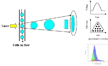
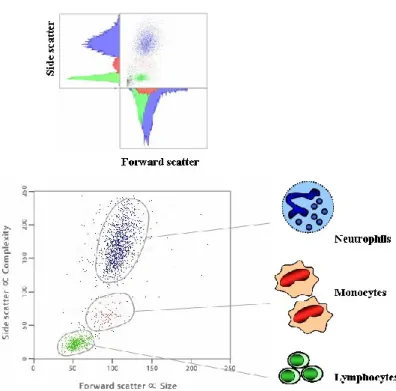
Flow cytometry - basic concepts
56
1.5.3.
Cell preparation and surface staining
60
1.6. Cytokine in vitro assay of CD4
+CD25
+and CD4
+CD25
-T cells
62
1.6.1.
CD4
+CD25
+and CD4
+CD25
-T cell isolation
62
1.6.2.
APC Preparation
64
1.6.3.
Detection of in vitro cytokine production
64
1.7. Gene expression analysis of Treg selective marker – Foxp3
66
1.7.1.
RNA extraction and reverse transcriptase reaction
66
1.7.2.
Molecular cloning of foxp3
67
1.7.2.1. PCR of foxp3 gene
67
1.7.2.2. Transformation of E. coli with recombinant plasmid DNA
68
1.7.2.3. Plasmid DNA extraction and restriction analysis
69
1.7.3.
Real-time PCR assay
70
1.8. Gene expression analysis of Toll-like receptor-2 on CD4
+CD25
+T cell subsets 75
1.8.1.
PCR of tlr2 gene
75
1.8.2.
Real-time PCR assay
75
2. Evaluation of the effect of TLR-2 modulation on Treg populations during L.
infantum in vivo infection
76
2.1. Experimental design
76
2.2. Animals and parasites
77
2.3. Sample collection
77
2.4. Parasite detection
78
2.5. Phenotypic characterization
78
2.5.1.
Isolation of total leukocytes
78
2.5.2.
Cell surface markers and intracellular staining of FOXP3
78
2.6. Detection of cytokine transcripts
79
2.7. Histopathology
81
3. Statistical analysis
82
CHAPTER III.
RESULTS
1. Phenotypic characterization of regulatory T cell populations during L. infantum
in vivo infection
85
1.1. Parasite detection
85
1.2. Phenotypic characterization of regulatory T cells
86
1.3. Detection of in vitro cytokine production
91
1.3.1.
CD4
+CD25
+T cell subset
91
1.3.2. CD4
+CD25
-T cell subset
93
1.4. Gene expression analysis of tlr2
95
2. Evaluation of the effect of TLR-2 modulation on Treg populations during L.
infantum in vivo infection
97
2.1. Parasite detection
97
2.2. Phenotypic characterization of regulatory T cells
98
2.3. Detection of cytokine transcripts
103
2.3.1. IFN-
103
2.3.2.
IL-4
105
2.3.3.
IL-10
108
2.3.4.
TGF-
110
2.4. Histopathology
112
CHAPTER IV. DISCUSSION 117
FINAL CONCLUSIONS 141
REFERENCES 145
3
1. The genus Leishmania
Leishmaniasis is a vector-borne disease caused by a pathogenic protozoan parasite belonging to the family Trypanosomatidae, genus Leishmania Ross, 1903. These parasites infect the mononuclear phagocytic system of the mammalian host and are transmitted by the bite of a female sand fly of the family Psychodidae, genus Phlebotomus Rondari, 1843 or Lutzomyia França, 1924. In man,
Leishmania spp. can cause a variety of symptoms that range from disfiguring cutaneous and
mucocutaneous lesions, widespread destruction of mucous membranes, to visceral disease affecting amongst others, haematopoietic organs.
2. Biology of Leishmania
2.1. Morphology and life cycle
Leishmania is a unicellular digenetic endoparasite that morphologically alternates between
intracellular aflagellated amastigotes within the vertebrate host and extracellular flagellated promastigotes within the intestinal tract of the phlebotomine sand fly. The amastigote stage is a round or ovoid-like organism, 2-6 m in diameter, containing a nucleus, a kinetoplast and an internal flagellum. The amastigotes multiply by binary fission within the acidic and hydrolase-rich environment of the parasitophorous vacuoles of macrophages. The promastigotes have an elongated cell body (about 10-20 m by 2.5-5 m) with a central nucleus, a kinetoplast and a long external flagellum that emerges from the anterior part of the cell and confers motility (Figure 1).
Most Leishmania species (subgenus Leishmania) are suprapylarian parasites: that is, their development is restricted to the midgut of the digestive tract of sand flies. Members of the subgenus Viannia are peripylarian parasites: they enter the hindgut before migrating forward into
4
the midgut. Infection initiates when the sand flies feed from an infected host and ingests blood containing macrophages infected with amastigotes. The infected blood meal passes to the posterior
abdominal gut where the infected macrophages are lysed and Leishmania amastigotes released. These begin to differentiate into several distinct developmental stages as they migrate anteriorly from the posterior hindgut or midgut to the stomodeal valve, which forms a junction with the foregut. Within 6–9 days, amastigotes transform into dividing procyclic promastigotes, relatively resistant to digestive enzyme activity, which in turn eventually attach to the lining of the midgut epithelium, rapidly multiply and differentiate into highly infectious metacyclic promastigotes. Each of the intermediate developmental promastigote stages is characterised by morphological and Figure 1. Transmission electron microscopy of Leishmania-infected host cells from hamster skin lesion showing amastigote and promastigote forms inside host cell vacuoles. Amastigote with the characteristic round shape, a centrally located nucleus, a bar-shaped kinetoplast, and large lipid inclusions. Promastigote form with the typical elongated body shape and a free flagellum. Arrowheads: parasite membrane; arrows: host parasitophorous vacuole membrane. N nucleus; K kinetoplast; FP: flagellar pocket (adapted from Corrêa and Soares, 2006).
5 functional changes aimed at ensuring its‘ survival in the sand fly vector. Depending on the parasite and vector species, there may be additional blood meals during maturation period, but most parasites can complete development within the timeframe of a single digestive cycle. The metacyclic promastigotes are freely motile and accumulate just behind the stomodeal valve, well positioned for egestion from the mouthparts of the sand fly upon uptake of a second blood meal and infection of a new host. Some of these promastigotes will survive, invade new cells and differentiate once again into amastigotes, hence completing its‘ life cycle. Once inside the skin, the promastigotes are phagocytosed by resident tissue macrophages, dendritic cells and neutrophils, transformed into amastigotes within the phagolysosome of macrophages. Replication of amastigotes can cause host cells to rupture and release parasites capable of invading other macrophages (Figure 2).
2.2. Pathogenecity
Disease progression is dependent on both the species of Leishmania involved and the genetics and immune status of the host. Leishmania pathogenesis involves key issues such as promastigote differentiation to the mammalian-infective amastigote stage, parasite responses and adaptations to life in host macrophages, genetic differences between major pathogenic species, and the intimate interface between the parasite and its‘ host cell (McConville et al., 2007). Parasite virulence factors and host mechanisms are inextricably linked to pathogenesis.
2.2.1. Parasite virulence factors
A successful completion of the life cycle of Leishmania parasites in the sand fly requires that they survive the digestive enzymes; avoid expulsion from the gut; and at a later stage, migrate anteriorly and break free from the midgut epithelium for transmission to the mammalian host. Several
6
molecules are central to vector-parasite interactions during critical stages of the Leishmania life cycle.
Figure 2. The Leishmania lifecycle. The promastigote form of Leishmania is transmitted into the skin by female phlebotomine sandflies. Once transmitted, the parasites are internalized by macrophages and dendritic cells in the dermis where they loose their flagella, transforming into the amastigote form. The amastigotes multiply, destroy the host cell and infect other phagocytic cells. The amastigotes disseminate through the lymphatic and vascular systems, eventually infiltrating the bone marrow, liver and spleen (reproduced from Handman, 2001).
7
Leishmania phosphoglycans share a common phosphorylated galactose-mannose disaccharide
structure -6Gal( 1-4)Man( 1)-PO4- and include glycosylphosphatidylinositol (GPI)-anchored molecules such as lipophosphoglycans (LPG), secreted extracellular phosphoglycans (PG) and secreted glycoproteins as acid phosphatase (AP) and proteophosphoglycans (PPG). Other GPI-anchored glycoconjugates include the small surface GPI lipids (GIPLs) and the surface protease or metalloproteinase gp63 (Yao et al., 2003).
Proteolytic enzymes secreted by the sand fly to digest the blood meal create a hostile environment for the parasites (Sacks et al., 2001; Volf et al., 2001; Ramalho-Ortigão et al 2003). Transitional L.
major forms transforming from amastigotes to promastigotes in midgut of Phlebotomus papatasi
are highly sensitive to proteolysis, and up to 50% of parasites can be killed in the first two days after blood feeding (Pimenta et al., 1997). To overcome this, Leishmania parasites have evolved specific abilities to modulate the activity of midgut digestive enzymes in their natural or competent vector species by inhibiting or delaying the peak of enzymatic activity. Glycoconjugates of
Leishmania appear to play a part in protecting Leishmania promastigotes from these enzymes.
Analysis of parasite mutants that are deficient in the synthesis of all PG were shown to be highly sensitive to the conditions in the early blood fed midgut (Sacks et al., 2000) although the addition of glycoconjugates from a L. major strain enhanced survival in P. papatasi (Schlein et al., 1990). These studies give emphasis on the importance of secreted phosphoglycans in protection of
Leishmania parasites from digestive enzymes.
Glycoconjugates also seem to be important virulence factors when Leishmania is in the vertebrate host. LPG is the largest and most abundant surface glycolipid of promastigotes and forms a dense glycocalyx on their surface. It has been implicated as an adhesion molecule that mediates the interaction with the midgut epithelium of the sand fly in the subgenus Leishmania, thus preventing their loss with the excreted blood meal (Pimenta et al., 1992). LPG variations have also been implicated in the specificity of various Leishmania to different Phlebotomus species and thus promote vectorial competence to the invertebrate hosts (Pimenta et al., 1994; Sacks et al., 1995;
8
Kamhawi et al., 2000). LPG undergoes several important modifications during the life cycle that are characteristic for each Leishmania species. During the acquisition of virulence features in the sand fly midgut, LPG elongates, the number of repeating units doubles from about 15 in procyclic promastigotes to about 30 in metacyclic forms (Descoteaux et al., 1999). These structural changes are required for the detachment of infectious metacyclic promastigotes from the sand fly midgut (Sacks et al., 2001).
LPG has also been implicated in many steps required for establishment of macrophage infections apart from its‘ survival in the insect vector (Sacks et al., 2000; Ilg et al., 2001). Studies demonstrated that Leishmania phosphoglycans-/- were unable to survive in activated macrophages but retained the ability to persist indefinitely in the mammalian host without inducing disease in non-activated macrophages (Spath et al., 2003). Later, the same outcome was reported in a model of L. major mutant specifically lacking LPG, revealing that the previous findings may be related to LPG (Spath et al., 2003).
By acting as an activator of the complement cascade and binding to serum proteins, LPG facilitates promastigote attachment to macrophages and contributes to the ability of metacyclic promastigote to resist complement-mediated lysis in the mammalian host (Puentes et al., 1988). This is accompanied by an absence of proinflammatory response and no oxidative burst thus enabling promastigotes silent entry and easy access to a safe haven, the inside of the macrophage. LPG serves as an excellent cover masking direct recognition of parasite molecules by macrophage receptors and therefore bypassing macrophage activation. In fact, attachment and internalization of
L. donovani promastigotes to bone marrow derived - macrophages failed to induce phosphorylation
of several kinases involved in innate macrophage function such as production of proinflammatory cytokines and nitric oxide (NO) (Privé et al., 2000). Failure to activate macrophages during the invasion process may contribute to the successful establishment of Leishmania within the mammalian host.
9 Another known function for LPG is related to its‘ ability in manipulating signalling pathways and subverting or attenuating normal macrophage function. To turn off host microbicidal functions,
Leishmania activates macrophage phosphotyrosine phosphatases and inhibits protein kinase C
(PKC) activity (Descoteaux et al., 1993; Nandan et al., 2000). Once inside the phagosome of the macrophage, Leishmania is now faced with the dangers of subsequent phagosome maturation involving interactions with endosomes and lysosomes, vacuoles containing antimicrobial proteins. In the case of phagosomes harbouring L. donovani promastigotes, interactions with late endosomes and lysosomes are inhibited (Desjardins et al., 1997). Inhibition of phagosome maturation is dependent on LPG repeating unit domain since phagosomes harbouring phosphoglycan-defective mutants quickly mature (Desjardins et al., 1997). The exact mechanism by which LPG exerts such an effect on the phagosome remains unclear; however, the insertion of LPG in cellular membranes (Tolson et al., 1990) may alter their fusogenic properties (Miao et al., 1995).
It is noteworthy to state that the diverse mechanisms used by promastigotes to invade macrophages, manipulate innate function and evade and survive host defense mechanisms may not be common to all Leishmania species. Different Leishmania species place different emphasis on the importance of canonical virulence determinants, including LPG (Turco et al., 2001). Overall, previous studies establish a role for LPG in many but not all of the steps previously identified in macrophage invasion and survival. In fact new evidence now suggests that LPG is detrimental to long-term survival in the vertebrate host, as it is associated with activation of dendritic cells (DC), natural killer (NK) cells and NKT cells (Becker et al., 2003; Amprey et al., 2004; Aebischer et al., 2005); this is perhaps the reason why it is rapidly downregulated (at least three orders of magnitude) (Turco et al., 1991) in amastigotes. Due to its structural characteristics and GPI-anchor, LPG has also been identified as a Leishmania ligand capable of activating some ―pathogen-associated molecular patterns‖ (PAMPs) recognising receptors, otherwise known as toll-like receptors (TLRs). This growing family of receptors are now known to be key players in the detection of pathogens
10
and the induction of anti-microbial immune response. The importance of Leishmania – TLR interactions will be discussed in further detail in section 7.3.
The process of differentiation of metacyclic promastigotes to amastigotes is one of the major developmental transitions in the Leishmania life cycle and a key event in establishing infection in the mammalian host. In vitro studies suggest that two environmental factors are sufficient to induce differentiation of promastigotes to amastigote-like forms (axenic amastigotes). A mild rise in temperature (33-37ºC) and a decrease in pH to 5.5 are the conditions that mimic the environment of macrophage phagolysosome. In vivo studies show that other factors, such as host serum components, may be required for differentiation (Bee et al., 2001). Several works indicate that
Leishmania in vitro differentiation can be triggered by pharmacological agents that induce protein
misfolding and promastigote heat shock response (Wiesgigl & Clos, 2001; Barak et al., 2005), by mitogen-activated protein (MAP) kinase signalling that are involved in modulating flagellum length (Wiese, 2007) and proteolytic systems involving protein turnover and degradation as key processes in autophagy and consequent promastigote-amastigote differentiation (Belesterio et al., 2007).
To be able to survive in host macrophages, amastigotes have thought to have acquired (1) amino acid permeases that allow them to scavenge host amino acids from the lumen of phagolysosome vacuole in competition with the host transporters (McConville et al., 2007), (2) the capacity of endocytosing and degrading host proteins using lysosomal proteinases (Besterio et al., 2007) and (3) lack of stage specific changes in mRNA levels and constitutive expression of enzymes involved in central carbon metabolism confer selective advantage by allowing these parasites to exploit variable nutrient conditions in the mammalian host.
Leishmania parasites are highly capable of manipulating host response in its favour. Depending on
parasite stage and species, Leishmania can evade humoral innate defenses, remodel intracellular compartments and pathways, and impair macrophage and DC mechanisms (Sacks et al., 2002a;
11 Engwerda et al., 2004; McMahon-Pratt et al., 2004). Parasites interfere with intracellular kinases and phosphatases activity, downregulate activating-type signalling pathways, upregulate suppressive-type signalling pathways, and affect transcription factors and gene expression (Buates et al., 2001; Bertholet et al., 2003; Rodriguez et al., 2004). In turn, macrophage responsiveness and secretion of cytokines, surface molecule expression, and generation of leishmanicidal mechanisms (reactive oxygen and nitrogen intermediates) are compromised (Bertholet et al., 2003; Engwerda et al., 2004; Ray et al., 2000). Parasite effects extend to dendritic cells, which are critical to antigen presentation, T-cell co-stimulation, and efficient development of acquired Th1 responses (Brandonísio et al., 2004). Effects on dendritic cells include inhibition of migration, maturation and activation, and of interleukin (IL-) 12 production (Brandonísio et al., 2004). More detailed immunological aspects of host-parasite interactions will be further discussed in section 7.
Different species of Leishmania often exhibit strong preference for specific sand fly vectors and can cause different types of diseases in the human host. Comparative genome analysis may help identify the molecular basis for these differences. To date three completed Leishmania genomes (L.
major, L. infantum, L. braziliensis) are in the public domain (Ivens et al., 2005; Peacock et al.,
2007) with sequencing of a fourth (L. mexicana) in progress (http://www.genedb.org/). These first four species were chosen to represent the main species complexes of the Leishmania subgenus together with the best-characterised species of the Viannia subgenus. Importantly, the three completed genomes represent species that usually give rise to distinct disease types. Smith (2007), details on the remarkable similarity of the three genomes (only ~200 of the ~8300 genes are differently distributed) as well as some striking differences. The genes that differ between the three species are good candidates for functional analysis to determine their roles in establishment of infection. The most divergent, L. braziliensis, possesses 47 genes that are absent from the other two species. In comparison, L. infantum has 27 species-specific genes while L. major has only 5. A number of the other differentially distributed genes are found in 2 out of the 3 species. Some of these species-specific sequences have already been analysed at the molecular level. Examples
12
include the L. infantum A2 gene that encodes an amastigote-specific repeat-containing protein previously characterised in L. donovani, the only Leishmania sequence to date that confers a change in virulence phenotype when introduced into L. major by genetic transfection (Zhang et al., 2003); and the HASP and SHERP genes, expressed from a single locus (absent in L. braziliensis) in infective stages of L. major and L. infantum, with their protein products localizing in the plasma membrane and intracellular membranes, respectively, in these species (Denny et al., 2000; Knuepfer et al., 2001). Despite the well-conserved synteny between the three model species, L.
braziliensis may have the capacity for more extensive genomic reorganisation, given the putative
retrotransposons and RNA interference (RNAi – post-transcriptional mechanism for gene silencing) machinery very recently found in this species (Peacock et al., 2007). The availability of RNAi in L. braziliensis may play a role in the biological differences and different disease outcomes observed between the two subgenera. This finding has major practical applications and provides an alternative tool to targeted gene replacement for silencing gene expression in these parasites and reducing the degree of host pathogenecity.
3. Origin, evolution and taxonomy
It has been differently proposed that the genus Leishmania first appeared either in the ‗Old World‘ (Africa, Asia and Europe) (Kerr et al., 2000; Momen et al., 2000) or in the ‗New World‘ (the Americas) (Noyes et al., 1997; Stevens et al., 2001). The ‗New World‘ origin is supported by the high genetic diversity of neotropical Leishmania species and by combined amino acid, DNA, and RNA polymerase-based trees, which root in America. This claim has received support by the description of a monoxenic insect flagellate from Costa Rica that branches at the root of the
Leishmania clade (Yurchenko et al., 2006). In fact, a recent revision of current taxonomy (Lukes et
al., 2007) proposes that the ancestor of the ‗New World‘ leishmaniasis evolved in South America in the Paleocene or Eocene, 46–36 Mya and then migrated via the Bering land bridge to Asia. The
13
Leishmania lineage would have, then, dispersed through Central and/or Southeast Asia during the
Miocene, 24–14 Mya (Fernandes et al., 1993), where a major diversification gave rise to L.
aethiopica, L. major, L. gerbilli, L. turanica, L. tropica, and the L. donovani complex (Croan et al.,
1997; Noyes et al., 1997). L. infantum would have then split from the early L. donovani lineage 1 Mya, and L. donovani soon thereafter invaded India and Africa. Closing the circle, after 500 years ago, L. infantum MON-1 European strains were transferred to South America, represented by the species formerly designated L. chagasi, considered synonymous with L. infantum (Maurício et al., 2000). The two main reservoir hosts of the L. donovani complex are humans and canids whose historical movements likely would have influenced the distribution of L. donovani and L. infantum.
Leishmania species must adapt to varied and heterogeneous environments during their life cycle.
Efficient biological mechanisms are essential for the long-term and short-term survival of the parasite. Leishmania parasites basically present a clonal population structure. The great diversity of
Leishmania species arises from different genetic processes such as asexual multiplication, gene
amplification, occasional genetic exchanges through interindividual recombination, intrachromosomic and intragenic recombination (Victoir et al., 2005). Concerning interindividual recombinations, some studies suggest genetic exchange in Leishmania despite the lack of evidence of a sexual stage. For example, in the ‗New World‘, hybrids between L. braziliensis and L.
peruviana, and L. guyanensis and L. braziliensis, have been described (Da-Cruz et al., 1992; Belli
et al., 1994; Dujardin et al., 1995; Bañuls et al., 1997) and in the ‗Old World‘ hybrids have been reported between L. major and L. arabica (Evans et al., 1987; Kelly et al., 1991) and between L.
infantum and L. donovani (Hide et al., 2006 and 2007) and between L. infantum and L. major
(Ravel et al., 2006). However, the occurrence of pseudo-recombination events and cross-species genetic exchanges is not frequent enough to disturb the propagation of clones stable in space and time. It is postulated that the asexual model is enough to ensure parasite fitness in various environmental conditions and that sexual recombination is not necessary for the production of a large repertoire of genotypes (Ayala et al., 1998; Victoir and Dujardin, 2002).
14
The classification of Leishmania was initially based on ecobiological criteria such as vectors, geographical distribution, tropism, antigenic properties and clinical manifestation (Bray, 1974; Lumsden, 1974; Pratt and David, 1981; Lainson and Shaw, 1987) - for example, L. guyanensis
(isolated in Guyana), L. peruviana (isolated in Peru), L. infantum (isolated from a child in Tunisia)
and L. gerbilli (isolated from gerbils). All members of the genus Leishmania are parasites of mammals. The two subgenera, Leishmania and Viannia, are separated on the basis of their location in the vector‘s intestine (Lainson and Shaw, 1987).
However, biochemical and molecular analysis showed that these criteria were often inadequate and thus other criteria such as the patterns of polymorphism exhibited by kinetoplast DNA (kDNA) markers, proteins or antigens came to be used to classify Leishmania (Kreutzer and Christensen, 1980; Arnot and Barker, 1981; Pratt and David, 1981; Barker et al., 1986). For epidemiological purposes, the most useful taxonomic technique is isoenzyme analysis or multi-locus enzyme electrophoresis (MLEE) (Miles et al., 1980; Evans et al., 1984; Rioux et al., 1990). MLEE detects different alleles of housekeeping genes by scoring the electrophorectic mobility of enzymes they encode. It is still considered the ‗gold standard‘ for Leishmania species identification and genetic diversity studies.
Use of molecular techniques led to the publication of a taxonomic scheme by the World Health Organization (WHO, 1990) (Figure 4). Today 30 Leishmania species are known and approximately 20 are pathogenic for humans. These species generally present different epidemiological and clinical characteristics related to different genetic and phenotypic profiles. The validity of the classification scheme, considered by some workers as too arbitrary, has been questioned several times. Debate has centred on L. panamensis, L. peruviana, L. chagasi, L.
infantum, L. archibaldi, L. garnhami, L. pifanoi, L. venezuelensis and L. forattinii (Bañuls et al.,
1999, 2000; Mauricio et al., 2000, 2001; Cupolillo et al., 2001). L. chagasi is accepted as a synonym of L. infantum (see Mauricio et al., 2000) and L. peruviana has been validated as an independent species (Bañuls et al., 2000). The other species listed above are still under discussion.
15 In addition, a recent study has shown to have identified a potential new species of Leishmania causing VL in Thailand (Sukmee et al., 2008). Through a combination of PCR-RFLP (restriction fragment length polymorphism) on the ssrRNA (small subunit ribosomal RNA) and mini-exon genes it was shown that the Leishmania spp. found is clearly distinguishable from other members of the genus Leishmania.
4. Epidemiology
Leishmaniasis is an important parasitosis, widespread on all the continents except Antartica. Human leishmaniasis constitutes a major public health problem and the burden is increasing (Desjeux, 2001, 2004). This disease can present different clinical features depending on the infecting species of Leishmania and the host‘s immunological status: cutaneous leishmaniasis (CL), a disease that can result in unaesthetic stigma if multiple lesions occur; mucocutaneous leishmaniasis (MCL; also known as espundia) a mutilating disease; diffuse cutaneous leishmaniasis (DCL) a long-lasting disabling disease due to a deficient cellular-mediated immune response; visceral leishmaniasis (VL; also known as kala-azar) a disease often fatal if untreated; and post-kala-azar dermal leishmaniasis (PKDL) a chronic cutaneous disease characterized by a macular, maculo-papular or nodular rash that develops after VL treatment. In 2002, the WHO estimated that the number of persons at risk to be around 350 million through 88 countries and the number of new cases to be 2 357 000 per year (WHO, 2002). In fact, 2 million new cases (1.5 million for CL and 500 000 for VL) are considered to occur annually, with an estimated 12 million people presently infected worldwide. It is noteworthy to mention that under-reporting is substantial - only around 600,000 infections are officially reported each year (WHO, 2002). Despite the scarcity of reliable data, there is little doubt that the case-load worldwide is considerably higher than official reported figures.
16
Figure 4. Taxonomy of Leishmania; underlined species are or have been questioned. (Based on the scheme published by the [WHO, 1990] with additions from the literature).
17 Of the 88 leishmaniasis-affected countries, 72 are classed as developing countries, including 13 of the least developed countries. Ninety percent of VL cases occur in just five countries — Bangladesh, India, Nepal, Sudan and Brazil. Ninety percent of CL cases occur in just seven countries — Afghanistan, Algeria, Brazil, Iran, Peru, Saudi Arabia and Syria. VL is of higher priority than CL as it is a fatal disease in the absence of treatment (Desjeux, 2004). Anthroponotic VL foci are of special concern as they are at the origin of frequent and large-scale tenacious epidemics with high case-fatality rates. Malnutrition is a well-known risk factor in the development of this form, and epidemics flourish under conditions of famine, complex emergencies and mass population movements. In Sudan, for example, a major decade-long epidemic of VL occurred from 1984 to 1994. Some studies estimate that the disease caused 100 000 deaths in a population of around 300 000 in the western upper Nile area of the country (Seaman et al., 1996). Despite the huge amount of research conducted on these pathogens in numerous scientific fields since the beginning of the last century, recent studies have also shown the reactivation of several other foci- in Italy, China, Brazil and Israel (Arias et al., 1996; Gradoni et al., 2003; Guan et al., 2003; Nasereddin et al., 2005) — and the emergence of new epidemic foci in Israel and Morocco (Jacobson et al., 2003; Al-Jawabreh et al., 2004; Guernaoui et al., 2005; Shani-Adir et al., 2005). Nevertheless, leishmaniasis is still considered as a neglected disease and is grossly underestimated, mainly due to lack of awareness of its serious impact on health.
5. Leishmaniasis
Leishmaniasis is a typical example of an anthropozoonosis although the majority of infections are originally zoonotic. The different epidemiological cycles are (i) a primitive or sylvatic cycle (human infection is accidental, transmission occurring in wild animal foci) e.g. L. braziliensis; (ii) a secondary or peridomestic cycle (the reservoir is a peridomestic or domestic animal, the parasite being transmitted to humans by anthropophilic sand flies), e.g. L. infantum; and (iii) a tertiary, strictly anthroponotic cycle, in which the animal reservoir has disappeared (or has not yet been
18
identified) and the sand fly vectors are totally anthroponotic, e.g. L. donovani. Nevertheless, many unknown factors remain. For example, the main animal reservoir of L. braziliensis is still unknown (Cupolillo et al., 2003). L. tropica was considered to be a strict anthroponosis, but several cases of canine infection have been described (Dereure et al., 1991; Banuls, 2007).
Cutaneous leishmaniasis, which accounts for more than 50% of new cases of leishmaniasis, is frequently self-healing in the ‗Old World‘ but, when the lesions are multiple and disabling with disfiguring scars, it creates a lifelong aesthetic stigma. It results in formation of skin ulcers at the site of the sand fly bite, usually on exposed parts of the body, the face, neck, arms and legs. Its‘ most severe form, recidivans leishmaniasis, is very difficult to treat CL, is caused by several species of Leishmania: L. major, L. tropica, L. aethiopica, and L. mexicana, but also by L.
braziliensis, L. panamensis, L. peruviana, L. amazonensis, L. guyanensis, L. naiffi and L. lainson
(Murray et al., 2005). Dermatropic L. infantum has also proven to cause localized CL (Belazzoug et al., 1985). Time to lesion resolution varies between species and between individuals.
Diffuse cutaneous leishmaniasis occurs in individuals with defective cell-mediated immune response. Its‘ severity is due to disseminated lesions that resemble those of lepromatous leprosy, which never heal spontaneously and is subject to relapse after treatment with any of the currently available drugs. Because of the devastating consequences to the patient, it is recognized as a special public health problem. DCL is caused by L. aethiopica, L. amazonensis and L. mexicana occurs in anergic hosts with poor immune responses. This form of disease is restricted to a few foci in Ethiopia, Kenya, Venezuela and the Dominican Republic, suggesting an important role for the genetics of the parasite as well as the genetics of the host in determining the disease phenotype.
In mucocutaneous leishmaniasis (―espundia‖) the initial skin lesions cure, but the late development of metastatic lesions can lead to extensive destruction of oral-nasal and pharyngeal cavities with hideous disfiguring lesions, mutilation of the face and great suffering for life. MCL may also arise after inadequate treatment of some Leishmania species, and if untreated can lead to severe
19 deformities or even death. Although mostly related to Leishmania species of the ‗New World‘ such as L. braziliensis but also more rarely by L. panamensis and L. guyanensis (Naiff et al., 1988; Saraiva et al., 1995; Osorio et al., 1998), MCL has been also reported in the ‗Old World‘ due to L.
donovani, L. major (Al-Gindan et al., 1983) and, in immmuno-suppressed patients, L. infantum
(Alvar et al., 1990).
Visceral leishmaniasis, also known as kala-azar, is the most severe and often fatal syndrome. VL is caused by parasites belonging to the L. donovani complex – L. donovani sensu stricto in East Africa and the Indian subcontinent and L. infantum in south Europe, North Africa and Latin America (Boelaert et al., 2000). There are two types of VL, which differ in their transmission characteristics: zoonotic VL is transmitted from animal to vector to human and anthroponotic VL is transmitted from human to vector to human. In the former, humans are occasional hosts and animals, mainly dogs, are the reservoir of the parasite. Zoonotic VL is found in areas of L. infantum transmission whereas anthroponotic VL is found in areas of L. donovani transmission. Following an incubation period that generally lasts between 2 and 6 months, VL patients present symptoms and signs of persistent systemic infection (including fever, fatigue, weakness, loss of appetite and weight loss), parasitic invasion of the blood and reticulo-endothelial system (that is, the general phagocytic system), such as enlarged lymph nodes, spleen (splenomegaly) and liver (hepatomegaly) and hypergammaglobulinaemia (mainly IgGs from polyclonal B cell activation). Fever is usually associated with rigor and chills and can be intermittent. Fatigue and weakness are worsened by anaemia, which is caused by the persistent inflammatory state, hypersplenism (the peripheral destruction of erythrocytes in the enlarged spleen) and sometimes by bleeding. If left untreated, the disease has a high mortality rate mainly due to immunosuppression and secondary infections. Some individuals develop PKDL (Zijlstra et al., 2003), which appears within a few years of the complete cure of VL. PKDL patients are considered a major source of parasites for new infections because of the large number of organisms in the skin accessible to sand fly bites.
20
Human susceptibility to L. infantum is low therefore asymptomatic infections are common in healthy populations. Clinical disease is associated with age (infants below 2 years are most affected), malnutrition and immunosuppression (e.g. HIV co-infection).
Table 1. Human-infective species of the Leishmania genus. The main species complexes and subgenus are shown in bold; the species. * Species that can also be associated with cutaneous leishmaniasis (adapted from Smith et al., 2007).
Old World species New World species Disease type
L. major complex L. mexicana complex Cutaneous
L. (L.) major L. (L.) mexicana L. (L.) tropica L. (L.) amazonensis L. (L.) aethiopica L. (L.) pifanoi L. (L.) venezuelensis L. (V.) (Viannia) subgenus L. (V.) braziliensis L. (V.) panamensis L. (V.) guyanensis L. (V.) perviana L. (V.) lansoni L. (V.) braziliensis Mucocutaneous
L. (L.) aethipoica L. (L.) amazonensis Diffuse cutaneous
L. (L.) pifanoi
L. donovani complex
L. (L.) donovani Visceral
21 VL has become a frequent infection in HIV-positive individuals in endemic areas and accelerates the onset of AIDS in these patients. Cases of HIV and visceral leishmaniasis co-infection have been reported in 35 countries worldwide (Chappuis et al., 2007). Both the cellular and humoral responses to L. infantum are diminished in co-infected patients (Moreno et al., 2000), leading to an increased parasite load in peripheral blood, lower sensitivity of serological tests, and a higher rate of treatment failure (Murray et al., 1999; Deniau et al., 2003). The impact of these co-infections was recognised as an alarming problem by international health authorities (Desjeux et al., 1995). Cases were reported showing a deadly overlapping between HIV pandemic and the zoonotic entity of VL (Gramiccia et al., 2005). In Europe, intravenous drug users have been identified as the main population at risk. The demonstration of unusual modes of anthroponotic transmission (i.e. by syringe exchange) (Cruz et al., 2002) and the high rate of relapses following antileishmanial treatments, were alarming features indicating a trend toward an even higher incidence. Highly active antiretroviral therapy (HAART) is used to modify the course of HIV infection. It has also been used to treat HIV-positive patients with VL and other infections. HAART appears efficient at preventing VL in individuals infected by Leishmania (de la Rosa et al., 2001), as reflected by the sharp decrease in the incidence of VL in Europe following the widespread use of HAART (Rosenthal et al., 2001; Lopez-Véléz et al., 2003). HAART is used to partially restore immune function but its efficacy in preventing VL relapses is disappointingly low (Mira et al., 2004).
6. Control strategies
The main control strategy includes: case finding and treatment, vector control, when feasible and, in zoonotic foci, animal reservoir control and vaccine development. It is impossible to devise a single control strategy for leishmaniasis control. A strategy combining these three approaches can help in eradication of the disease.

![Figure 4. Taxonomy of Leishmania; underlined species are or have been questioned. (Based on the scheme published by the [WHO, 1990] with additions from the literature)](https://thumb-eu.123doks.com/thumbv2/123dok_br/19195711.951699/46.892.208.698.159.1055/figure-taxonomy-leishmania-underlined-questioned-published-additions-literature.webp)