Replication in Human Thymocytes
Helena Nunes-Cabaço, Paula Matoso, Russell B. Foxall, Rita Tendeiro,* Ana R. Pires, Tânia Carvalho, Ana I. Pinheiro, Rui S. Soares, Ana E. Sousa
Instituto de Medicina Molecular, Faculdade de Medicina, Universidade de Lisboa, Lisbon, Portugal ABSTRACT
A unique HIV-host equilibrium exists in untreated HIV-2-infected individuals. This equilibrium is characterized by low to
un-detectable levels of viremia throughout the disease course, despite the establishment of disseminated HIV-2 reservoirs at levels
comparable to those observed in untreated HIV-1 infection. Although the clinical spectrum is similar in the two infections,
HIV-2 infection is associated with a much lower rate of CD4 T-cell decline and has a limited impact on the mortality of infected
adults. Here we investigated HIV-2 infection of the human thymus, the primary organ for T-cell production. Human thymic
tis-sue and suspensions of total or purified CD4 single-positive thymocytes were infected with HIV-2 or HIV-1 primary isolates
us-ing either CCR5 or CXCR4 coreceptors. We found that HIV-2 infected both thymic organ cultures and thymocyte suspensions,
as attested to by the total HIV DNA and cell-associated viral mRNA levels. Nevertheless, thymocytes featured reduced levels of
intracellular Gag viral protein, irrespective of HIV-2 coreceptor tropism and cell differentiation stage, in agreement with the low
viral load in culture supernatants. Our data show that HIV-2 is able to infect the human thymus, but the HIV-2 replication cycle
in thymocytes is impaired, providing a new model to identify therapeutic targets for viral replication control.
IMPORTANCE
HIV-1 infects the thymus, leading to a decrease in CD4 T-cell production that contributes to the characteristic CD4 T-cell loss.
HIV-2 infection is associated with a very low rate of progression to AIDS and is therefore considered a unique naturally
occur-ring model of attenuated HIV disease. HIV-2-infected individuals feature low to undetectable plasma viral loads, in spite of the
numbers of circulating infected T cells being similar to those found in patients infected with HIV-1. We assessed, for the first
time, the direct impact of HIV-2 infection on the human thymus. We show that HIV-2 is able to infect the thymus but that the
HIV-2 replication cycle in thymocytes is impaired. We propose that this system will be important to devise immunotherapies
that target viral production, aiding the design of future therapeutic strategies for HIV control.
T
he thymus is the primary organ for T-cell production, and
despite the age-associated decline, thymic function is
main-tained until late in life (
1
,
2
). Thymic activity is vital in clinical
settings requiring de novo T-cell generation, such as HIV infection
(
1
,
3
,
4
). Accordingly, impairment of thymic output impacts the
rate of HIV-1 disease progression, while the degree of
immuno-logical reconstitution achieved after antiretroviral therapy has
been shown to rely on thymus recovery (
1
,
3
,
4
). Moreover, a
functional cure for HIV infection is thought to entail a diverse
T-cell repertoire, which can be generated only by the thymus.
HIV-1 targets the thymus in both children and adults, resulting
in severe disruption of the thymic microenvironment, as
demon-strated by the morphological changes and thymocyte depletion
reported in the thymuses of HIV-1-infected individuals (
5
,
6
).
Several studies based on HIV-1 infection of the human thymus
either in vitro (
7–9
) or in vivo using the SCID/hu mouse model
(
10
,
11
) have indicated that both direct infection of thymic cells
and indirect viral effects upon the microenvironment play a role
in HIV-1-associated thymic pathology. Furthermore, viral entry,
viral replication kinetics, and the cytopathicity of HIV-1 in human
thymocytes have been shown to be highly dependent on viral
tro-pism, due to the predominance of CXCR4 (X4) versus CCR5 (R5)
expression in the human thymus (
9
,
12
,
13
). Thymic disruption
has also been described in nonhuman primate models of simian
immunodeficiency virus infection (
14
).
Here we addressed, for the first time, the direct impact of
HIV-2 infection on the human thymus. This is particularly
rele-vant because HIV-2-infected individuals feature low rates of CD4
T-cell decline and disease progression (
15–17
). Moreover, they
typically have low to undetectable levels of viremia, with this being
observed even in AIDS patients with
⬍200 CD4 T cells/l (
18
,
19
).
The low levels of circulating virus account for the reduced
hori-zontal and vertical transmission observed in HIV-2 infection (
20
,
21
), as well as for its geographical confinement to West Africa and
connected countries, such as Portugal. Despite the high
preva-Received 20 October 2014 Accepted 26 November 2014 Accepted manuscript posted online 3 December 2014
Citation Nunes-Cabaço H, Matoso P, Foxall RB, Tendeiro R, Pires AR, Carvalho T, Pinheiro AI, Soares RS, Sousa AE. 2015. Thymic HIV-2 infection uncovers posttranscriptional control of viral replication in human thymocytes. J Virol 89: 2201–2208.doi:10.1128/JVI.03047-14.
Editor: G. Silvestri
Address correspondence to Helena Nunes-Cabaço, [email protected], or Ana E. Sousa, [email protected].
* Present address: Rita Tendeiro, Division of Infection and Immunity, UCL Institute of Immunity and Transplantation, London, United Kingdom.
Supplemental material for this article may be found athttp://dx.doi.org/10.1128 /JVI.03047-14.
Copyright © 2015, American Society for Microbiology. All Rights Reserved.
doi:10.1128/JVI.03047-14
on January 26, 2015 by Jose Moniz Pereira
http://jvi.asm.org/
lence of HIV-2 in several regions of West Africa, such as in Guinea
Bissau (8% in adults and up to 20% in people over 40 years of age)
(
22
), there is no significant impact on the mortality of infected
adults. HIV-2 thus constitutes a unique naturally occurring model
of attenuated HIV disease valuable for the study of HIV
pathogen-esis.
In spite of the low to undetectable HIV-2 plasma loads,
HIV-2-and HIV-1-infected patients at equivalent stages of CD4 T-cell
depletion feature comparable levels of cell-associated viral burden
(
18
,
23
,
24
), indicating the establishment of disseminated HIV-2
reservoirs. They also feature similar levels of T-cell activation (
19
),
suggesting distinct control of viral replication in the presence of
cell activation in HIV-2 and HIV-1 infections.
Our previous data support the preservation of thymic function
in HIV-2 infection, as estimated by signal joint (sj)/
 T cell
recep-tor excision circle (TREC) quantification (
3
) in circulating T cells
of HIV-2-infected patients (
25
). However, there are no studies on
the direct impact of HIV-2 infection on the human thymus either
in vivo, due to the difficulty of obtaining thymic tissue from
HIV-2-infected patients, or in vitro. We show here that HIV-2 is able to
infect the human thymus but that this is associated with limited
viral replication, irrespective of viral coreceptor tropism and
mocyte differentiation stage. HIV-2 infection of the human
thy-mus thus offers a novel approach to investigate the mechanisms
underlying the establishment of HIV reservoirs and the control of
viral replication.
MATERIALS AND METHODS
Ethical statement. Thymic tissue specimens (from individuals ranging from newborns to children 4 years old) were obtained during routine thymectomy performed during pediatric corrective cardiac surgery at the Hospital de Santa Cruz, Carnaxide, Portugal, after the parents provided written informed consent. Buffy coats from healthy donors were provided by the Instituto Português do Sangue e da Transplantação after they pro-vided written informed consent. The study was approved by the Ethical Boards of the Faculty of Medicine of the University of Lisbon and of the Hospital de Santa Cruz, Carnaxide, Portugal.
HIV stocks. The viruses used are described inTable 1. Viral stocks were propagated in pools of isolated peripheral blood mononuclear cells (PBMCs) stimulated for 3 days with phytohemagglutinin (PHA; 5g/ml; Sigma) and maintained with human recombinant interleukin-2 (IL-2; 10 U/ml; from Maurice Gately, Hoffmann-La Roche Inc., through the NIH AIDS Reagent Program), as described previously (28). Virus in cell-free
culture supernatants was quantified by measuring reverse transcriptase (RT) activity using a Lenti-RT activity kit (Cavidi).
HIV infection of thymocyte suspensions. Total thymocytes were re-covered through tissue dispersion and separation on a Ficoll-Paque Plus (GE Healthcare) density gradient. The CD4 single-positive (CD4SP) pop-ulation was sorted from total thymocytes as CD3high CD8-negative
(CD8neg) cells (purity,⬎98%), using a FACSAria high-speed cell sorter (BD Biosciences). Viral stocks were ultracentrifuged for 30 min at 50,000⫻ g and 4°C (Beckman L8 ultracentrifuge), resuspended in com-plete medium (RPMI 1640 with 10% fetal bovine serum [FBS], 2 mM
L-glutamine, 50 U/ml penicillin-streptomycin, and 50g/ml gentamicin
[all from Gibco/Invitrogen] plus 3g/ml Polybrene [Sigma]), and added to the cells at 0.3 ng RT/106thymocytes. Total and CD4SP thymocytes
were cultured for 3 to 4 h at 108and 3.5⫻ 107cells/ml, respectively, in the
absence or presence of virus. After infection, the cells were washed and cultured at 107cells/ml in complete medium supplemented with IL-2 (20 U/ml), IL-4 (20 ng/ml; R&D), and IL-7 (10 ng/ml; R&D) at 37°C. A quarter of the medium was replaced every 3 to 4 days. On day 10, the thymocyte number per well was determined using 10-m latex beads (Coulter), and the cells were analyzed by flow cytometry or stored as pellets at⫺80°C. Viral production in supernatants was quantified at day 10 postinfection of CD11cnegCD14negCD123negT-cell receptor
␥␦-neg-ative (TCR␥␦neg) thymocytes by measuring RT activity using SYBR green
product-enhanced RT (SG-PERT), as described previously (29,30). HIV infection of TOCs. Thymic tissue blocks (diameter, 1 to 2 mm) were placed on Millicell organotypic inserts (Millipore) in a 6-well plate con-taining 1 ml of thymic organ culture (TOC) medium (complete medium with 15% FBS, 10 mM HEPES, 1 mM sodium pyruvate, and 1% minimal essential medium nonessential amino acids [all from Gibco/Invitrogen]). TOCs were placed at 37°C in 5% CO2overnight, and half of the medium was replaced
prior to infection. HIV infection was performed by placing a 5-l drop con-taining 3 ng RT of virus on top of each TOC. A third of the medium was replaced every 2 to 3 days. On day 10 or 11 postinfection, TOCs were placed in 4% formaldehyde or mashed. Thymocytes were analyzed by flow cytometry or stored as cell pellets at⫺80°C. The role of Env proteins was assessed by culturing the TOCs for 7 days on Isopore membranes (Millipore) placed in TOC medium containing 1g/ml of purified recombinant glycoproteins: gp105ROD(MRC EVA621), gp120Ba-L(catalog no. 4961; NIH), and gp120IIIB
(MRC EVA607) or anti-CD4 monoclonal antibody (MAb; BD Biosciences) as a control.
Flow cytometry. Surface staining was performed for 20 min at room temperature and always included fixable viability dye (eBioscience) for dead cell exclusion. Thymocytes were fixed, permeabilized, and stained using an intracellular staining kit (eBioscience), as described previously (31). The anti-human MAbs used were (clone designations are given in parentheses) CD3 (UCHT1), CD4 (RPA-T4), CD8␣ (RPA-T8), CD11c (3.9), CD14 (61D3), CD16 (eBioCB16), CD19 (HIB19), CD123 (6H6), and TCR␥␦ (B1.1) from eBioscience. The following anti-Gag MAbs were used: KC57 (Beckman Coulter), anti-p24 (MAb Kal-1; Dako), anti-p24 (MAb AG3.0 or 183-H12-5C; NIH), and anti-p27 (MAb ARP396/397; P. Szawlowski, NIBSC, Centre for AIDS Reagents, Medical Research Council [MRC]). Alexa Fluor 488 goat anti-mouse IgG (H⫹L) antibody (Molec-ular Probes) was used for secondary detection. Cells were acquired using a LSRFortessa cell analyzer (BD Biosciences), and data were analyzed with FlowJo software (TreeStar).
Immunohistochemistry. Fixed TOCs were embedded in paraffin and cut into 3-m sections (Minot microtome Leica RM2145). Epitope re-trieval was performed at pH 9 (Leica Biosystems buffer) for 15 min using a microwave (800 W). Samples were stained with the appropriate anti-Gag primary antibodies, incubated with a peroxidase-diaminobenzidine detection system (EnVision; Dako), and counterstained with Harris’s he-matoxylin (BioOptica). Images were acquired using a Leica DM2500 mi-croscope.
Quantification of total HIV DNA and gag mRNA by real-time PCR. Total HIV DNA was quantified in cell lysates prepared by treating cell TABLE 1 HIV isolates used in the study
Virus Major coreceptor(s) used Source
Primary isolates
HIV-192US660 R5 NIH
a HIV-192HT599 X4 NIH a,b HIV-260415K R5 NIH a,c HIV-220.04 d X4 Nuno Taveira Lab-adapted strains HIV-1NL4-3 X4 MRCe HIV-2ROD10 R5, X4 MRCe a
From the Multicenter AIDS Cohort Study, NIH AIDS Reagent Program, Division of AIDS, NIAID, NIH.
b
Provided by Neal Halsey.
cProvided by Feng Gao and Beatrice Hahn. d
Previously represented as PTHCC20/2004 (26) and 19/2004 (27).
eNIBSC, Centre for AIDS Reagents, Medical Research Council, United Kingdom.
on January 26, 2015 by Jose Moniz Pereira
http://jvi.asm.org/
pellets with 100g/ml proteinase K in 10 mM Tris-HCl for 1 h at 56°C, followed by 10 min of enzyme inactivation at 95°C. For gag mRNA quan-tification, 200 ng of total RNA, purified using a mirVana microRNA iso-lation kit (Ambion), was used to synthesize cDNA using oligo(dT)20and
SuperScript III RT (Invitrogen) according to the manufacturer’s instruc-tions. Real-time PCR was performed using Platinum Taq plus carboxy-X-rhodamine (ROX) or TaqMan gene expression master mix (Applied Biosystems) with the primers and probes described in Table S1 in the supplemental material. Standard curves were generated from serial dilu-tions of cDNA prepared from the mRNA of 3 different thymuses (for GAPDH [glyceraldehyde-3-phosphate dehydrogenase] quantification) or of plasmids containing the amplicons of HIV-1 gag and CD3␥ (a kind gift from Rémi Cheynier) (32) or of HIV-2 gag. A plasmid carrying HIV-2 gag was generated by inserting a sequence including the sequences for the long terminal repeat/gag from HIV-2ROD10into the pGEM-T Easy vector
(Pro-mega). Quantification was performed using an Applied Biosystems 7500 Fast real-time PCR system.
Statistical analysis. Statistical analysis was performed using Graph-Pad Prism (v5.01) software (GraphGraph-Pad Software Inc.). Data from two samples were compared using the Wilcoxon matched-pairs test. Data from more than two samples were compared using the Friedman test or the Kruskal-Wallis test with Dunn’s multiple-comparison posttest. P val-ues of⬍0.05 were considered significant.
RESULTS
HIV-2 infects the human thymus. We investigated HIV-2
infec-tion of the human thymus in vitro using both TOCs, which
pre-serve the thymic microenvironment and have been shown to be
permissive to HIV-1 infection (
7
,
8
), and thymocyte suspensions.
TOCs and thymocyte suspensions were infected with HIV-2 or
HIV-1 primary isolates with selective R5 or X4 coreceptor
speci-ficity and cultured for 10 days. RT activity was used to normalize
the viral input, as HIV-1 and HIV-2 RT enzymes were shown to
possess similar specific activities (
33
).
We found that HIV-2 was able to infect both thymic tissue and
total thymocyte suspensions, as indicated by the levels of total HIV
DNA, which includes unintegrated and integrated proviral DNA,
in HIV-2-infected samples (
Fig. 1A
). Furthermore, we observed
that HIV-2 infection of both thymic tissue and total thymocyte
suspensions was less coreceptor dependent than HIV-1 infection.
Next we sorted purified mature CD4SP thymocytes, which
constitute a target of HIV-1 (
11
), and cultured them for 10 days
after infection with the above-mentioned viruses. In contrast to
HIV-2 infection of TOCs and thymocyte suspensions, HIV-2
in-fection of CD4SP thymocytes resulted in coreceptor-dependent
levels of total HIV DNA (
Fig. 1A
). The limited ability of R5-tropic
HIV-2 to infect the CD4SP subset was in contrast to our
observa-tions in TOCs and total thymocyte suspensions (
Fig. 1A
),
suggest-ing that R5-tropic HIV-2 targeted other human thymocyte
popu-lations.
We next assessed the levels of ongoing viral transcription in
HIV-2-infected TOCs and thymocytes by measuring viral gag
mRNA. Both R5- and X4-tropic HIV-2 isolates generated gag
mRNA levels comparable to those found upon infection with the
X4-tropic HIV-1 isolate (
Fig. 1B
), indicating efficient integration
and transcription of HIV-2 in TOCs and thymocyte cultures. Of
note, HIV-2 gag mRNA levels showed coreceptor dependency in
the case of purified CD4SP thymocytes but not in TOCs or
thy-mocyte suspensions (
Fig. 1B
), in line with the total HIV DNA
levels that were observed in the same samples.
In summary, our results indicated that HIV-2 is able to
suc-cessfully enter, reverse transcribe, and integrate in human
thymo-cytes in TOCs and in suspension and that viral transcription is
ongoing in these systems.
Limited viral production in HIV-2 infection of human
thy-mocytes. Next we assessed viral production in HIV-2-infected
thymic tissue using immunohistochemistry. The Gag protein was
detected in R5- and X4-tropic HIV-2-infected TOCs after culture
(
Fig. 2A
), using validated antibodies (see Fig. S1 in the
supplemen-tal material) (
34
). Gag protein expression was both cell associated
and extracellular (
Fig. 2B
), with the latter likely corresponding to
virus produced by HIV-infected cells during TOC, as no Gag
staining was observed on day 1 (
Fig. 2A
). Of note, Gag protein
levels were often indistinguishable in X4- and R5-tropic HIV-1
TOC infections (
Fig. 2A
), despite the differences in the levels of
the cell-associated viral burden (
Fig. 1
), indicating a lack of
corre-lation between the amount of virus detected by
immunohisto-chemistry and the levels of thymocyte viral production. Therefore,
although our immunohistochemistry data confirmed the ability
of HIV-2 to productively infect the human thymus, it was not
possible to infer the specific contribution of thymocytes versus
that of the thymic stroma to the viral protein detected in the
his-tological analysis.
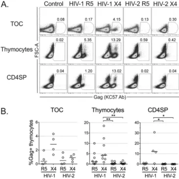
We thus evaluated viral production at the single-cell level by
flow cytometry. Notably, we found very low levels of intracellular
Gag protein in cells isolated from HIV-2-infected TOCs or in
thymocyte suspensions, irrespective of the virus used (
Fig. 3
) and
the time point analyzed (see Fig. S2 in the supplemental material).
In contrast, Gag protein expression was clearly detected in cells
FIG 1 HIV-2 infects human thymic tissue and isolated thymocytes. TOCs were infected with R5- or X4-tropic HIV-2 or HIV-1 primary isolates and cultured for 10 or 11 days on Millipore inserts. Single-cell suspensions of total thymocytes or CD4SP thymocytes, sorted as CD3highCD8negcells, were also
infected with the above-mentioned viruses and cultured for 10 days. Graphs show total HIV DNA (A), as assessed by quantitative real-time PCR, or viral
gag mRNA (B), as assessed by real-time RT-PCR (the level of mRNA
expres-sion was normalized to the level of GAPDH expresexpres-sion). Each dot represents a single thymus. Lines indicate median values. *, P⬍ 0.05; **, P ⬍ 0.01.
on January 26, 2015 by Jose Moniz Pereira
http://jvi.asm.org/
infected with HIV-1, particularly with the X4-tropic isolate (
Fig.
3
), excluding the possibility that the low level of Gag detection in
HIV-2 infections was due to sample processing. Of note,
intracel-lular Gag protein was also barely detected upon thymocyte
infec-tion with the lab-adapted strain HIV-2
ROD(see Fig. S2 in the
sup-plemental material), despite the high levels of total HIV DNA
detected (181,499 copies/10
6cells in the example presented). Low
Gag levels upon HIV-2 infection were particularly evident in
CD4SP thymocytes, which featured extremely low levels of
intra-cellular Gag after infection with 2 primary isolates or
HIV-2
ROD(
Fig. 3
; see also Fig. S2 in the supplemental material).
The antibody used (KC57) has previously been shown to be
specific for both HIV-2 Gag and HIV-1 Gag (
35
). We further
confirmed the ability of the antibody to efficiently detect HIV-2
Gag by flow cytometry in activated PBMCs infected with the
HIV-2 strains used in the current study (see Fig. S3 in the
supple-mental material). Moreover, the level of KC57 staining was
com-parable to that obtained with other available anti-Gag antibodies
in HIV-2-infected PBMCs or thymocytes (see Fig. S2 and S3 in the
supplemental material), confirming the low level of viral
produc-tion in HIV-2-infected thymocytes.
The low levels of intracellular Gag protein translated into
re-duced viral production in HIV-2 infections, as indicated by the
lower levels of virus in the supernatant of HIV-2-infected
thymo-cyte cultures than in the supernatant of their HIV-1-infected
counterparts (RT activities, 5.97
⫾ 3.23 pg/ml for R5-tropic
HIV-1, 1,318
⫾ 583.5 pg/ml for X4-tropic HIV-1, 0.31 ⫾ 0.15
pg/ml for R5-tropic HIV-2, and 8.54
⫾ 3.40 pg/ml for X4-tropic
HIV-2; n
⫽ 3).
Overall, we found that HIV-2 infection of human thymocytes
occurred without significant viral production per cell, despite
ev-idence of cell-associated viral burden and active ongoing viral
transcription.
Impact of HIV-2 infection on thymocyte populations.
Fi-nally, we assessed the impact of HIV-2 on the distribution of
thy-mocyte populations in infected TOCs and thythy-mocyte
suspen-sions. TOCs infected with X4-tropic viruses, whether they were
HIV-2 or HIV-1, featured a dramatic decrease in CD4
⫹thymo-cytes compared to the amounts in TOCs infected with R5-tropic
viruses (
Fig. 4A
and
B
). This was also observed in thymocyte
sus-pensions infected with X4-tropic HIV-1 but not with X4-tropic
HIV-2, infection with which did not result in significant
altera-tions of the thymocyte phenotype in culture (
Fig. 4C
and
D
). One
possible explanation for these results is that downregulation of the
FIG 2 Viral production in HIV-2-infected TOCs. TOCs infected with R5- or X4-tropic HIV-2 or HIV-1 primary isolates were analyzed for viral production by immunohistochemistry using anti-Gag antibodies (brown) and hematox-ylin counterstain (blue). (A) Viral production at days 1 and 11 after infection with HIV-2 (top) or HIV-1 (bottom), as assessed using an anti-p27 MAb (ARP396/397 from MRC) or an anti-p24 MAb (Kal-1 from Dako), respec-tively. (B) Extracellular (arrow) and cytoplasmic (arrowhead) Gag expression in R5- or X4-tropic HIV-2-infected TOCs at day 11 (determined with the ARP396/397 anti-p27 antibody).
FIG 3 Limited replication of HIV-2 in human thymocytes. Viral production at the single-cell level was determined by flow cytometry using the anti-Gag antibody (Ab) KC57 at 10 days postinfection of TOCs, total thymocytes, or CD4SP thymocytes with R5- or X4-tropic HIV-1 and HIV-2 primary isolates. Representative dot plots of intracellular Gag expression (numbers show the proportion of cells inside the gate) (A) and frequency of productively infected KC57-positive thymocytes (B) under each condition are shown. Flow cyto-metric analysis was performed after exclusion of dead cells and aggregates. Each dot represents a single thymus. Lines indicate median values. *, P⬍ 0.05; **, P⬍ 0.01. FSC, forward scatter.
on January 26, 2015 by Jose Moniz Pereira
http://jvi.asm.org/
CD4 coreceptor is distinctly induced by HIV-2 and HIV-1.
Ac-cordingly, our analysis of productively infected cells indicated that
HIV-2 was less efficient than HIV-1 at downregulating CD4
(
Fig. 4E
).
The effects on the subset distribution were mirrored by the
cytopathic effect of the respective virus: both X4-tropic HIV-2 and
X4-tropic HIV-1 but not R5-tropic viruses were cytopathic in
TOCs (
Fig. 4F
), while X4-tropic HIV-1 had a small effect on cell
viability in thymocyte suspensions (
Fig. 4G
). The low level of cell
recovery in X4-tropic HIV-2-infected TOCs, together with the
imbalance in cell subsets and the apparent low impact of HIV-2
infection on CD4 downregulation, suggested that X4-tropic
HIV-2 infection of TOCs directly or indirectly led to the death of
CD4
⫹thymocytes. This occurred despite the lack of impact of
X4-tropic HIV-2 infection on total thymic cell suspensions,
sup-porting the contribution of other cell populations present in the
whole tissue.
Given our previous data supporting the immunosuppressive
properties of the HIV-2 envelope proteins in peripheral blood
lymphocytes mediated, at least in part, by monocytes (
36–38
), we
also evaluated whether the alterations observed in X4-tropic
HIV-2-infected TOCs were induced by the direct action of viral
enve-lope proteins. We cultured TOCs in the presence of recombinant
HIV-2 and HIV-1 Env proteins binding to either X4 (HIV-2
gp105
RODand HIV-1 gp120
IIIB) or R5 (HIV-1 gp120
Ba-L). In our
system, Env proteins did not induce CD4
⫹thymocyte depletion
or subset imbalances (
Fig. 5
), indicating that the effects observed
may require viral entry and/or postentry steps.
Altogether, our data support a cytopathic effect of X4-tropic
HIV-2 on the human thymus that was not observed in thymocyte
suspensions, suggesting a dependency on the stromal
compart-ment. Importantly, R5-tropic HIV-2 had no significant cytopathic
effect on either TOCs or thymocyte suspensions.
DISCUSSION
We addressed here, for the first time, the ability of HIV-2 to infect
the human thymus. We showed that HIV-2 is able to infect thymic
tissue and thymocyte suspensions in the absence of exogenous
stimulation, although with a very low level of viral production per
thymocyte, supporting the existence of posttranscriptional
con-trol in viral replication.
HIV-2 infection of the human thymus was confirmed by the
levels of cell-associated HIV DNA and gag mRNA measured
fol-lowing HIV-2 infection of either TOCs or thymocyte suspensions.
FIG 4 Distinct cytopathic impact of HIV-2 infection on TOCs and thymocytes. HIV-infected TOCs (A and B) or thymocyte suspensions (C and D) were cultured for 10 days. Representative dot plots of CD4 and CD8 expression in cells in TOCs (A) and in total thymocyte suspensions (C), with the graphs showing the mean frequency of thymocyte subsets in all thymuses analyzed (n⫽ 4 for panel B and n ⫽ 8 for panel D). TOC analysis was performed in CD14negCD16neg
CD19negCD123negcells. Dead cells and aggregates were excluded from the flow cytometric analysis. Numbers in dot plots represent the proportion of cells inside
quadrants. (E) Frequency of CD4negcells within Gag-positive (KC57 antibody-positive) thymocytes in HIV-infected thymocyte suspensions. (F) Fold change in
the frequency of live cells in TOCs relative to that in the uninfected controls, as assessed by flow cytometry. (G) Fold change in the number of live thymocytes in culture relative to that in the uninfected control. Lines indicate median values. *, P⬍ 0.05.
on January 26, 2015 by Jose Moniz Pereira
http://jvi.asm.org/
The high levels of total HIV DNA observed were consistent with
those from previous studies where unintegrated plus integrated
proviral DNA were quantified (
39
,
40
). HIV-1 infection of human
thymocytes was shown to be coreceptor dependent (
9
,
12
), and
our data recapitulated these results. Conversely, we showed that
HIV-2 infection of TOCs and thymocyte suspensions was much
less dependent on coreceptor tropism than HIV-1 infection in
terms of both cell-associated HIV DNA and viral RNA, which
could be related to the broader coreceptor usage reported for
HIV-2 than for HIV-1 (
41
).
On the other hand, HIV-2 cytopathicity in human thymic
tis-sue was coreceptor dependent, with X4-tropic virus inducing
sig-nificant T-cell death in TOCs, which was not observed for
R5-tropic HIV-2. This was in line with the findings of a previous study
of HIV-2 infection of lymphoid tissue, where lymphocyte
deple-tion also occurred with X4- but not R5-tropic isolates (
42
).
Im-portantly, we showed that X4-tropic HIV-2 cytopathicity in TOCs
was not due to the direct action of HIV-2 Env, indicating a
re-quirement for viral entry and/or postentry steps for CD4
⫹thymo-cyte depletion in the human thymus.
The observed cytopathicity caused by X4-tropic HIV-2 in
tis-sue but not in thymocyte suspensions could be related to the
in-fection of components of the thymic stroma that are present in
TOCs. Thymic stromal cells, including dendritic cells,
macro-phages, and even thymic epithelial cells, have been reported to be
permissive to HIV-1 infection (
43–45
). It is not known whether
HIV-2 infects the thymic stroma, and due to alterations in tissue
morphology that occurred during the culture process, we were not
able to directly infer the type of infected cells from our
immuno-histochemistry data. However, in contrast to the HIV-1 genome,
the HIV-2 genome encodes the lentiviral accessory protein Vpx,
which targets for degradation the restriction factor sterile alpha
motif and histidine/aspartic acid domain-containing protein 1
(SAMHD1), a factor that has been shown to limit the productive
infection of HIV-1 in myeloid cells (
46
). Moreover, additional cell
targets may be considered, given the broader coreceptor usage
reported for HIV-2 (
41
). We are currently addressing the
possi-bility that infection of thymic stroma components might have
distinct consequences in HIV-1 and HIV-2 infections.
We had previously reported that thymic function, estimated
via sj/
TREC measurement, was better preserved in
HIV-2-in-fected than HIV-1-inHIV-2-in-fected patients (
25
). It is likely that most of
these individuals were infected with R5-tropic HIV-2 (
25
).
Im-portantly, we showed here that R5-tropic HIV-2 infection did not
significantly impact TOCs or thymocyte suspensions in terms of
cytopathicity or the subset distribution or induce CD4
downregu-lation in the latter. Nevertheless, the elevated levels of total HIV
DNA and viral mRNA support the potential establishment of
HIV-2 reservoirs in human thymocytes. It would thus be
impor-tant to investigate whether, as reported for HIV-1 (
32
), CD4
⫹recent thymic emigrant T cells from HIV-2-infected patients may
constitute viral reservoirs.
Our data suggest that, in our system, distinct regulation of the
replicative cycle of HIV-2 and HIV-1 occurs at the
posttranscrip-tional/translational level. The discrepant levels of Gag protein in
the two infections markedly contrasted with the similarly high
levels of total HIV DNA and viral mRNA documented. HIV-2 and
HIV-1 have been reported to differ significantly in their
mecha-nisms of translation initiation (
47
). For instance, HIV-2 was
de-scribed to have a lower translational efficiency than HIV-1 due to
differences in the 5= untranslated region (5= UTR) of viral genomic
RNA (
48
,
49
). In agreement with our observations, in the presence
of equivalent T-cell-associated viral gag mRNA levels, the levels of
Gag protein production were reported to be lower in T-cell lines
and macrophages infected with HIV-2 than those infected with
HIV-1 (
49
). Importantly, and in relation to our data, this was not
due to the lower stability of the Gag protein or to the higher rate of
viral particle release (
49
). Interestingly, the authors raised the
pos-sibility that host factors may differentially regulate the initiation of
Gag translation in HIV-2 and HIV-1 (
49
). HIV-2 infection of the
human thymus provides, to our knowledge, the first model of
HIV-2 posttranscriptional regulation in ex vivo T cells using
pri-mary isolates, providing a unique opportunity to study the
molec-ular factors and mechanisms involved in the regulation of HIV
translation.
Human thymocytes represent an ideal system for the study of
HIV latency due to their ability to become infected in the absence
of external activation (
50
). Models using peripheral CD4
⫹T cells
require cellular activation followed by quiescence induction (
51
),
thus inducing molecular modifications that may impact the
pro-cesses under study. The system described here, utilizing HIV-2
infection of the human thymus in the absence of exogenous
stim-ulation, thus provides an important cellular model for the study of
latency and reservoir generation in HIV pathogenesis.
Our results regarding HIV-2 infection also highlight the potential
importance of posttranscriptional control of viral production,
possi-bly through the regulation of translation, in viral pathogenesis.
Fur-ther studies using this model will enable the discovery of potential
molecular targets to be used as the basis for new immunotherapies
aimed at achieving a functional cure for HIV infection.
ACKNOWLEDGMENTS
This work was supported by the Fundação para a Ciência e a Tecnologia (FCT) and by the Programa Operacional Ciência e Inovação 2010 (POCI2010), grant PTDC/SAU-MII/66248/2006 to A.E.S. H.N.-C., R.B.F., R.T., and R.S.S. received scholarships from FCT cofinanced by POCI2010.
We thank Miguel Abecasis and Rui Anjos for human thymus sample collection; Rémi Cheynier, Andreia Amaral, and Íris Caramalho for dis-cussion; Francisca Matos and nurses and staff at the Hospital de Santa Cruz for technical assistance; Nuno Taveira, Rémi Cheynier, and Nicolas Manel for reagents; the patients; and the families.
We declare no conflicting financial interests. FIG 5 The X4-tropic HIV-2 envelope per se does not impact the thymocyte
distribution in TOCs. TOCs were cultured for 7 days with medium only (con-trol) or in the presence of the recombinant envelope protein HIV-2 gp105ROD,
HIV-1 gp120Ba-L, or HIV-1 gp120IIIB(all at 1g/ml) or anti-CD4 MAb. The
dot plots show a representative example (from one of three independent ex-periments with different thymuses) of the frequency of subsets in TOCs, as determined by CD4 and CD8 expression. Numbers correspond to the propor-tion of cells inside gates.
on January 26, 2015 by Jose Moniz Pereira
http://jvi.asm.org/
REFERENCES
1. Douek DC, McFarland RD, Keiser PH, Gage EA, Massey JM, Haynes BF, Polis MA, Haase AT, Feinberg MB, Sullivan JL, Jamieson BD, Zack JA, Picker LJ, Koup RA. 1998. Changes in thymic function with age and during the treatment of HIV infection. Nature 396:690 – 695.http://dx.doi .org/10.1038/25374.
2. Nunes-Cabaço H, Sousa AE. 2013. Repairing thymic function. Curr Opin Organ Transplant 18:363–368.http://dx.doi.org/10.1097/MOT.0b013e3283615df9. 3. Dion ML, Poulin JF, Bordi R, Sylvestre M, Corsini R, Kettaf N, Dalloul
A, Boulassel MR, Debre P, Routy JP, Grossman Z, Sekaly RP, Cheynier R. 2004. HIV infection rapidly induces and maintains a substantial sup-pression of thymocyte proliferation. Immunity 21:757–768.http://dx.doi .org/10.1016/j.immuni.2004.10.013.
4. Li T, Wu N, Dai Y, Qiu Z, Han Y, Xie J, Zhu T, Li Y. 2011. Reduced thymic output is a major mechanism of immune reconstitution failure in HIV-infected patients after long-term antiretroviral therapy. Clin Infect Dis 53:944 –951.http://dx.doi.org/10.1093/cid/cir552.
5. Haynes BF, Hale LP, Weinhold KJ, Patel DD, Liao H-X, Bressler PB, Jones DM, Demarest JF, Gebhard-Mitchell K, Haase AT, Bartlett JA. 1999. Analysis of the adult thymus in reconstitution of T lymphocytes in HIV-1 infection. J Clin Invest 103:453– 460.http://dx.doi.org/10.1172 /JCI5201.
6. Joshi VV, Oleske JM, Saad S, Gadol C, Connor E, Bobila R, Minnefor AB. 1986. Thymus biopsy in children with acquired immunodeficiency syndrome. Arch Pathol Lab Med 110:837– 842.
7. Bonyhadi ML, Su L, Auten J, McCune JM, Kaneshima H. 1995. Devel-opment of a human thymic organ culture model for the study of HIV pathogenesis. AIDS Res Hum Retroviruses 11:1073–1080.http://dx.doi .org/10.1089/aid.1995.11.1073.
8. Choudhary SK, Choudhary NR, Kimbrell KC, Colasanti J, Ziogas A, Kwa D, Schuitemaker H, Camerini D. 2005. R5 human immunodefi-ciency virus type 1 infection of fetal thymic organ culture induces cytokine and CCR5 expression. J Virol 79:458 – 471.http://dx.doi.org/10.1128/JVI .79.1.458-471.2005.
9. Gurney KB, Uittenbogaart CH. 2006. Human immunodeficiency virus persistence and production in T-cell development. Clin Vaccine Immunol 13:1237–1245.http://dx.doi.org/10.1128/CVI.00184-06.
10. Bonyhadi ML, Rabin L, Salimi S, Brown DA, Kosek J, McCune JM, Kaneshima H. 1993. HIV induces thymus depletion in vivo. Nature 363: 728 –732.http://dx.doi.org/10.1038/363728a0.
11. Stanley SK, McCune JM, Kaneshima H, Justement JS, Sullivan M, Boone E, Baseler M, Adelsberger J, Bonyhadi M, Orenstein J. 1993. Human immunodeficiency virus infection of the human thymus and dis-ruption of the thymic microenvironment in the SCID-hu mouse. J Exp Med 178:1151–1163.http://dx.doi.org/10.1084/jem.178.4.1151. 12. Pedroza-Martins L, Gurney KB, Torbett BE, Uittenbogaart CH. 1998.
Differential tropism and replication kinetics of human immunodeficiency virus type 1 isolates in thymocytes: coreceptor expression allows viral en-try, but productive infection of distinct subsets is determined at the postentry level. J Virol 72:9441–9452.
13. Berkowitz RD, Alexander S, Bare C, Linquist-Stepps V, Bogan M, Moreno ME, Gibson L, Wieder ED, Kosek J, Stoddart CA, McCune JM. 1998. CCR5- and CXCR4-utilizing strains of human immunodeficiency virus type 1 exhibit differential tropism and pathogenesis in vivo. J Virol 72:10108 –10117.
14. Dutrieux J, Fabre-Mersseman V, Charmeteau-De Muylder B, Rancez M, Ponte R, Rozlan S, Figueiredo-Morgado S, Bernard A, Beq S, Couëdel-Courteille A, Cheynier R. 2014. Modified interferon-␣ subtypes production and chemokine networks in the thymus during acute simian immunodeficiency virus infection, impact on thymopoiesis. AIDS 28: 1101–1113.http://dx.doi.org/10.1097/QAD.0000000000000249. 15. Marlink R, Kanki P, Thior I, Travers K, Eisen G, Siby T, Traore I, Hsieh
CC, Dia MC, Gueye EH. 1994. Reduced rate of disease development after HIV-2 infection as compared to HIV-1. Science 265:1587–1590.http://dx .doi.org/10.1126/science.7915856.
16. Poulsen AG, Aaby P, Larsen O, Jensen H, Nauclér A, Lisse IM, Chris-tiansen CB, Dias F, Melbye M. 1997. 9-year HIV-2-associated mortality in an urban community in Bissau, West Africa. Lancet 349:911–914.http: //dx.doi.org/10.1016/S0140-6736(96)04402-9.
17. Clavel F, Mansinho K, Chamaret S, Guetard D, Favier V, Nina J, Santos-Ferreira MO, Champalimaud JL, Montagnier L. 1987. Hu-man immunodeficiency virus type 2 infection associated with AIDS in
West Africa. N Engl J Med 316:1180 –1185.http://dx.doi.org/10.1056 /NEJM198705073161903.
18. Popper SJ, Sarr AD, Gueye-NDiaye A, Mboup S, Essex ME, Kanki PJ. 2000. Low plasma human immunodeficiency virus type 2 viral load is independent of proviral load: low virus production in vivo. J Virol 74: 1554 –1557.http://dx.doi.org/10.1128/JVI.74.3.1554-1557.2000. 19. Sousa AE, Carneiro J, Meier-Schellersheim M, Grossman Z, Victorino
R. 2002. CD4 T cell depletion is linked directly to immune activation in the pathogenesis of HIV-1 and HIV-2 but only indirectly to the viral load. J Immunol 169:3400 –3406. http://dx.doi.org/10.4049/jimmunol.169.6 .3400.
20. Kanki PJ, Travers KU, Mboup S, Hsieh CC, Marlink RG, Gueye-NDiaye A, Siby T, Thior I, Hernandez-Avila M, Sankalé JL. 1994. Slower heterosexual spread of HIV-2 than HIV-1. Lancet 343:943–946. http://dx.doi.org/10.1016/S0140-6736(94)90065-5.
21. O’Donovan D, Ariyoshi K, Milligan P, Ota M, Yamuah L, Sarge-Njie R, Whittle H. 2000. Maternal plasma viral RNA levels determine marked differences in mother-to-child transmission rates of HIV-1 and HIV-2 in the Gambia. MRC/Gambia Government/University College London Medical School Working Group on Mother-Child Transmission of HIV. AIDS 14:441– 448.
22. Poulsen AG, Aaby P, Gottschau A, Kvinesdal BB, Dias F, Mølbak K, Lauritzen E. 1993. HIV-2 infection in Bissau, West Africa, 1987-1989: incidence, prevalences, and routes of transmission. J Acquir Immune Defic Syndr 6:941–948.
23. Soares R, Foxall R, Albuquerque A, Cortesao C, Garcia M, Victorino RMM, Sousa AE. 2006. Increased frequency of circulating CCR5⫹CD4⫹ T cells in human immunodeficiency virus type 2 infection. J Virol 80: 12425–12429.http://dx.doi.org/10.1128/JVI.01557-06.
24. Soares RS, Tendeiro R, Foxall RB, Baptista AP, Cavaleiro R, Gomes P, Camacho R, Valadas E, Doroana M, Lucas M, Antunes F, Victorino RMM, Sousa AE. 2011. Cell-associated viral burden provides evidence of ongoing viral replication in aviremic HIV-2-infected patients. J Virol 85: 2429 –2438.http://dx.doi.org/10.1128/JVI.01921-10.
25. Gautier D, Beq S, Cortesao CS, Sousa AE, Cheynier R. 2007. Efficient thymopoiesis contributes to the maintenance of peripheral CD4 T cells during chronic human immunodeficiency virus type 2 infection. J Virol 81:12685–12688.http://dx.doi.org/10.1128/JVI.01131-07.
26. Borrego P, Marcelino J, Rocha C, Doroana M, Antunes F, Maltez F, Gomes P, Novo C, Barroso H, Taveira N. 2008. The role of the humoral immune response in the molecular evolution of the envelope C2, V3 and C3 regions in chronically HIV-2 infected patients. Retrovirology 5:78. http://dx.doi.org/10.1186/1742-4690-5-78.
27. Marcelino JM, Borrego P, Nilsson C, Família C, Barroso H, Maltez F, Doroana M, Antunes F, Quintas A, Taveira N. 2012. Resistance to antibody neutralization in HIV-2 infection occurs in late stage disease and is associated with X4 tropism. AIDS 26:2275–2284.http://dx.doi.org/10 .1097/QAD.0b013e328359a89d.
28. Soares RS, Matoso P, Calado M, Sousa AE. 2011. Strategies to quantify unspliced and multiply spliced mRNA expression in HIV-2 infection. J Virol Methods 175:38 – 45.http://dx.doi.org/10.1016/j.jviromet.2011.04 .012.
29. Pizzato M, Erlwein O, Bonsall D, Kaye S, Muir D, McClure MO. 2009. A one-step SYBR green I-based product-enhanced reverse transcriptase assay for the quantitation of retroviruses in cell culture supernatants. J Virol Meth-ods 156:1–7.http://dx.doi.org/10.1016/j.jviromet.2008.10.012.
30. Vermeire J, Naessens E, Vanderstraeten H, Landi A, Iannucci V, Van Nuffel A, Taghon T, Pizzato M, Verhasselt B. 2012. Quantification of reverse transcriptase activity by real-time PCR as a fast and accurate method for titration of HIV, lenti- and retroviral vectors. PLoS One 7:e50859.http://dx.doi.org/10.1371/journal.pone.0050859.
31. Nunes-Cabaço H, Caramalho Í Sepúlveda N, Sousa AE. 2011. Differ-entiation of human thymic regulatory T cells at the double positive stage. Eur J Immunol 41:3604 –3614.http://dx.doi.org/10.1002/eji.201141614. 32. Fabre-Mersseman V, Dutrieux J, Louise A, Rozlan S, Lamine A, Parker
R, Rancez M, Nunes-Cabaço H, Sousa AE, Lambotte O, Cheynier R. 2011. CD4⫹ recent thymic emigrants are infected by HIV in vivo, impli-cation for pathogenesis. AIDS 25:1153–1162.http://dx.doi.org/10.1097 /QAD.0b013e3283471e89.
33. Hizi A, Tal R, Shaharabany M, Loya S. 1991. Catalytic properties of the reverse transcriptases of human immunodeficiency viruses type 1 and type 2. J Biol Chem 266:6230 – 6239.
34. Fernandes SM, Pires AR, Ferreira C, Tendeiro R, Correia L, Paulo SE,
on January 26, 2015 by Jose Moniz Pereira
http://jvi.asm.org/
Victorino RMM, Sousa AE. 2014. Gut disruption in HIV-2 infection despite reduced viremia. AIDS 28:290 –292. http://dx.doi.org/10.1097 /QAD.0000000000000114.
35. Duvall MG, Lore K, Blaak H, Ambrozak DA, Adams WC, Santos K, Geldmacher C, Mascola JR, McMichael AJ, Jaye A, Whittle HC, Rowland-Jones SL, Koup RA. 2007. Dendritic cells are less susceptible to human immunodeficiency virus type 2 (HIV-2) infection than to HIV-1 infection. J Virol 81:13486 –13498.http://dx.doi.org/10.1128/JVI.00976-07.
36. Cavaleiro R, Sousa AE, Loureiro A, Victorino RMM. 2000. Marked immunosuppressive effects of the HIV-2 envelope protein in spite of the lower HIV-2 pathogenicity. AIDS 14:2679 –2686. http://dx.doi.org/10 .1097/00002030-200012010-00007.
37. Cavaleiro R, Brunn GJ, Albuquerque AS, Victorino RMM, Platt JL, Sousa AE. 2007. Monocyte-mediated T cell suppression by HIV-2 enve-lope proteins. Eur J Immunol 37:3435–3444.http://dx.doi.org/10.1002/eji .200737511.
38. Cavaleiro R, Baptista AP, Foxall RB, Victorino RMM, Sousa AE. 2009. Dendritic cell differentiation and maturation in the presence of HIV type 2 envelope. AIDS Res Hum Retroviruses 25:425– 431.http://dx.doi.org/10 .1089/aid.2008.0247.
39. MacNeil A, Sarr AD, Sankalé JL, Meloni ST, Mboup S, Kanki P. 2007. Direct evidence of lower viral replication rates in vivo in human immu-nodeficiency virus type 2 (HIV-2) infection than in HIV-1 infection. J Virol 81:5325–5330.http://dx.doi.org/10.1128/JVI.02625-06.
40. Suspene R, Meyerhans A. 2012. Quantification of unintegrated HIV-1 DNA at the single cell level in vivo. PLoS One 7:e36246.http://dx.doi.org /10.1371/journal.pone.0036246.
41. Mörner A, Björndal Å, Albert J, KewalRamani VN, Littman DR, Inoue R, Thorstensson R, Fenyö EM, Björling E. 1999. Primary human immu-nodeficiency virus type 2 (HIV-2) isolates, like HIV-1 isolates, frequently use CCR5 but show promiscuity in coreceptor usage. J Virol 73:2343– 2349.
42. Schramm B, Penn ML, Palacios EH, Grant RM, Kirchhoff F, Goldsmith MA. 2000. Cytopathicity of human immunodeficiency virus type 2 (HIV-2) in human lymphoid tissue is coreceptor dependent and
compa-rable to that of HIV-1. J Virol 74:9594 –9600.http://dx.doi.org/10.1128 /JVI.74.20.9594-9600.2000.
43. Schmitt N, Nugeyre M-T, Scott-Algara D, Cumont M-C, Barré-Sinoussi F, Pancino G, Israël N. 2006. Differential susceptibility of human thymic dendritic cell subsets to X4 and R5 HIV-1 infection. AIDS 20:533–542. http://dx.doi.org/10.1097/01.aids.0000210607.63138.bc.
44. Rozmyslowicz T, Murphy SL, Conover DO, Gaulton GN. 2010. HIV-1 infection inhibits cytokine production in human thymic macrophages. Exp Hematol 38:1157–1166.http://dx.doi.org/10.1016/j.exphem.2010.08 .009.
45. Braun J, Valentin H, Nugeyre M-T, Ohayon H, Gounon P, Barré-Sinoussi F. 1996. Productive and persistent infection of human thymic epithelial cells in vitro with HIV-1. Virology 225:413– 418.http://dx.doi .org/10.1006/viro.1996.0617.
46. Laguette N, Sobhian B, Casartelli N, Ringeard M, Chable-Bessia C, Ségéral E, Yatim A, Emiliani S, Schwartz O, Benkirane M. 2011. SAMHD1 is the dendritic- and myeloid-cell-specific HIV-1 restriction factor counteracted by Vpx. Nature 474:654 – 657.http://dx.doi.org/10 .1038/nature10117.
47. Ricci EP, Soto-Rifo R, Herbreteau CH, Decimo D, Ohlmann T. 2008. Lentiviral RNAs can use different mechanisms for translation initiation. Biochem Soc Trans 36:690.http://dx.doi.org/10.1042/BST0360690. 48. Strong CL, Lanchy J-M, Dieng Sarr A, Kanki PJ, Lodmell JS. 2009. A
5=UTR-spliced mRNA isoform is specialized for enhanced HIV-2 gag translation. J Mol Biol 391:426–437.http://dx.doi.org/10.1016/j.jmb.2009.06.046.
49. Soto-Rifo R, Limousin T, Rubilar PS, Ricci EP, Decimo D, Moncorge O, Trabaud MA, Andre P, Cimarelli A, Ohlmann T. 2012. Different effects of the TAR structure on HIV-1 and HIV-2 genomic RNA translation. Nucleic Acids Res 40:2653–2667.http://dx.doi.org/10.1093/nar/gkr1093. 50. Brooks DG, Kitchen SG, Kitchen C, Scripture-Adams DD, Zack JA. 2001. Generation of HIV latency during thymopoiesis. Nat Med 7:459 – 464.http://dx.doi.org/10.1038/86531.
51. Marini A, Harper JM, Romerio F. 2008. An in vitro system to model the establishment and reactivation of HIV-1 latency. J Immunol 181:7713– 7720.http://dx.doi.org/10.4049/jimmunol.181.11.7713.