Beatriz Figueiras dos Santos
Licenciada em BioquímicaRegulation of neuronal morphology: Exploring
the function of Ral GTPase in nerve thickness
and in structural plasticity
Dissertação para obtenção do Grau de Mestre em
Genética Molecular e Biomedicina
Orientadora: Doutora Rita Teodoro,
Investigadora Principal, CEDOC, FCM-UNL
Júri:
Presidente: Prof. Doutora Paula Gonçalves
Arguente: Prof. Doutora Lara Carvalho
Vogal: Prof. Doutora Rita Teodoro
ii
iii
Regulation of neuronal morphology: Exploring the function of Ral GTPase in nerve thickness and in structural plasticity
“Copyright” Beatriz Figueiras dos Santos, FCT/UNL e UNL
A Faculdade de Ciências e Tecnologia e a Universidade Nova de Lisboa têm o direito, perpétuo e sem limites geográficos, de arquivar e publicar esta dissertação através de exemplares impressos reproduzidos em papel ou de forma digital, ou por qualquer outro meio conhecido ou que venha a ser inventado, e de a divulgar através de repositórios científicos e de admitir a sua cópia e distribuição com objetivos educacionais ou de investigação, não comerciais, desde que seja dado crédito ao autor e editor.
v
Part of the results of this thesis were presented in the following meetings:Beatriz Santos, Joana Rodrigues and Rita O. Teodoro. Is Ral GTPase regulating nerve thickness via JNK? Drostuga 2017. Tomar, Portugal, Setember 8th-9th 2017 [poster]
vii
Agradecimentos
Em primeiro lugar, gostaria de agradecer à minha orientadora, Dra. Rita Teodoro,
por me ter recebido e me ter dado a oportunidade de realizar este trabalho no seu laboratório. Agradeço também toda a disponibilidade, o apoio, e todo o conhecimento que me proporcionou ao longo deste trabalho. Foi um grande ano em que aprendi muito e cresci muito tanto a nível pessoal como profissional. Obrigada!Quero também agradecer às minhas colegas que fazem parte do laboratório Neuronal Growth
and Plasticity Lab, Cátia, Joana e Andreia por toda a ajuda ao longo deste percurso, também
companheirismo, amizade e por tornarem os meus dias mais divertidos.
Nunca esquecendo todos os membros dos grupos CH e CM, pelo apoio, convívio e por me proporcionarem um ambiente saudável e sempre cheio de alegria.
A todos os meus amigos que me acompanharam ao longo deste percurso, pela amizade, pela paciência, por me apoiarem e estarem ao meu lado tanto nos bons como nos maus momentos.
Gostaria de agradecer especialmente à minha família pelo apoio incondicional durante este meu percurso que foi repleto de altos e baixos. Muito obrigada pela dedicação, o carinho, a amizade, a motivação e por acreditarem sempre em mim. Sem vocês isto não teria sido possível. Obrigada!
ix
Palavras-chave
Ral GTPase, JNK, Plasticidade estrutural, Espessura do nervo, Drosophila, FasII, Células da Glia, Junção Neuromuscular
Resumo
Os neurónios são o tipo de célula morfologicamente mais diversificada, sendo o seu desenvolvimento e manutenção essenciais para o correto funcionamento do sistema nervoso. A estrutura primária de um neurónio é estabelecida durante o crescimento das dendrites, do axónio e na formação da sinapse, contudo está sujeita a modificações subsequentes em resposta a atividade sináptica. A Ral é uma GTPase, membro da superfamília da Ras, conhecida por desempenhar um papel importante em vários processos biológicos, como por exemplo a regulação da plasticidade estrutural no compartimento pós-sináptico. Neste trabalho, procuramos compreender o envolvimento da Ral GTPase na regulação da plasticidade estrutural pré-sináptica, ao analisar o seu papel na formação de novos botões sinápticos em resposta à atividade sináptica.
Um aspeto importante no desenvolvimento do sistema nervoso é a organização dos axónios em feixes nervosos. Ao saírem do sistema nervoso central, os axónios de vários neurónios são agrupados para inervar diferentes músculos, de uma maneira estereotipada. Com este estudo, mostramos que a Ral GTPase de Drosophila regula a grossura e a organização do nervo. Os mutantes de Ral têm feixes nervosos mais grossos e níveis de uma molécula de adesão celular, diminuídos, a Fasciclin II, sugerindo que, possivelmente, existe um defeito na fasciculação axonal. A Ral GTPase mostrou ser um regulador positivo ou negativo da sinalização JNK, dependendo do contexto celular, enquanto a sinalização JNK demonstrou estar envolvida na remoção do axónio através da desestabilização da proteína de adesão celular FasII. Com este estudo esperamos entender se a Ral regula a grossura do nervo via JNK através da modulação da adesão celular e se a sua função é necessária nos neurónios e/ou na glia. As células da glia também fazem parte do sistema nervoso e desempenham um papel importante na regulação do desenvolvimento e da função neuronal.
Os nossos resultados sugerem que Ral não interage com a sinalização JNK nos neurónios ou nas células da glia para regular a grossura do nervo. No entanto, a função da Ral na glia parece desempenhar um papel importante na regulação da grossura do nervo. Sendo assim, é fundamental compreender como as células da glia regulam a grossura do nervo e quais são as vias envolvidas neste processo, uma vez que os defeitos na morfologia neuronal e da glia estão envolvidos em várias doenças neurodegenerativas.
xi
Keywords
Ral GTPase, JNK, Structural plasticity, Nerve thickness, Drosophila, FasII, Glial Cells, Neuromuscular junction
Abstract
Neurons are the most morphologically diverse cell type whose development and maintenance are essential for proper function of the nervous system. The primary shape of a neuron is established during axon and dendrite outgrowth and synapse formation, but is subject to subsequent modifications by physiological events. Ral is a small GTPase, member of the Ras superfamily that is known to play an important role in a plethora of biological processes such as the regulation of structural plasticity in the postsynaptic compartment. Here, we aim to understand the involvement of Ral GTPase in the regulation of presynaptic structural plasticity, by studying its role in the formation of new synaptic boutons in response to activity.
An important aspect of nervous system development concerns how axons are organized into nerve bundles. When exiting the Central Nervous System (CNS), axons from several neurons are bundled together to innervate different muscles in a stereotyped manner. Here, we show that Drosophila Ral GTPase regulates nerve thickness and organization. Ral mutants have thicker nerve bundles and decreased levels of Fasciclin II, a cell adhesion molecule, suggesting that possibly, there is a defect in axonal fasciculation. Ral GTPase has been shown to be a positive or negative regulator of JNK signaling, depending on the cellular context, while JNK signaling has been shown to be involved in axon pruning by destabilization of the cell adhesion protein FasII. We want to understand if Ral regulates nerve thickness via JNK, via cell adhesion modulation, and whether its function is required in neurons and/or glia. Glial cells are an integral part of the nervous system and play an important role in the regulation of neuronal development and function.
Our results suggest that Ral does not interact with JNK signaling in neurons or in glial cells to regulate nerve thickness. However, the role of Ral in glia appears to play a role in the regulation of nerve thickness. Thus, it is critical to understand how glial cells regulate nerve thickness and what are the pathways involved in this process since defects in neuronal and glia morphology are a hallmark of several neurodevelopmental and neurodegenerative disorders.
xiii
Table of Contents
Chapter 1. Introduction ... 1
1.1. Drosophila melanogaster as a model system ... 3
1.1.1. Drosophila life cycle ... 4
1.1.2. Drosophila Neuromuscular Junction... 4
1.2. Neuronal Growth and Development ... 5
1.3. Regulation of Synaptic Growth and Plasticity ... 6
1.4. Glial Cells ... 7
1.5. Molecular Players involved in Neuronal Growth and Plasticity ... 9
1.5.1. Ral GTPase ... 9
1.5.2. JNK signaling ... 12
1.6. Aims of the work ... 16
Chapter 2. Materials and Methods ... 17
2.1. Genetic tools ... 19
2.1.1. UAS-Gal4 System... 19
2.2. Fly stocks and husbandry ... 20
2.3. Larval dissection and fixation ... 21
2.4. Immunocytochemistry protocol ... 22
2.5. Acute Induction of Activity-Dependent Structural Plasticity ... 23
2.5.1. Stimulation Protocol ... 23
2.5.2. Identification of new activity-dependent boutons ... 24
2.6. Image acquisition and image analysis ... 24
2.6.1. Quantification of nerve thickness... 24
2.6.2. Quantification of nerve area ... 24
2.6.3. Quantification of fluorescence levels ... 25
Chapter 3. Results and Discussion ... 27
3.1. Activity-Dependent Structural Plasticity ... 29
3.1.1. Involvement of Ral and JNK signalling pathway in the regulation of microtubules ... 33
3.2. Contribution of Ral to the regulation of nerve thickness ... 36
3.2.1. Is Ral regulating nerve thickness through downregulation of FasII? ... 37
3.2.2. Involvement of Ral in the regulation of nerve thickness via JNK signalling pathway .... 39
3.2.3. Is Ral regulating nerve thickness, through modulation of cell adhesion via JNK? ... 40
3.3. Is Ral required in glia in the regulation of nerve thickness? ... 44
Chapter 4. Conclusion and Future Perspectives ... 49
xv
Index of Figures
Figure 1.1. Schematics of the Drosophila melanogaster life cycle ………...4
Figure 1.2. Schematic representation of the development of the Neuromuscular Junction (NMJ) in muscles 6 and 7 …...………...7
Figure 1.3. Schematic representation of the glial cell types that are part of Drosophila peripheral nerve….8 Figure 1.4. Activated Ral protein interact with several effectors implicated in numerous cellular processes……….10
Figure 1.5. Schematic representation of the exocyst complex ………...……….11
Figure 1.6. Schematic representation of different JNK outputs in response to different types of stimuli...15
Figure 2.1. Schematic representation of the UAS/Gal4-based system for transgene expression…………..19
Figure 2.2. Drosophila NMJ dissection………... 21
Figure 2.3. Schematic representation of the stimulation paradigms used ………...23
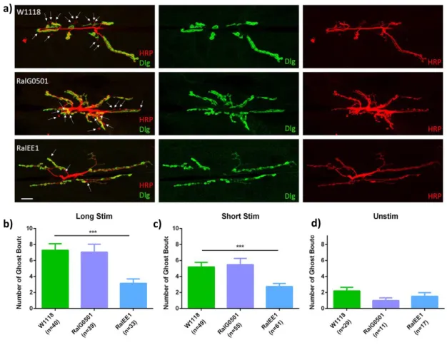
Figure 3.1. Analysis of activity-dependent bouton formation in ral mutants………30
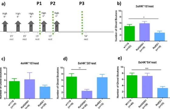
Figure 3.2. Analysis of time-lapse stimulation protocols in ral mutants………....31
Figure 3.3. Analysis of the relative frequency distribution of activity-dependent boutons……….32
Figure 3.4. Ral expression levels in ral mutants………...33
Figure 3.5. Analysis of the presence of futsch in terminal boutons………34
Figure 3.6. ral mutants have thicker nerve bundles……….36
Figure 3.7. ral mutants have defects in neuronal fasciculation in the nerve bundle………...37
Figure 3.8. ral mutants have decreased levels of cell adhesion molecule FasII in the nerve bundle………...38
Figure 3.9. ral mutants have the same levels of activated JNK (p-JNK) as the control………...40
Figure 3.10. The neuronal expression of bskDN in ral mutants does not rescue nerve thickness nor the nerve area………...41
Figure 3.11. It is possible that JNK is not required in neurons to regulate Ral-dependent nerve thickness………..43
Figure 3.12. Schematic representation of activated JNK and JNK dominant-negative protein………...44
xvi
Figure 3.14. The glial expression of bskDN in ral mutants does not rescue nerve thickness nor the area………46 Figure 3.15. It is possible that JNK is not required in glial cells to regulate Ral-dependent nervexvii
Index of tables
Table 1.1. Components of JNK signaling pathway and its mammalian homologs……….13
Table 2.1. Detailed list of Drosophila stocks used throughout this project………..20
Table 2.2. Composition of the solutions used for dissection and stimulation assays………..21
Table 2.3. Primary antibodies used in immunofluorescence assays………..22
xix
Abbreviations
μm – Micrometer
AMPA – α-amino-3-hydroxy-5-methyl-4-isoxazolepropionic acid
AP-1 – Activator protein-1
AZ – Active Zones
ATP – Adenosine Triphosphate BBB – Blood-Brain Barrier
BDSC – Bloomington Drosophila Stock Center Bsk – Basket
CAM – Cell Adhesion Molecule CNS – Central Nervous System
DABCO – 1,4- Diazabicyclo[2.2.2]octane
Dlg – Discs large
DN – Dominant Negative
ERK – Extracellular Signal Regulated Kinase FasII – Fasciclin II
FOXO – Forkhead Box O GAP – GTPase activating protein GDI – GDP dissociation inhibitor
GDP – Guanosine Diphosphate
GEF – Guanine exchange factor
Gsk3β – Glicogen synthase kinase 3β GTP – Guanosine Triphosphate Hep – Hemipterous
HL3.1 – Hemolymph-Like solution 3.1 HRP – Horseradish Peroxidase
JIP1 – c-Jun-amino-Terminal-interacting Protein 1 JNK – c-Jun NH2-terminal kinase
Jra – Jun-Related Antigen
Kay – Kaya
MAP1B – Microtubule-Associated Protein 1 MAPKs – Mitogen Activated Protein Kinase Mkk4 – MAP Kinase Kinase 4
Msn – Misshapen
NGS – Normal Goat Serum NMJ – Neuromuscular Junction
p-JNK – Phosphorylated JNK PBS – Phosphate-buffered saline
PBST – Phosphate-buffered saline with Triton-X PFA – Parafolmaldehyde
xx
PG – Perineural Glia
PNS – Peripheral Nervous System PSD-95 – Postsynaptic density-95
Puc – Puckered
RalBP1 – Ral Binding Protein1 ROS – Reactive Oxygen Species RT – Room Temperature
SAPs – Synapse-associated proteins
SCG10 – Superior Cervical Ganglion 10 Protein SNARE – Soluble NSF-attachment protein
SPAKs – Stress-Activated Protein Kinases
SPG – Subperineural Glia
UAS – Upstream Activating Sequences WG – Wrapping Glia
1
3
The development and function of the nervous system depend on the proper establishment of neuronal connections. Neurons are highly polarized cells whose morphology determines many aspects of function of the nervous system (Chung & Barres 2009; Poulain & Sobel 2010). In response to changes in the environment and in synaptic activity, neurons can alter both pre- and postsynaptic elements of the synapse (Pfenninger 2009). Defects in synaptic morphology and activity-dependent plasticity are a hallmark of several neurodevelopmental and neurodegenerative disorders. Thus, it is critical to understand the mechanisms that are regulating the shape of neurons and how they change in response to environmental perturbations. It is known that Ral GTPase is involved in postsynaptic plasticity at theDrosophila melanogaster (from now on called Drosophila) neuromuscular junction (NMJ), regulating the
subsynaptic reticulum (SSR) growth in an activity-dependent manner (Teodoro et al. 2013). However, little is known about the involvement of Ral GTPase in the regulation of presynaptic structural plasticity. Therefore, we want to study whether Ral also plays a role in the pre-synaptic side, contributing to the formation of activity-dependent synaptic boutons. Besides playing a role in structural plasticity, we found that Drosophila Ral GTPase regulates nerve thickness at the neuromuscular junction but the mechanisms and the pathways involved in this process are not understood. Defects in axon bundling can lead to serious problems in the transduction of information between neurons (Banerjee & Bhat 2008). Our objective is to uncover if and how Ral GTPase is involved in presynaptic structural plasticity and the mechanism behind the regulation of nerve thickness, using the Drosophila NMJ as a model synapse.
1.1.
Drosophila melanogaster as a model system
Drosophila melanogaster, also known as the fruit fly, started to be used as model organism in the
early 1900s by Thomas Morgan and his co-workers. It is one of the most studied organisms in biological research, with research in Drosophila having contributed to significant discoveries in biological processes, including development (Lawrence 1992), signaling (Cadigan & Peifer 2009), cell cycle (Lee & Orr-Weaver 2003), nervous system development, function and behavior (Bellen et al. 2010; Weiner 1999), which altogether have contributed to the understanding of several developmental and neurological disorders (Bier 2005; Markow 2015). Drosophila is a good model system for studying various aspects of cellular biology, mainly because many of the genetic pathways that are associated with basic developmental processes are conserved during evolution, showing several similarities with higher eukaryotes. Furthermore, the fruit fly genome has approximately 75% of homology with known genes associated to human diseases, making it a powerful organism for the study of human genetics (Reiter et al. 2001; Bier 2005; Adams et al. 2000). Drosophila is an inexpensive animal model to maintain, has a short life cycle and is easy to manipulate, enabling many experiments in a short period of time, thus promoting the rapid advancement of research. In addition, it has a simple and accessible anatomy, a vast and powerful set of genetic tools, and is accessible to various experimental techniques (Bier 2005; Collins & DiAntonio 2007).
4
1.1.1.
Drosophila life cycle
A major advantage of working with Drosophila is its short life cycle lasting approximately 10 days at 25°C, and this generation time doubles when kept at 18ºC. Drosophila life cycle consists of four stages: embryo, larva, pupa and adult fly (Figure 1.1). When fertilized, females can store the sperm for the fertilization of several eggs to be laid over the next few days. At 25°C, the embryo development in the egg occurs in approximately 21 hours, after which it hatches as a larva (Prokop 2013; Weigmann 2003).
The larval stage is composed of three distinct phases of development called instars. In this stage, the larva eats and grows from 1st to 3rd instar entering then in pupariation, a stage where the larva becomes an immotile pupa and metamorphosis occurs. During the pupal stage, allorgans of the larva are degenerated and then restructured into adult structures. Adult flies emerge from pupal cases about 10 days after egg laying (at 250C). (Prokop 2013; Weigmann 2003).
1.1.2.
Drosophila Neuromuscular Junction
Drosophila NMJ is a well characterized model system for the study of neuronal development,
plasticity and function due to its stereotypical structure from animal to animal, for being one of the best studied synapses, and of course, given the availability of a wide variety of molecular, genetic and experimental techniques. The fruit fly larval NJM is a well-characterized and simple system, constituted by 32 motor neurons and 30 identified muscle cells in each hemisegment that are repeated and bilaterally symmetric (Menon et al. 2013; Collins & DiAntonio 2007). During development, each motor neuron innervates a specific muscle cell leading to synapses with stereotyped arborization and with a
Figure 1.1. Schematics of the Drosophila melanogaster life cycle. At 25°C Drosophila melanogaster life cycle lasts 9-10 days. After 1 day of embryonic development, the hatched larvae (1st instar) spend 1 day until reaching the 2nd instar. 1 day later, the larvae proceed to the 3rd larval stage (3rd instar) which lasts for 2-3 days. In the following 5 days, the pupal stages takes place, where the organs degenerate and restructure into their adult shapes (metamorphosis). 10 days after egg laying an adult fly emerges from the pupal case. (From Weigmann et al. 2003; Prokop 2013).
5
relatively constant and quantifiable number of synaptic boutons, but that are different for each muscle cell. Synaptic boutons are composed by many active zones that represent the presynaptic releasing sites, where synaptic vesicles are clustered. Boutons are surrounded by a postsynaptic muscle membrane containing clusters of glutamate receptors, which translate presynaptic activity in postsynaptic depolarization, leading to activation of postsynaptic signaling cascades (Collins & DiAntonio 2007). Given that these NMJs are highly stereotyped, it makes it possible to compare the same NMJ from larva to larva. Also, it is accessible to various experimental techniques such as electrophysiology, calcium imaging, immunocytochemistry, electron microscopy and live imaging, providing a great advantage for structure and molecular anatomy studies of the synapses. Besides its stereotyped circuitry, Drosophila NMJ also shows robust plasticity, adapting structurally and functionally in response to changes in the environment, neuronal activity and gene function (Menon et al. 2013; Collins & DiAntonio 2007; Featherstone & Broadie 2000). The Drosophila NMJ synapses use glutamate as neurotransmitter, which closely resembles the vertebrate central nervous system (CNS) synapses. In Drosophila, larval NMJ synapses express ionotropic glutamate receptors that are similar to AMPA-Type glutamate receptors in the mammalian brain. Also, the postsynaptic scaffold protein, Discs large (Dlg) is identical to those found in mammalian postsynaptic densities, belonging to the family of PSD-95 and SAPs. Altogether, given the cellular and molecular similarities, Drosophila NMJ synapses are an excellent model to study excitatory glutamatergic synapses (Menon et al. 2013).1.2.
Neuronal Growth and Development
Nervous system function depends on proper establishment of complex neuronal networks determined during development (Chung & Barres 2009; Poulain & Sobel 2010). Neurons are the fundamental unit of function of the nervous system and are highly polarized cells composed by a soma, dendrites, axons and axon terminals containing synapses. During morphogenesis, neurons start to extend their axons and dendrites that are structurally and functionally different. The axon is usually a thin and long process that conducts nerve impulses through long distances, delivering specific signals to multiple cells, while dendrites are characterized by its branched projections that are important to receive and integrate synaptic inputs (Chung & Barres 2009; Poulain & Sobel 2010; Polleux & Snider 2010). When exiting the central nervous system (CNS), motor axons from several neurons are bundled together forming long nerve fibers that will establish a connection with a target cell (Araújo & Tear 2003). After reaching its target, the growth cone (the specialized structure at the tips of extending axons) contacts with dendrites of other neuron or with another cell type, like a muscle cell (neuromuscular junction) and starts to differentiate the presynaptic terminal (Chung & Barres 2009). Synapses are essential for the proper communication between neurons and other cells. Regulated formation of the synapses requires bidirectional signals between pre- and post-synaptic cells which results in the development of specialized structures important for neurotransmitter release and detection (Südhof 2012; Collins & DiAntonio 2007). After synapse formation, the continuous growth leads to addition of new synaptic branches and of synaptic boutons, which are round varicosities where synapses are located. Synaptic boutons are composed of active zones (AZ) containing neurotransmitter-filled vesicles
6
and the necessary machinery for their release (Südhof 2012;Harris & Littleton 2015). Opposed to that, are present other neuron or specialized cells containing postsynaptic structures with specific receptors for the neurotransmitter released by the presynaptic cell, like the muscles in case of NMJs. When an action potential reaches the presynaptic terminal, voltage-gated Ca2+ channels are opened which allows for Ca2+ ions to bind to synaptotagmins present in the synaptic vesicles, triggering neurotransmitter release to the synaptic cleft, activating the postsynaptic receptors and downstream postsynaptic cascades (Shen & Cowan 2010; Südhof 2012). Proper regulation of these various steps is necessary to avoid neuronal related disorders.1.3.
Regulation of Synaptic Growth and Plasticity
During development, neurons undergo significant remodeling processes in order to generate appropriate axonal and synaptic connections. Excitatory synapses in the vertebrate nervous system use glutamate as their primary neurotransmitter. This type of synapses shows robust plasticity, a process characterized by modifications in terminal connections in response to neuronal activity (Menon et al 2013; Harris & Littleton 2015). There are two main types of plasticity, structural and functional plasticity that are thought to be involved in learning and memory. Usually, functional plasticity is related to changes in the strength of synaptic transmission while structural plasticity is associated with changes in synaptic morphology, like alterations in the number, size and shape of synaptic elements (Shen & Cowan 2010; Griffith & Budnik 2006). In response to changes in the environment, neurons can alter both pre- and postsynaptic structures of the synapse, requiring regulated membrane trafficking and exocytosis for membrane addition (Pfenninger 2009). During the process of synaptic remodeling, coordinated dynamics and organization of actin and microtubule cytoskeleton are essential for controlling shape changes in the synapse. They are key players in the support of active transport of membranes, organelles and macromolecules required for development (Poulain & Sobel 2010; Menon et al. 2013). Regulation of these critical processes has been associated with the ability of neurons to strengthen synapses and is essential to prevent defects in neuronal structure and function. Drosophila has been an excellent model for studies of the synapse, given that the its NMJ is glutamatergic resembling the vertebrate central nervous system, and because NMJs are organized into branched arbors that are composed of chains of synaptic boutons with stereotyped morphology that is genetically determined but where its synaptic structure and function can be modified by extrinsic factors, such as the environment, or changes in neuronal activity (Menon et al. 2013; Collins & DiAntonio 2007). Like in vertebrates, Drosophila larvae synapses of the NMJ are composed by a presynaptic terminal containing active zones with pools of vesicles filled with neurotransmitters, opposed to clusters of neurotransmitters receptors in the membrane of the postsynaptic cell. The postsynaptic membrane, called subsynaptic reticulum (SSR) is formed by numerous folds and invaginations of the membrane that grows in an activity-dependent manner (Teodoro et al. 2013). During larval development, muscles and synaptic boutons grow from 1st to 3rd instar increasing the muscle area about 100 times and the number of synaptic boutons in 10 times. The addition of new boutons can occur throught different mechanisms
7
including, asymmetric budding of preexisting bouton, symmetric division of a bouton and de novo formation of a bouton from the axonal membrane (Zito et al. 1999). (Figure 1.2.)In addition to neurons, glial cells have varied functions that are important for proper development and function of both vertebrates and invertebrates nervous systems.
1.4.
Glial Cells
Glial cells are part of the CNS and peripheral nervous system (PNS) in most animals. They play important roles in the regulation of neuronal development and function (Stork et al. 2012; Brink et al. 2012). Glia are involved in many processes in the development of the nervous system, like modulation of neural stem cell proliferation (Ebens et al. 1993), regulation of the differentiation of neural precursors guiding axon pathfinding (Hidalgo & Booth 2000; Sepp et al. 2001), ensheathing axon fascicles (nerves) and individual axons supplying trophic support for neurons (Barres 2008; Booth et al. 2000), function as primary immune cells by engulfing neurons and debris that are eliminated during development (Watts et al. 2004), and promoting synapse formation and maturation (Barres 2008).
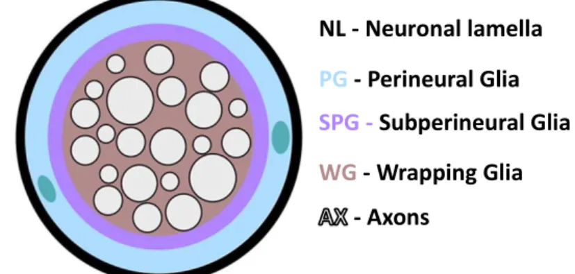
The development of the nervous system in vertebrates and flies has several similarities. Due to its simplicity and the availability of robust molecular genetic tools for developmental studies, Drosophila proved to be an ideal model organism to study glia development (Freeman 2015; Parker & Auld 2004). There are different subtypes of glia in both central and peripheral nervous systems of Drosophila. The outer layer of cells is composed by perineural glia (PG), which are thought to be responsible for secreting a dense lamella that functions as a physical and chemical barrier covering the CNS and the peripheral nerves. This layer is discontinuous, however, below it there is another layer of a different subtype of glia, the subperineural glial cells (SPGs). These flattened cells cover the entire surface of the CNS that only contacts with the most superficial layer of neuronal cell bodies in the cortex and form septate junctions between them creating a blood-brain barrier (BBB). Closely associated with neurons, there are more specialized subtypes of glia as cortex glia, ensheathing glia, and astrocytes. In Drosophila, the ensheathment, support, modulation of the function and development of peripheral sensory neurons,
Figure 1.2. Schematic representation of the development of the Neuromuscular Junction (NMJ) in muscles 6 and 7. Drosophila larva grows from 1st (left) to 3rd instar (right), increasing the muscle size which is accompanied by the addition of new branches and synaptic boutons. (Adapted from Menon et al. 2013).
8
motor neuron axons and terminals are carried out by various glial subtypes. Like vertebrates, Drosophila peripheral nerves are covered by perineural glia and subperineural glia creating a BBB similar to the CNS, but also present other types of glia such as wrapping glia that ensheath motor and sensory axons (Figure 1.3.) (Freeman 2015; Limmer at al. 2014). Proper ensheathment of axons in Drosophila separate the axons from the hemolymph. This partitioning is very important because while vertebrates present a highly vascularized nervous system, Drosophila nervous system floats in the hemolymph that contains high concentration of potassium and other ions that could interfere with action potential propagation (Blauth et al. 2010; Banerjee et al. 2006; Banerjee & Bhat 2008). Also, defects in nerves and axon fasciculation and ensheathment could lead to diminished conduction of electrical impulses, affecting the transmission of information and thus, neuronal development (Banerjee & Bhat 2008).In the NMJ, subperineural glia interact with motor neuron synaptic contacts on muscles where they play an essential role in neurotransmitter recycling, in the modulation presynaptic growth by engulfing synaptic debris during development, and secreting molecules that modulate retrograde signaling between the muscle and the pre-synapse, contributing for NMJ growth (Freeman 2015; Ou et al. 2014). Glial cells have been raising an increased interest in recent years, since disruption of these cells can have many implications in the function of the nervous system, whose disruption may lead to the development of neurodegenerative diseases such as Alzheimer’s disease, amyotrophic lateral sclerosis and multiple sclerosis (Kurosinski and Gotz 2002; Barres 2008; Banerjee & Bhat 2008; Blauth et al. 2010; Parker & Auld 2004; Danjo et al. 2011).
NL - Neuronal lamella
PG
- Perineural Glia
SPG -
Subperineural Glia
WG
-
Wrapping
Glia
-
Axons
Figure 1.3. Schematic representation of the glial cell types that are part of Drosophila peripheral nerve. Covered by the neuronal lamella (NL) it is represented the outermost layer of glial cells which is composed by perineural glia (PG). Immediately below, there is another layer of glial cells, called subperineural glial (SPG) that is responsible for creating the blood brain barrier. Closely associated with the axons (AX) there is a layer composed of wrapping glia that enwrap the axons (WG). In green are represented two PG nuclei (From Xie & Auld 2011).
9
1.5.
Molecular Players involved in Neuronal Growth and Plasticity
During development and in response to changes in the environment, neurons undergo significant remodeling processes in order to generate appropriate axonal and synaptic connections. Many genetic pathways contribute to this process: below we summarize the ones relevant for this project.
Ral GTPase regulates numerous biological processes by interacting with effectors such as Ral Binding Protein-1 (RalBP1) and the exocyst complex. Also, it has been shown that Ral influences several pathways including the c-Jun NH2-terminal kinase (JNK) signaling pathway. Together, Ral and the JNK pathway seem to be involved in responses to stress and cell shape changes. Besides this, JNK has been shown to be involved in axonal pruning by regulating the levels of the cell adhesion molecule Fasciclin II (Bornstein et al 2015), and in synaptic plasticity (Collins et al. 2006; Coffey 2014). Moreover, Ral GTPase has been shown to be involved in postsynaptic structural plasticity (Teodoro et al. 2013) and also, appears to have a role in nerve thickness regulation (unpublished results and this thesis).
Because Ral GTPase and JNK were shown to act together in the regulation of some remodeling processes, we want to understand if these two molecular players play a role in the regulation of presynaptic structural plasticity and in the regulation of nerve thickness.
1.5.1.
Ral GTPase
Ral is a small GTPase member of the Ras superfamily, and like other small GTPases plays an important role in several biological processes, such as the regulation of vesicle and membrane transport (Figure 1.4). This protein is ubiquitously expressed in tissues, but is specially enriched in places like the brain and platelets (van Dam & Robinson 2006). It was shown that Ral is located in cellular compartments such as the plasma membrane, secretory granules and synaptic vesicles (Moskalenko et al. 2002; Sugihara et al. 2002; van Dam & Robinson 2006; Shirakawa & Horiuchi 2015). The cellular localization and activity of Ral can be regulated by post-translational events since it has distinct phosphorylation sites (Shirakawa & Horiuchi 2015; Gentry et al. 2014). In mammals, Ral GTPase has two isoforms, RalA and RalB, which share 82% of homology between them, however, in invertebrates such as Drosophila melanogaster and the nematode Caenorhabditis elegans, only one Ral gene is present, which is more similar to the RalA isoform. Ral gene orthologues are not present in the yeast genome, indicating that Ral appeared in multicellular organisms throughout evolution (Shirakawa & Horiuchi 2015). Similarly to other small GTPases, Ral also functions as a molecular switch, meaning that they have two inter-convertible forms, an active form (GTP-bound) and an inactive form (GDP-bound), cycling between them. In its active form Ral GTPase interacts with several effector proteins, triggering downstream pathways involved in actin cytoskeletal rearrangement, membrane trafficking, gene transcription, kinase cascade activation, cell survival, apoptosis and other biological processes (Shirakawa & Horiuchi 2015; Gentry et al. 2014; Sugihara et al. 2002; Carmena 2012). The cycling rate between the active and the inactive forms of Ral is very slow and weak, like in other small GTPases,
10
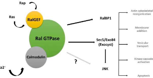
and can be significantly enhanced by guanine exchange factors (GEFs) and GTPase activating proteins (GAPs), respectively. Thus, RalGEFs stimulate GTPase activity by releasing GDP and promoting GTP binding (activating Ral), whereas GAPs allow the hydrolysis of the bound GTP (inactivating Ral). Besides cycling between GTP and GDP form, GTPases can also change its cellular localization, from cytoplasm to membrane or the other way around, aided by GDP-dissociation inhibitors (GDIs) and GDI-dissociation factors (GDFs). Once at the membrane and in its active form (GTP-bound), GTPases can interact with their specific effectors (van Dam & Robinson 2006; Shirakawa & Horiuchi 2015; Gentry et al. 2014; Segev 2011). In Drosophila, Ral GTPases have specific GEFs and GAPs, that are downstream of Ras proteins and are indirectly activated by them. Rap1 and Ras lead to the activation of specific RalGEFs that subsequently activate fruit fly Ral proteins. In addition, Ral has a calmodulin (CaM) binding site, so it can be directly activated by Ca2+/Calmodulim binding, in a Ras/GEF-independent manner. CaM is a conserved sensor of Ca2+-dependent signaling pathways involved in the regulation of numerous biological processes. The presence of high intracellular calcium levels induces conformational changes in calmodulin proteins, allowing them to interact with the target proteins, regulating their functions. Ral GTPase has both RalGEFs and calmodulin binding sites. The activation of Ral by these two proteins leads to an interaction with various effectors and, therefore the initiation of downstream signaling events (Figure 1.4.) (van Dam & Robinson 2006; Wang & Roufogalis 1999; Feig 2003; Park 2001).Figure 1.4. Activated Ral protein interacts with several effectors implicated in numerous cellular processes. Ral can be activated by RalGEFs (represented in yellow) which in turn are activated by Ras and Rap1, and by calmodulin binding (represented in grey) in response to calcium influx. Upon activation, Ral binds to several effectors such as RalBP1, exocyst subunits Sec5/Exo84 and JNK which are involved in multiple cell processes, such as membrane addition, actin cytoskeleton rearrangements, membrane trafficking and many other cellular responses (Shirakawa & Horiuchi 2015; Gentry et al. 2014; Sugihara et al. 2002; Carmena 2012).
11
Among all Ral effectors, there are two that are particularly well known: Ral Binding Protein-1 (RalBP1) and the Sec5 and Exo84 subunits of the octameric Exocyst complex. RalBP1 was one of the first to be identified as a Ral effector (Figure 1.4). It contains a GAP catalytic domain that activates the Cdc42 and Rac small GTPases, inducing actin cytoskeleton rearrangements like filopodia and lamellipodia formation, respectively. RalBP1 is also involved in receptor-mediated endocytosis (i. e. EGF receptor, insulin receptor) through interaction with two endocytic proteins, Reps1 and Reps2/POB1 (Shirakawa & Horiuchi 2015; van Dam & Robinson 2006; Gentry et al. 2014).Another well characterized Ral GTPase effector is the exocyst (Figure 1.4.). The exocyst is an octameric complex, which is composed of eight subunits, Sec3, Sec5, Sec6, Sec8, Sec10, Sec15, Exo70 and Exo84, firstly identified in yeast and conserved to humans. Ral can bind directly to Sec5 and Exo84 subunits promoting the assembly of the complex, however these subunits have a competitive behavior due to the overlapping of their Ral binding sites. Together, Ral and the exocyst, are involved in intracellular trafficking and in regulation of several exocytic pathways, by tethering exocytic vesicles (i. e. Golgi-derived vesicles) to specific sites in the plasma membrane, before the assembly of the SNARE complex (van Dam & Robinson 2006) (Figure 1.5.).
In absence of Ral, the assembly and stability of exocyst complex is reduced, affecting the capacity to activate many biological cascades and therefore the regulation of diverse cellular processes. In mammals, the disengagement of the exocyst, after Ral activation, is achieved through Sec5 phosphorylation at the Ral-binding site (process catalyzed by protein kinase C (PKC)), resulting in the dissociation of the exocyst subunit from active Ral. Following dissociation, Sec5 is dephosphorylated, being available for the transport of other vesicles (Chen et al 2011). Whether this inactivation mechanism is conserved in invertebrates is currently unknown. The distribution of the exocyst within a cell can be highly dynamic, however it is located in limited regions of the plasma membrane, resulting in polarized growth and secretion of essential proteins into extracellular space. The exocyst complexis also present
Figure 1.5. Schematic representation of the exocyst complex. a) Illustration of the structure of the assembled yeast exocyst complex, in “Y” conformation, composed by eight subunits (Adapted from Munson & Novick 2006) b) Schematic representation of the exocyst complex attached to a secretory vesicle. Activated Ral can bind directly to its exocyst effectors, Sec5 and Exo84; Exocyst complex binds to vesicles that have specific identity due to the presence of Rab proteins (Adapted from Liu & Guo 2012).
12
in the nervous system, at the ends of neuronal growth cones, axons, during neurite branching and in sites of synaptogenesis, promoting membrane addition (Heider & Munson 2012; Liu & Guo 2012). In fact, together Ral and the exocyst can modulate postsynaptic growth in Drosophila NMJ in an activity-dependent manner. In a previous study, Rita Teodoro and her collaborators demonstrated that, in response to activity, activated Ral in the muscle induces recruitment of Sec5 to the NMJ, which trough membrane addition promotes SSR growth (Teodoro et al. 2013). So, it is possible that the exocyst is involved in synaptic plasticity through its involvement in the regulation of tethering, docking and fusion of vesicles to specific places in plasma membrane (van Dam & Robinson 2006; Teodoro et al. 2013).Besides, RalBP1 and the exocyst, Ral also influences other pathways, like JNK, that belongs to the mitogen activated protein kinase (MAPKs) superfamily (Gentry et al. 2014) (Figure 1.4). JNK is activated in response to different extracellular stimuli, including growth factors, cytokines, and cellular stress inducers, inducing the phosphorylation of the transcription factor c-Jun (Kim & Choi 2010).
Ral can be a positive or negative regulator of the JNK signaling pathway, depending on the extracellular stimuli (Massaro et al. 2009). In mammals, Ral has been shown to activate JNK (van den Berg et al. 2013; Essers et al. 2004), or to be a negative regulator of JNK (Balakireva et al., 2006; Beraud et al. 2014), while in Drosophila, Ral activity appears to antagonize the JNK pathway (Swamoto et al. 1999; Balakireva et al. 2006). Massaro et al. (2009) have shown that JNK signaling can be activated or inhibited by the same disruption depending on the origin of the stress, and this may explain these seemingly antagonistic results. Together, Ral and JNK pathway have been involved in responding to ROS-signaling, cell shape changes and apoptosis. Because Ral and JNK signaling could be involved in human diseases, such as cancer, it is critical to understand how they interact with each other in response to different stimuli (Shirakawa & Horiuchi 2015).
1.5.2.
JNK signaling
JNK is a member of a large evolutionarily conserved MAPK family, which also integrates the extracellular signal regulated kinase (ERK) and p38 subfamilies. MAPKs are serine-threonine kinases that allow the amplification and integration of signals from a wide range of stimuli, regulating genomic and physiological responses due to changes in the environment (Weston & Davis 2002; Kim & Choi 2010). MAPKs cascades are composed by at least three components, including a MAPK kinase kinase (MAP3K), which phosphorylates and activates a MAPK kinase (MAP2K), and a MAPK that is phosphorylated and activated by MAP2K. The activation of this family of proteins is known to phosphorylate various substrate proteins including transcription factors, that are involved in the regulation of several cellular activities such as cell proliferation, differentiation, inflammatory responses, morphogenesis and apoptosis (Kim & Choi 2010; Weston & Davis 2002; Stronach 2005).
MAPKs can be activated either by interactions between kinase components, or through the aid of a scaffold protein which induces the formation of a signaling complex composed of specific kinases. The scaffold proteins facilitate the activation of MAPK signaling pathways, like the kinase suppressor of
Ras-13
1 (KSR) and MEK partner 1 (MP1) for the ERK pathway, JNK-interacting proteins (JIPs) are scaffold proteins for JNK signaling and β-Arrestin 2 serves as scaffold protein for ERK and JNK signaling pathways (Kim & Choi 2010; Weston & Davis 2002).JNKs, also known as stress-activated protein kinases (SPAKs), are activated by inflammatory cytokines such as tumor necrosis factor (TNF-α) and interleukins, or in response to cellular stress due to environmental insults (UV irradiation, oxidative stress) (Kim & Choi 2010). This subfamily of the MAPK signaling cascade transfers phosphate groups to serine/threonine residues that are flanked by carboxyl-terminal prolines (Ser/Thr-Pro) (Coffey 2014; Wang et. al 2005; Kim & Choi 2010).
In the nervous system, JNK signaling also contributes for brain morphogenesis, axon polarization, extension, synaptic plasticity, dendrite development and memory formation (Rallis et. al 2010; Coffey 2014; Weston & Davis 2002). The duration and intensity of JNK signaling, the type of stimuli, the use of different scaffolding proteins to connect certain components can induce distinct outputs. Also, the use of different upstream kinases that recognize unique signals in a specific cell type, developmental stage or spatial position contributes for the diversity of cellular responses by the JNK pathway (Stronach 2005). In Drosophila, JNK pathway is composed of one JNK, Basket (Bsk), that is regulated by two JNK kinases (JNKK) Hemipterous (Hep) and MAP kinase kinase 4 (Mkk4), which in turn are controlled by six JNKK kinases (JNKKKs). Also, JNKKKs are regulated by a single upstream JNKKKK, Misshapen (Msn) (Stronach 2005; Rallis et al. 2010). In Drosophila Bsk activation is accomplished by phosphorylation on two predicted residues, threonine 181 and tyrosine 183, through the JNKKs, Hep and MKK4 (Rallis et al. 2010). In table 1.1. are summarized the JNK cascade components in Drosophila and their homologs in mammalians.
Activated Bsk phosphorylates the Activator protein-1 (AP-1) complex, composed of the transcription factors kayak (kay) the homolog of mammalian c-Fos and Jra (Jun-related antigen) the mammalian homolog of c-Jun. In flies, these transcription factors act either as heterodimers or Fos homodimers (Rallis et al. 2010; Stronack 2005). The strength and duration of JNK signaling is regulated
Table 1.1. Components of JNK signaling pathway and its mammalian homologs. (Adapted from Stronach 2005)
14
by one of its targets genes, puckered (puc) which encodes a JNK-specific MAPK phosphatase, inducing a negative feedback loop (Wang 2005).In Drosophila JNK/Bsk is involved in a plethora of cellular processes, including dorsal closure (Zeitlinger et al., 1997), imaginal disc development (Agnes et al., 1999), wound healing (Ramet et al., 2002), apoptotic regulation (Ryoo et al., 2004), innate immunity (Delaney et al., 2006), can prolong life span and protect against oxidative stress (Wang et al., 2003). Also, in the nervous system, JNK/Bsk contributes to axon degeneration or overextension depending on the level of its inactivation at
Drosophila NMJ (Rallis et al. 2010), regulates synaptic plasticity, growth (Collins et al., 2006; Sanyal et
al., 2002) and axonal transport (Horiuchi et al. 2007). In most of these responses Bsk phosphorylates the AP1 complex (Rallis et al. 2010; Ciapponi et al., 2001). More recently, a new role for Bsk has been suggested: Bsk has been shown to be required for axon pruning, through the reduction of membrane levels of the adhesion molecule Fasciclin II (Bornstein et al 2015).
Another interesting work revealed that Ral GTPase regulates developmental cell shape changes, by acting as a negative regulator of JNK in Drosophila. In this study, it was reported that the loss of bristles and hairs caused by dominant-negative form of Ral was genetically suppressed by loss of function of Hep and Bsk. Also, a constitutively active form of Ral caused defects in the process of dorsal closure during embryogenesis and inhibited the JNK phosphorylation in S2 cells (Swamoto et al. 1999). In other independent study, it was suggested that Ral activity suppresses the JNK activation and induces p38 mitogen-activated protein (MAP) kinase activation. They also proposed that a molecular basis of Ral action on JNK could be mediated by the exocyst complex, influencing developmental regulatory programs (Balakireva et al. 2006). In mammals, it has been shown that Ral is activated in response to cellular stress such as reactive oxygen species (ROS) which in complex with the kinase scaffold protein JIP1 (c-Jun-amino-terminal-interacting protein 1) activates the JNK cascade resulting in FOXO (forkhead box O) activation and its nuclear translocation (van den Berg et al. 2013; Essers et al. 2004). FOXOs are transcription factors that are involved in various cellular processes including cell cycle regulation and apoptosis. Such as Ral, JNK interacts with a huge variety of signaling pathways, and its regulation could differ according to the type of stimuli, development stage, cell-type and even with the components of the cascade, generating a wide range of outputs (Figure 1.6.). Thus, it is important to decipher what is the molecular mechanism of action between Ral and JNK in the different cellular contexts to prevent pathological conditions.
15
JNK also plays a role in the regulation of microtubule cytoskeleton which is a major component of neurons and is essential for numerous cellular and developmental processes, such as neuronal migration, polarity, and differentiation (Kapitein & Hoogenraad 2015). In mammals, JNK appears to stabilize microtubules by phosphorylation of substrates such as SCG10 (superior cervical ganglion 10 protein) and MAP1B (Microtubule-associated protein 1) (Chang et al. 2003; Tararuk et. al 2006; Kawauchi et al. 2003). Another study revealed that microtubule stability requires concomitant inhibition of GSK3β and activation of JNK (Ciani & Salinas 2007).In Drosophila, JNK activity has been associated with regulation of FOXO through Toll-6 receptor, and this pathway has been associated to microtubule dynamics (McLaughlin et al. 2016). In addition, FOXO has been shown to negatively regulate microtubule stability, by analyzing Futsch distribution in larvae NMJ (Nechipurenko & Broihier, 2012). Futsch is the Drosophila homolog of MAP1B that binds to tubulin, making it an excellent marker for stable and dynamic microtubules (Roos et al. 2000; Hummel et al. 2000). Together, these studies demonstrate that JNK can regulate microtubules through the interaction with different substrates and signaling pathways.
a)
b)
c)
d)
e)
f)
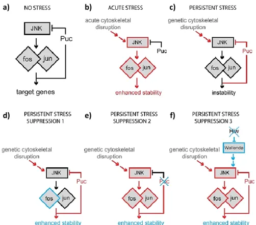
Figure 1.6. Schematic representation of different JNK outputs in response to different types of stimuli. a) In the absence of cellular stress the JNK-Fos pathway is not required for synapse stability, being
present in a stable state. b) Upon acute cellular stress, such as cytoskeletal disruption, JNK and Fos are activated (represented in red), inducing synapse stabilization. c) During persistent cellular stress, the phosphatase Puckered induces a negative feedback preventing Fos reactivation, which favors synapse instability or disassembly. d) Fos overexpression (experimental manipulation) leads to synaptic stability in the presence of persistent cellular stress.
e) Also during persistent cellular stress, loss of puckered (experimental manipulation), induces synaptic stability,
possibly due to enhanced Fos activity. f) The loss of Highwire (experimental manipulation), induces upstream MAPK signaling, leading to synaptic stability through Fos activity. (from Massaro et al. 2009).
16
As mentioned above, JNK signaling pathway is involved in many biological processes, and its deregulation has been implicated in the pathogenesis of many human diseases such as cancer, Alzheimer's disease (AD), Parkinson’s disease (PD) and other neurodegenerative diseases. So, the understanding of how JNK regulates these numerous activities, is a critical step towards the development of new therapies for these diseases (Kim & Choi 2010).1.6.
Aims of the work
From previous studies, Teodoro et al. (2013) found a novel pathway that regulates neuronal morphology in response to activity through the engagement of Ral and the Exocyst complex, regulating postsynaptic membrane growth at the synapse in response to neuronal activity. Here, we want to ask whether Ral also participates in presynaptic structural plasticity. For this, we will manipulate the levels of synaptic activity by inducing acute structural plasticity and testing whether activity-dependent bouton formation remains intact.
In parallel, we observed that Ral mutants have widespread thicker nerves, suggesting a role for this GTPase in the regulation of nerve bundle structure and possibly function. We will dissect the mechanisms and pathways involved in the regulation of nerve thickness, and how Ral GTPase regulates this trait. Because Ral has been shown to regulate JNK signaling, positively or negatively, depending on the cellular context of activation of this cascade, we will test if this pathway is involved in the regulation of nerve thickness.
Like neurons, glial cells are essential for proper development of the nervous system, regulating many aspects of neuronal development, morphology and function. When exiting the CNS, axons from several neurons are bundled in fascicles that, together with glial cells, form the nerve bundle. So, we wanted to understand the contribution of these cells and the involvement of Ral and JNK signaling pathway to the regulation on nerve thickness.
With this work, we expect to have a better understanding about the mechanism of synaptic bouton formation and the pathways that might be involved in this process. In addition, we expect to understand the contribution of neurons and glial cells, as well as the mechanisms and pathways involved in the regulation of axon bundling/nerve thickness. Defects in axon bundling can lead to serious problems in the transduction of information between neurons consecutively affecting the development of the nervous system.
17
19
2.1.
Genetic tools
Drosophila melanogaster has the advantage of having a great diversity of genetic tools that
provides the ability to study Drosophila, using various experimental techniques. Here are reviewed some tools that were used in this project.
2.1.1. UAS-Gal4 System
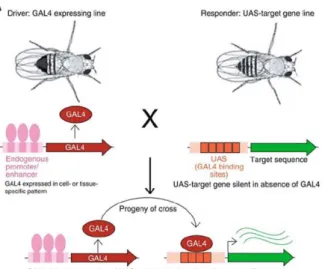
One of the main and most important genetic tools used in Drosophila is the UAS/Gal4 system, which allows the control of gene expression in a temporal and tissue-restricted manner. The transcriptional activation factor Gal4 was firstly identified in Saccharomyces cerevisiae and then used by Andrea Brand and Norbert Perrimon in 1993 to develop a method to induce gene expression regulated by Gal4. This system consists of two components present in separate fly lines, a Gal4 protein that binds directly to an Upstream Activating Sequences (UAS), which are Gal4 binding sites on DNA that are upstream of the gene of interest (Duffy 2002; Brand & Perrimon 1993) (Figure 2.1.). To activate transcription, lines under UAS control are mated with flies containing a Gal4 transgene, which is expressed in a certain pattern, called the driver. Thus, the progeny will express the gene of the interest in a pattern dictated by Gal4 expression, allowing tissue specific expression of the gene. In the absence of a Gal4 line the target gene is silent (Brand & Perrimon 1993).This system can also be regulated by temperature, since the minimal Gal4 activity is at 16°C and the maximal activity is reached at 29°C, although at this point there is a balance between the activity and minimal effects on fertility due to high temperature (Duffy 2002). One of the greatest advantages of this system is the existence of thousands of Gal4 lines available, allowing the expression of specific modified forms of a gene (e.g. dominant negative, constitutively active), inducing targeted mutations and knockdown of specific genes anywhere in the fly, in vivo (Elliot and Brand, 2008; Caygill & Brand 2016).
Figure 2.1. Schematic representation of the UAS/Gal4-based system for transgene expression.
20
2.2.
Fly stocks and husbandry
In this project, all fly stocks were maintained at room temperature (RT) in vials or bottles containing standard fly food (mixture of water, corn meal, agar, sugar, yeast and fungicides). When carrying out experiments, virgin females were collected and crosses were performed, maintaining them at 25°C in an appropriate atmosphere with controlled humidity.
All the Drosophila stocks used in this work were obtained from Bloomington Drosophila Stock Center or generated in our laboratory and are described in Table 2.1.
Table 2.1.Detailed list of Drosophila stocks used throughout this project.
BDSC: Bloomington
Drosophila Stock Center
Name Description/Genotype Stock
W1118
Wild-type W[1118]
BDSC#5905
RalG0501
P-element disruption of ral locus; Genetic null w[67c23] P{w[+mC]=lacW}Rala[G0501]/FM7c
BDSC#12283
RalEE1
Point mutation in amino acid Ser154 predicted to be the nucleotide binding site
Rala[EE1]/FM7i, P{ActGFP}JMR3
BDSC#25095
nSyb-Gal4/TM6b
Expresses Gal4 in all neurons n-Syb-Gal4/TM6b
Rita Teodoro Lab
UAS-BSKDN
Amino acid replacement; Expresses a dominant-negative form of bsk under UAS control
w*; P{UAS-bsk.K53R}20.1a
BDCS#9311
Repo-Gal4
Expresses Gal4 in glia w1118; P{GAL4}repo/TM3, Sb1
BDSC#7415
RalG0501;;nSyb-Gal4
Ral mutant with Gal4 being expressed in all neurons
RalG0501/ FM7i, P{ActGFP}JMR3;;nSyb-Gal4/TM3,Ser, Act-GFP
Rita Teodoro Lab
RalEE1;;nSyb-Gal4
Ral mutant with Gal4 being expressed in all neurons
RalEE1/ FM7i, P{ActGFP}JMR3;;nSyb-Gal4/TM3,Ser, Act-GFP
Rita Teodoro Lab
RalG0501;;Repo-Gal4
Ral mutant with Gal4 being expressed in glia
RalG0501/ FM7i, P{ActGFP}JMR3;;Repo-Gal4/TM3,Ser, Act-GFP
Rita Teodoro Lab
RalEE1;;Repo-Gal4
Ral mutant with Gal4 being expressed in all glial cells
RalEE1/ FM7i, P{ActGFP}JMR3;;Repo-Gal4/TM3,Ser, Act-GFP
21
2.3.
Larval dissection and fixation
Drosophila third instar larvae of the appropriate genotypes were selected and dissected in a drop
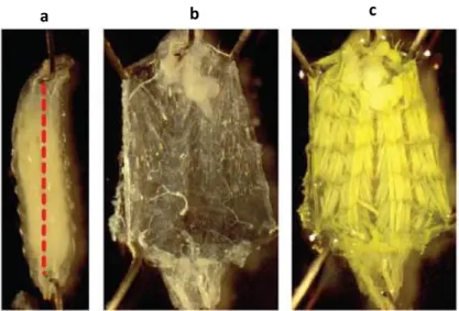
of PBS 1x (Phosphate Buffer Saline) or of HL3.1 (hemolymph-like solution – composition described in table 2.2) using Sylgard plates (Brent et al. 2009). Larvae are placed with the dorsal side up and, using forceps (Student Dumont #5 Forceps - Fine Science Tools), a pin is placed in the anterior end of the larva, near the mouth hooks, followed by another pin inserted in the posterior end of the larva, between the posterior spiracles. Using ultra-fine clipper scissors (Fine Science Tools), a horizontal incision is made at the posterior end of the larvae and then a vertical cut is made from the incision, along the dorsal midline until the anterior pin. With the forceps, the organs are taken out and the tips of the larvae stretched, vertically and horizontally, followed by the placement of the pins, as shown in figure 2.2. After dissection, larvae are fixed in a solution of 4% of paraformaldehyde (PFA) in 1x PBS for 20 min at RT or with Bouin’s fixative (Sigma-Aldrich) for 5 min at RT.
Table 2.2. Composition of the solutions used for dissection and stimulation assays. (From Feng et al. 2004)
Components HL3.1 Low Ca2+/K+ HL3.1 High Ca2+/K+
NaCl 5M 70 mM 40 mM KCl 1M 5 mM 90 mM CaCl2 1M 0.1 mM 1.5 mM MgCl2 1M 4 mM 4 mM NaHCO3 1M 10 mM 10 mM Trehalose 0.1 M 5 mM 5 mM Sucrose 1M 115 mM 115 mM HEPES0.1 M 5 mM 5 mM
Figure 2.2. Drosophila NMJ dissection. The larvae are pinned in the anterior region near the mouth hooks, and in the posterior end between the spiracles. A horizontal cut is made close the posterior end of the larvae followed by a vertical incision (dotted line) (a) exposing the muscles and nerves (b). After dissection, larva is fixed with Bouin’s fixative (c). From Frank et al. 2014.
22
2.4.
Immunocytochemistry protocol
After fixation, larvae are washed and permeabilized by incubating 3 times for 15 minutes with PBT [PBS 1x with 0.3% Triton-X (Sigma-Aldrich)]. Larvae are then incubated in blocking solution [PBT, 5% normal goat serum (NGS, Life Technologies)], followed by primary antibody incubation (Table 2.3.) diluted in blocking solution and PBT at 4°C, overnight. Larvae were washed 3 times for 15 minutes with PBT and blocked for 30-60 min in PBT/5% NGS, followed by incubation with secondary antibody (Table 2.4.) for 2h, diluted in blocking solution at RT. At the end of the incubation with secondary antibody, the larvae are washed again 3 times for 15 minutes with PBT and then placed in 50% glycerol solution (Invitrogen) for 10 min to exchange from a water based to a glycerol based medium. Samples are then mounted on microscope slides using mounting media DABCO (1,4- Diazabicyclo[2.2.2]octane, Sigma-Aldrich) and stored at 4°C, until its observation.
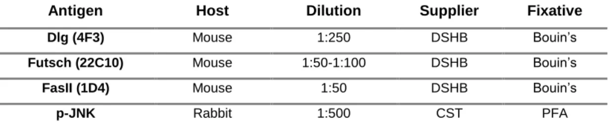
Table 2.3. Primary antibodies used in immunofluorescence assays. DSHB: Developmental Studies Hybridoma Bank; CST: Cell Signaling Technology
Antigen Host Dilution Supplier Fixative
Dlg (4F3) Mouse 1:250 DSHB Bouin’s
Futsch (22C10) Mouse 1:50-1:100 DSHB Bouin’s
FasII (1D4) Mouse 1:50 DSHB Bouin’s
p-JNK Rabbit 1:500 CST PFA
Table 2.4. Secondary antibodies used in immunofluorescence assays. * - Conjugated atibodies Antibody Dilution Supplier
Alexa Fluor 488 anti-mouse 1:500 Jackson Immuno Research
HRP Cy3 * 1:500 Jackson Immuno Research
Alexa Fluor 647 anti-rabbit 1:500 Jackson Immuno Research
HRP A488 * 1:500 Jackson Immuno Research
Texas Red-X-phalloidin * 1:500 Roche
23
2.5.
Acute Induction of Activity-Dependent Structural Plasticity
2.5.1. Stimulation Protocol
During development and in response to activity neurons can alter the size and shape of both pre- or post-synaptic compartments. Structural plasticity of nerve terminals can involve addition, removal or remodeling of synaptic components, including the number and order of branches, the number and size of synaptic boutons and the number of active zones. Also, the postsynaptic membrane can change in size, structure and can alter the number and localization of postsynaptic molecules such as receptors and scaffolding proteins (Griffith & Budnik 2006).
In order to assay new bouton formation two different protocols were used that are known to promote morphological changes at the synapse, namely, they induce the formation of activity-dependent boutons, schematized in figure 2.3. (Ataman et al. 2008; Vasin et al. 2014). Both of protocols are composed of pulses of high K+ and high Ca2+, intercalated with a resting phase where normal K+ and low Ca2+ are added to the dissected larvae. This leads to muscle contraction, inducing the formation new synaptic boutons. The high K+ depolarizations were achieved using 90 mM K+ and 1 mM Ca2+ in HL3 solution, and the resting with 0.1 mM Ca2+ and 5mM K+ in HL3 solution. The solutions were the ones described in Feng et al. 2004. Prior to the stimulation, third instar larvae were partially dissected, until the vertical incision without stretching the larvae to allow body wall contractions. During the last resting time, in both protocols, the organs are removed and larvae were stretched vertically and horizontally. After that, the larvae are fixed, followed by the immunocytochemistry protocol.
Figure 2.3. Schematic representation of the stimulation paradigms used. a) Long stimulation - 5
stimulations of 3 of 2 minutes, one of 4 minutes and a last of 6 minutes, all interspaced by 15 minutes of rest (Adapted from Ataman et al 2008). b) Short stimulation - 3 stimulations of 2 minutes interspaced by 10 minutes of rest (Apated from Vasin et al. 2014). At the end of both protocols, larvae are fixed and proceed for immunocytochemistry.