·
·
This article was published in Food Engineering Reviews, 7(4), 491-513, 2015
http://dx.doi.org/10.1007/s12393-015-9116-0
Edible Bio-Based Nanostructures: Delivery, Absorption and Potential
Toxicity
Joana T. Martins • O´ scar L. Ramos • Ana C. Pinheiro • Ana I. Bourbon • He´lder D. Silva • Melissa C. Rivera • Miguel A. Cerqueira • Lorenzo Pastrana • F. Xavier Malcata • A´ frica Gonza´lez-Ferna´ndez • Anto´nio A. Vicente
J. T. Martins · O´ . L. Ramos · A. C. Pinheiro · A. I. Bourbon · H. D. Silva · M. C. Rivera · M. A. Cerqueira A. A. Vicente
Centre of Biological Engineering (CEB), University of Minho, Campus de Gualtar, 4710-057 Braga, Portugal
e-mail: joanamartins@deb.uminho.pt O´ . L. Ramos F. X. Malcata
Laboratory for Process Engineering, Environment, Biotechnology and Energy (LEPABE), Department of Chemical Engineering, Faculty of Engineering, University of Porto, Rua Dr. Roberto Frias, 4200-465 Porto, Portugal
L. Pastrana
Biotechnology Group, Department of Analytical Chemistry and Food Science, University of Vigo, As Lagoas s/n, 32004 Ourense, Spain
A´ . Gonza´lez-Ferna´ndez
Immunology, Institute of Biomedical Research (IBIV), Biomedical Research Center (CINBIO), Universidade de Vigo, Campus Lagoas Marcosende, 36310 Vigo, Pontevedra, Spain
Abstract
The development of bio-based nanostructures as nanocarriers of bioactive compounds to specific body sites has been presented as a hot topic in food, pharmaceutical and nanotechnology fields. Food and pharmaceutical industries seek to explore the huge potential of these nanostructures, once they can be entirely composed of biocompatible and non-toxic materials. At the same time, they allow the incorporation of lipophilic and hydrophilic bioactive compounds protecting them against degradation, maintaining its active and functional performance. Nevertheless, the physicochemical properties of such structures (e.g., size and charge) could change significantly their behavior in the gastrointestinal (GI) tract. The main challenges in the development of these nanostructures are the proper characterization and understanding of the processes occurring at their surface, when in contact with living systems. This is crucial to
understand their delivery and absorption behavior as well as to recognize potential toxicological effects. This review will provide an insight into the recent innovations and challenges in the field of delivery via GI tract using bio-based nanostructures. Also, an overview of the approaches followed to ensure an effective deliver (e.g., avoiding physiological barriers) and to enhance stability and absorptive intestinal uptake of bioactive compounds will be provided. Information about nanostructures’ potential toxicity and a concise description of the in vitro and in vivo toxicity studies will also be given.
Keywords Nanoparticles · Bioactive compounds · Gastrointestinal tract · Intestinal
absorption · Absorption enhancers
Introduction
Food industry is constantly looking for novel technologies to improve the nutritional value, taste, flavor, shelf life and food safety of their food products. However, addition of micronutrients and bioactive compounds to food products is a major technological challenge. Many of these bioactive ingredients are chemically and physically vulnerable to production conditions and to digestion process. These may lead to detrimental effects on food properties, particularly changing food sensory properties and limiting bioactive compounds efficiency (i.e., bioaccessibility and bioavailability). In fact, a considerable number of bioactive com- pounds showed irrelevant or unsatisfactory therapeutic effects when orally administered in a free state once they were rapidly degraded losing their bioactivity [47].
In recent years, nanotechnology has become an answer to many of these problems, as it offers the ability to develop delivery structures (at submicron/subcellular size) for bioactive compounds’ (e.g., vitamins and antioxidants). This enables protecting bioactive compounds against degradation and controlling delivery at specific sites in the body [169].
Gastrointestinal (GI) tract is a particularly attractive targeting site due to its large surface area, in addition to its high potential to absorb (e.g., nutrients, bioactive com- pounds) and deliver it through the bloodstream [160]. One of the advantages of site-specific delivery to the GI tract will likely be for treatment of local conditions of the gut since there is a direct contact with the material being ingested. Such gut health conditions includes chronic inflammatory diseases (e.g., Crohn’s disease and ulcerative colitis), gastric and duodenal ulcers, GI infections and gastric and colon cancers [127]. However, GI tract delivery of bioactive ingredients is a great challenge owing to peculiar physiological barriers and physicochemical proper- ties, such as metabolism and gastrointestinal instability. An ideal GI tract delivery nanostructure system should be capable of (1) maintaining its integrity until it reaches the site of absorption, (2) attaching itself to the GI mucosa through specific interactions, releasing the bioactive compound at the target absorption site, and (3) holding inside the GI tract independently of its environmental conditions [53]. Each one of these issues is of utmost importance for an efficient bioactive compound delivery strategy. Recent advances in nanotechnology-based delivery systems showed potential for achieving these goals. Many of the polymeric materials used in the development of nanostructures for bioactive compounds delivery are natural, non-toxic, biocompatible and biodegradable such as proteins, polysaccharides and lipids [9, 46]. With the development of novel technologies, including permeability enhancers, it is expected that the number of bioactive compounds safely and efficiently delivered into the GI tract mucosa will increase.
At the same time, it is noteworthy that materials manipulation at the nanoscale can lead to the formation of novel structures with potential toxic characteristics. Their dimension allows penetrating biological tissues, which could cause the disruption of their normal function or cell death. Examples of toxic effects include tissue inflammation and modification of cellular redox balance [6]. Current knowledge on the toxicity of macro- and microstructures may not be reliable in predicting toxic forms of nanostructures, and thus further studies to evaluate ‘‘nanotoxicity’’ are mandatory.
The present review will focus in the current research on bio-based nanostructures for bioactive compounds delivery that could improve GI tract interactions and absorption. The review will be divided in five main sections:
1. An overview of the main challenges and characteristics of the GI system and how those may influence the passage and integrity of nanostructures will be presented. In addition, the potential transport mechanisms of these nanostructures once they reach the intestine will be also discussed; 2. The bio-based nanostructures that are currently being developed and their main physicochemical characteristics will also be highlighted. Each different nanostructure has been further subdivided according to its material, structural design and function. Consequently, strategies to improve these nanostructures in order to fulfill their primary function of transporting bioactive compounds during transit and absorption processes in the GI system will be addressed; 3. The separation, purification and characterization techniques of bio-based nanostructures following digestion and absorption will be highlighted. These techniques can be of great help for researchers providing a new insight on the behavior and integrity of developed nanostructures during their passage through the GI tract;
4. A thorough understanding of the interactions and potential risks of bio-based nanostructures with biological systems (e.g., cells of the immune system) using in vitro and in vivo models will be also addressed;
5. Lastly, a brief section on regulation will mention the problem of missing regulatory definitions and global consistency concerning nanostructures.
Delivery Challenges in GI Tract
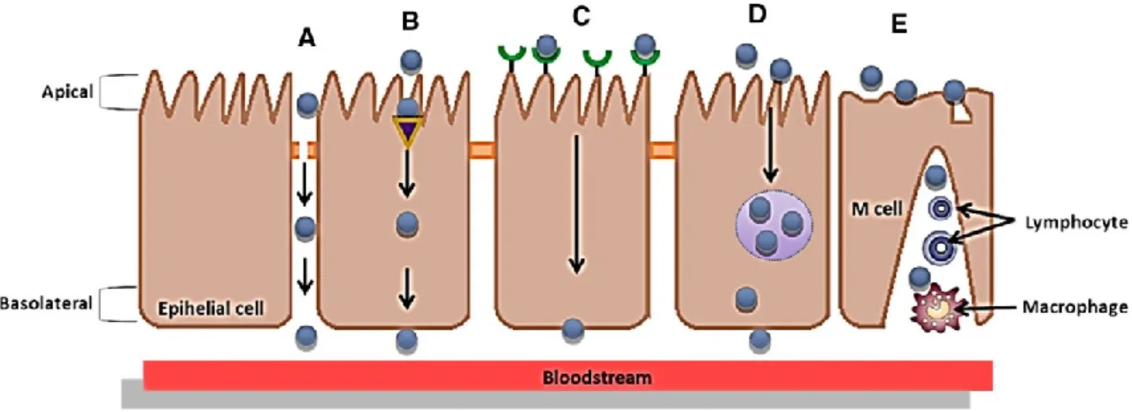
Overall, the main GI tract function is to digest (through a dissolving and breaking down process) food into molecular forms that can be absorbed, i.e., that are able to cross the intestinal epithelium. During the passage through the GI tract, nanostructures find several physiological and morphological barriers (e.g., enzymes present in the gut lumen and in brush border membrane, the mucus layer and epithelial cell lining) that play a key role in the bioactive compounds delivery (Fig. 1) [47]. Also, at the nanometer scale, the biological fate of the delivery systems and bioactive compounds incorporated within may be altered [104]. In contrast to microparticles that are too large to pass through epithelium and must release the bioactive compounds in the GI tract, nanostructures can be taken up and cross the intestinal barrier [31].
The uptake of nanostructures in the GI tract and the consequent bioactive compounds delivery depends on diffusion and accessibility through mucus, initial contact with the gut epithelium and various uptake and translocation processes [13, 117].
GI Barriers
The GI system comprises the GI tract (mouth, pharynx, esophagus, stomach, small intestine and large intestine) and accessory organs (salivary glands, liver, gallbladder and pancreas) that secrete substances into the GI tract via connecting ducts [166].
Immediately after the ingestion, nanostructures are mixed with saliva being influenced by digestive enzymes, pH, ionic strength and temperature changes. Therefore, even at this early stage of digestion, the initial size and interfacial characteristics of bio-based nanostructures can be changed.
After reaching the stomach, ingested nanostructures are mixed with enzymes such as gastric lipases that initiate lipid digestion and proteases that initiate protein digestion. The nanostructures are exposed to a highly acidic medium (pH 1–3) and to a peristalsis process. This process consists in an advancing walls contractile wave of a flexible conduit forcing their contents to move forward [77]. At this stage, original interfacial characteristics (e.g., charge and thick- ness) of ingested nanostructures may be changed, as well as their size (i.e., may no longer be at nanometric scale) due to pH, ionic composition changes and digestive enzymes [104].
Then, ingested structures move to the small intestine, where most of absorption occurs. At this stage, the nanostructures are mixed with bile salts, phospholipids, pancreatin, colipase and bicarbonate, and mixture pH in- creases, being almost neutral. The digestive enzymes pre- sent may hydrolyze the components that constitute the nanostructures (e.g., pancreatic lipase hydrolyzes triacylglycerols into monoacylglycerols and free fatty acids, and proteases hydrolyze proteins into peptides and amino acids). Within the small intestine, besides the digestion process, other phenomena can occur (e.g., particle aggregation or competitive adsorption process) resulting in changes in particle size and interfacial characteristics of nanostructures (Fig. 1) [127]. After digestion, the small molecules produced move from the lumen of the GI tract across a layer of epithelial cells into blood or lymph [166]. The small intestine is di- vided into three segments, i.e., duodenum, jejunum and ileum, designed to maximize absorption. Human intestinal epithelium is composed of villi that increase the total absorptive surface area to about 300–400 m2 [47]. Villi are covered by enterocytes (absorptive) and goblet cells (mucus secreting), which are interspersed with follicle-associated epithelium (FAE). FAE is the interface between the luminal environment and the lymphoid tissue associated to the gut composing Peyer’s patches [124]. These lymphoid regions are covered with M cells which play a significant role in absorption of nanostructures and bioactive com- pounds, since they are relatively less protected by mucus and have a high transcytotic capacity [124]. The mucus layers, which are mainly composed of gel-forming mucins (glycoproteins), are considered to be significant barriers to nanostructures penetration [82]. Mucus is continuously secreted to protect epithelial surface against pathogens by rapidly removing foreign particles from the GI tract and to lubricate the epithelium as material passes through. Therefore, nanostructures residence time decreases being unable to penetrate the mucus layer [47].
In addition to the GI mucus layer, the microbiota within the gut can also play an important role on the interaction with nanostructures. GI microbiota is responsible for a series of activities such as protection against potential pathogens, digestion of polysaccharides (e.g., chitosan and pectins), enzymatic hydrolysis, biosynthesis of vitamins (K and B), modulation of immune system and regulation of fat storage [10, 78]. Adherence of microbiota to gut wall can result in biofilm formation due to exopolymers secretion and subsequent gut protection from contact with nanostructures [113].
Transport Mechanisms
GI tract barriers, in particular intestinal epithelium, highly limit absorption and consequent efficient delivery of nanostructures (i.e., entering bloodstream). Intestinal epithelium acts as a selective barrier that tightly mediates transport from intestinal lumen into the bloodstream [161]. There are two main transport mechanisms of biomolecules across intestinal epithelium: (1) between cells via tight junctions (TJs)—paracellular route and (2) through intestinal membrane cells—transcellular route. Transport by paracellular route is mainly passive, whereas transcellular pathway includes passive diffusion, active carrier-mediated and endocytosis transport mechanisms (Fig. 2) [92].
Each pathway will depend on absorbing molecule physicochemical characteristics (e.g., size, charge and inter- facial chemistry), physical (e.g., TJs and lipid composition) and biochemical barriers (e.g., presence of enzymes, efflux and influx transporters) of cell membrane, which are able to metabolize or expel the biomolecules from the cell [160]. In the scope of studying transport mechanisms of nanostructures, two types of intestinal cells are mainly considered: enterocytes and M cells, which are the primary intestinal cells for the transport of a wide range of nanostructures [31, 61]. It is probable that both paracellular and transcellular routes contribute to the absorption of a single biomolecule or of more complex systems, i.e., bio-based nanostructures [57, 176].
In the literature, it is possible to find several research reports where bio-based nanostructures were able to cross the intestinal barrier via paracellular transport mechanism (Fig. 2), e.g., glyceride-based colloidal nanosystems, as delivery system for doxorubicin [76], nanohydrogel as carrier of salmon calcitonin [163], nanoparticles self- assembled by chitosan and PGA for delivery of tea catechins [156].
On the other hand, bio-based nanostructures can be absorbed by simple transcellular mechanism (passive transport) or by active carrier-mediated transport (Fig. 2). Certain nanostructures can also enter the cell through endocytosis (phagocytosis or pinocytosis) [74]. Guri et al.[58] showed that curcumin-loaded solid lipid nanoparticles were able to rapidly permeate through Caco-2 cell mono- layer using simple diffusion mechanism. Feng et al. [50] demonstrated that clathrin-dependent endocytosis played an important role in transcellular transport of chitosan/carboxymethyl chitosan nanohydrogels carrying the anticancer compound, doxorubicin hydrochloride. Internalization of nanostructures by pinocytosis includes receptor-mediated endocytosis (RME) [136]. Teng et al.[158] reported that curcumin transport using folic acid/soy protein nanoparticles was enhanced. They assume that folic acid could have targeted folate receptor protein, thus improving curcumin REM transport. Other well-known receptor is lactoferrin (Lf) receptor, expressed on apical surface of intestinal cells. Zhang et al. [180] studied the absorption mechanism of gambogic acid-Lf nanoparticles (GL-NPs), where Lf receptor seems to play an important role in the GL-NPs transport through the membrane.
After providing an overview of GI tract challenges that need to be addressed when encapsulating bioactive com- pounds, the next section will describe how food-grade nanostructures can be designed to meet these challenges.
Features of Bio-Based Nanostructures for GI Tract Delivery
Effects of Nanostructures’ Physicochemical Properties on Absorption
Absorption, metabolism, distribution and excretion processes depend on nanostructures’ composition and physicochemical properties such as size, shape, charge and hydrophobicity. Nanostructures physicochemical characteristics determine their fate once they have entered epithelium cell. They may be (1) digested by cellular enzymes into their constituent parts which may be absorbed; (2) transported directly out of the cell, and into the blood or lymph systems; or (3) accumulated within specific locations in the cell [104].
Nanostructures particle size significantly affects their absorption and biodistribution. It has been found that particle size can affect efficiency and pathway of cellular uptake once it influences structure adhesion and interaction with cells [98]. Nanostructures have shown to increase GI uptake and the level of translocation to lymphatic organs, when compared to microstructures [32]. For example, biodegradable structures of different sizes were produced using PLGA and bovine serum albumin as a model protein. Nano- and microstructures uptake was evaluated in situ with a rat intestinal loop model. Compared with larger particle sizes, uptake efficacy of 100 nm particles by the intestinal tissue was found to be 15- to 250-fold higher, depending on the type and location of tissue collected. Also, histological evaluation of tissue sections showed that 100 nm particles diffused through submucosal layers, while microparticles were predominantly localized in epithelial layer tissue [32].
It has been demonstrated that cells can take up nanostructures with various shapes including spheres, rods, tubes and sheets. However, there is some controversy about the influence of particle shape on nanostructures translocation. Thus, efforts should be done to clarify this effect [63]. When comparing spherical and rod-like nanostructures cellular uptake, it was found that spherical nanostructures were more easily endocytosed by HeLa cells [21]. On the other hand, Alemdaroglu et al. [5] investigated whether the shape of micelle aggregates influences the internalization. They observed that rod-like polymeric particles were taken up 12 times more efficiently than their spherical counter- parts, although they were composed with the same constituents.
Nanostructures cellular uptake can be divided in two steps: first, a binding step on the cell membrane and second, an internalization step. Nanostructures’ binding to cell membrane seems to be dependent on nanostructure surface charge. However, there is not an agreement on the optimal charge for translocation. Cationic nanostructures have been shown to be absorbed more readily than anionic ones. The interactions between anionic membrane and cationic nanostructures facilitate their binding to cell membrane and consequently their uptake. It has been shown that cationic polymers such as chitosan can form nanostructures that maintain a prolonged contact time with the intestinal layer. This leads to a larger absorptive surface and subsequently, enhanced absorption rates [95]. Contrarily, Patil et al. [118] showed that nanostructures with the highest negative charge values have the highest cellular uptake compared with other formulations with less negative or positive surface charge. The authors concluded that the high cellular uptake of negatively charged nanostructures is related firstly to the nanostructures non-specific adsorption to cell membrane, and secondly, to nanostructures’ clusters formation.
Particle absorption is also believed to depend on nanostructure surface hydrophobicity. Although there is not a very clear tendency, uptake of nanostructures pre- pared from hydrophobic polymers seems to be higher than that of structures with more hydrophilic surfaces [70]. Norris and Sinko [114] suggested that increasing hydrophobicity leads to an increase in permeability
through mucin, but also to a decrease in the translocation across the cell interior, which has a more hydrophilic environment.
Bio-Based Nanostructures Delivery Systems
Multifunctional nanostructures using biomaterials with distinctive architectures have been designed and evaluated for bioactive compound delivery applications [68]. Biopolymer architecture, composition and stability are important factors which will influence bioactive compound delivery carriers’ effectiveness. Moreover, biopolymer nanostructure dimensions change its functional attributes in foods and GI tract. For instance, nanostructure architecture influences physicochemical properties of foods (e.g., stability), encapsulation characteristics (e.g., loading and re- lease), and behavior within the Gl tract (e.g., interaction with the environment and degradation) [2]. Therefore, appropriate nanostructures need to be designed rationally according to specific applications and needs.
Most of the publications focus on the synthesis and study of core–shell structures [23]. These structures are obtained from polymers of different sizes and shapes and can be functionalized with stimuli-responsive polymers (e.g., proteins) [110]. The most common shape of food- grade nanostructures suitable for encapsulation is spheroid (Fig. 3). Nanoemulsions, for instance, tend to be approximately spherical, and may have a variety of internal organization, such as homogeneous or dispersion structures, depending on the material and preparation method used [68].
Multilayer nanostructured systems could also be produced with multiple layers of emulsifiers and/or polyelectrolytes by the layer-by-layer (LbL) technique (Fig. 3). Additionally, hollow multilayered nanostructures can be obtained using colloidal templates (e.g., polystyrene nanoparticles). After LbL deposition procedure, the tem- plate could be dissolved using an acid or a solvent. These nanostructures can be applied as delivery systems of bioactive compounds, being those compounds either entrapped inside the core or adsorbed on the surface of nanocapsule [15].
In this section, a selected group of bio-based nanostructures (i.e., nanoemulsion, nanohydrogel and nanocapsules) that have been most commonly used in bioactive compounds GI tract delivery applications are reviewed.
Nanoemulsions
Oil-in-water nanoemulsions are a mixture of two immiscible liquids, where a thin interfacial layer is created due to the adsorption of the emulsifier molecules surrounding the oil droplets. The emulsifier promotes the dispersion of the oil phase in the continuous phase either using low-energy or high-low-energy techniques [1, 146]. In water-in-oil nanoemulsions, the oil is the continuous phase, where the emulsifier surrounds the water droplets (disperse phase). The emulsifiers can also be dispersed in the continuous phase or in the dispersed phase, depending on their hydrophilic–lipophilic balance [75].
Due to the emulsifier ability to decrease the interfacial tension between two immiscible liquids, emulsifiers play a major role in the formation of the nanoemulsions, creating an interfacial layer able to protect the oil droplets [146]. Thickening agents such as starch, flour and gums can also be used to stabilize emulsions. These agents act by increasing the viscosity of the medium, which helps stabilizing the suspension of droplets of the dispersed phase preventing them from
moving around and coalescing [34]. Nanoemulsion normally presents a core–shell structure and may be manufactured from a great variety of food- grade ingredients (Fig. 3). For example, the core may be formed from nonpolar components, including monoacylglycerols, diacylglycerols, triacylglycerols, waxes, mineral oils, oil-soluble vitamins and nutraceuticals [103]. The shell is typically formed from one or more surface-active components, including small molecule surfactants, phospholipids, proteins (e.g., whey proteins) and polysaccharides (e.g., chitosan) [135]. The materials that comprise the core and shell may be more or less digestible within different regions of the human GI tract, which plays an important role in determining their biological fate.
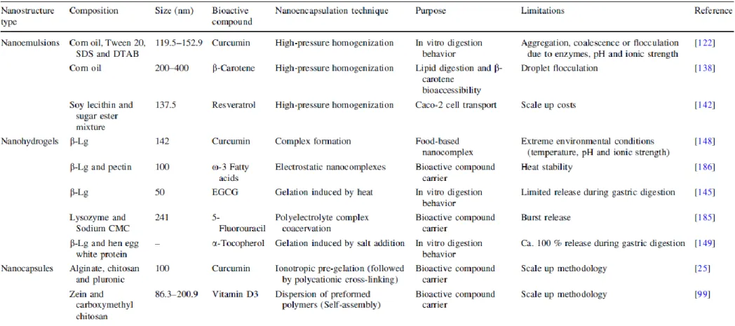
Nanoemulsion technology can be used to encapsulate, protect and deliver lipophilic bioactive compounds, such as essential oils (e.g., x-3-rich oils), antioxidants (e.g., quercetin), antimicrobials (e.g., thymol) and vitamins (e.g., vitamin A) [139]. Nevertheless, hydrophilic and amphiphilic bioactive compounds can also be incorporated [11]. This technology allows improving the solubility and bioavailability of these compounds, also preventing the degradation against light and oxidation using only food- grade ingredients, by simple process operations (Table 1). By controlling the composition and structure of nanoemulsions, it is possible to create different rheological properties and release profiles in response to environmental triggers [105].
The main challenges in nanoemulsion technology are: (1) proper selection of the emulsifier, due to the limited number of food-grade emulsifiers that can be used in its formation, restricting the ability to create nanoemulsions with different release characteristics; (2) their physical instability under environmental stresses, such as freeze-thawing, pH, salt, heating, dehydration and chilling; (3) limited control over oxidation of the bioactive compounds due to the very thin interfacial layer; and (4) ability to provide better protection and stability for encapsulated bioactive compounds during the GI passage (Table 1) [38, 105]. One strategy aimed at overcoming these limitations is to create one or more layers of a polyelectrolyte surrounding the nanoemulsion through the LbL technique [59]. This strategy may offer potential advantages due to: (1) the ability to control the order and location of multiple polymer layers with nanoscale precision; (2) the ability to define the concentrations of incorporated materials simply by varying the number of polyelectrolyte layers [153]; and (3) possibility to use a wide range of polymers, which increase its potential application. Biopolymers (such as proteins and polysaccharides) are of high interest because of their unique characteristics: naturally available, nontoxic and biocompatible. Consequently, an exceptionally broad range of biopolymer components, morphological characteristics, functional responses, variety of interactions and versatility in assembly approaches enhance the use of LbL technique [29]. LbL technique consists in the deposition of polyelectrolytes on charged surfaces due to strong electrostatic attractions be- tween the surface and the charged polyelectrolytes, thus building a ‘‘new’’ layer. This technique also allows that further layers can be built by simple addition of oppositely charged polyelectrolytes in solution, promoting the ad- sorption of the polyelectrolytes on the top of the first layer. Repetition of this adsorption steps leads to the formation of multilayers (Fig. 3) [59]. The number of layers will be de- fined by the final application of these systems [87, 122]. Charged nanoemulsions can be used as substrate, allowing the development of a new system (i.e., multilayer nanoemulsion) [105]. Salminen and Weiss [137] showed, through the production of a whey protein–pectin multilayer nanoemulsion, that it is possible to improve its stability to different environmental stresses: salt (0–500 mM NaCl) and heat (40–90 ºC, 30 min). These authors demonstrated that emulsions saturated with biopolymer complexes exhibited good stability (no aggregation) to salt (up to 200
mM) and heat (up to 90 ºC) at pH 3.5–4.5. However, the biopolymer complex layer adsorbed to the emulsion interface was not able to prevent instability after freeze-thawing cycles (-20 ºC, 22 h). Li and McClements [86] built an alginate layer on Tween 20 and -lactoglobulin nanoemulsions. This layer reduced lipid digestion inhibiting lipase access to the lipid in the nanoemulsion.
The modification of nanoemulsion composition and structural characteristics such as particle size, emulsifier and oil and polymer type can change the digestion rate. Therefore, the effects of these characteristics are being widely studied [87, 122]. Li et al. [87] examined the influence of chitosan (intermediate layer), pectin and alginate (outer layer) layers in the digestibility of the multilayer system using an in vitro digestion model. They evaluated the effect of composition and structure of the biopolymer on the digestion rate, accessing the release of free fatty acids from the nanoemulsions and multilayer nanoemulsions (first and second layer). Nanoemulsions were digested between 30 and 40 min, which show that lipase adsorbed to the nanoemulsions promotes the lipolysis of triacylglycerols. For the multilayer nanoemulsions the digestion rate was significantly slower, where only 40 % of the lipids where digested after the same time (i.e., 40 min). The authors hypothesized that biopolymers layers protected the lipid droplets restricting the access of the lipase to triacylglycerols, retarding the lipolysis reaction and there- fore delaying the lipid digestibility.
Pinheiro et al. [122] studied the effect of different charged emulsifiers—Tween 20 (non-ionic), sodium dodecyl sulfate (anionic) and dodecyltrimethylammonium bromide (cationic)—in the behavior of curcumin nanoemulsions during in vitro digestion. Nanoemulsions were produced by high-pressure homogenization, and an in vitro digestion model was used. All emulsifiers used formed stable nanoemulsions within the nanometric scale. Nevertheless, during simulated digestion, all nanoemulsions increased their sizes, being this attributed to aggregation, coalescence or flocculation due to the action of digestive enzymes, as well as to changes in pH and ionic strength. The emulsions produced with the cationic emulsifier were the least stable. The positive charge of the cationic emulsifier may have promoted the adsorption of anionic lipase and anionic bile salts to the oil–water interface. The emulsifier type had also impact in curcumin bioavailability. Nanoemulsions stabilized with the non-ionic emulsifier increased curcumin bioavailability during digestion time, which can be due to the formation of digestion products (ability to form mixed micelles). On the other hand, the use of cationic emulsifier allowed achieving only a very low bioavailability during simulated intestinal conditions, i.e., emulsions became unstable, leading to phase separation.
Nanohydrogels
Nanohydrogels are three-dimensional hydrophilic or amphiphilic biopolymer nanosized networks that can swell and hold a large amount of water. However, they are prevented from dissolving due to their chemically or physically cross-linked structure [19] (Fig. 3). The swelling ability is attributed to the presence of hydrophilic moieties groups (e.g., hydroxyl, carboxyl, ethers, amines and sulfates) in the polymers forming the nanohydrogel structure, which is responsible for the soft and pliable characteristic of such nanostructure [120].
Bio-based nanohydrogels can be prepared from several polysaccharides (e.g., alginate, chitosan, pectin, pullulan and dextran) and proteins (e.g., whey proteins and collagen) with different techniques; being the most commonly used method the gelation process [164]. Gelation is a
process that typically encompasses two stages: (1) partial unfolding of the native structure leading to dissociation of intramolecular bonds that can be induced by several environmental conditions (e.g., temperature) and (2) formation of new bonds, leading to a progressively larger embranchment of molecules. The continuous cross-linking increased the size of the ramified polymer chains (in nanometer range) decreasing their solubility, which results in the formation of a gel structure [133].
Their reduced size enables a controlled release of bioactive compounds and improved bioavailability of those compounds with poor absorption rates. Moreover, they specified delivery to the associated tissues, reducing the GI mucosa irritation caused by continuous contact with some compounds, and assured their stability in the GI tract [116]. In addition, nanohydrogels allow overcoming some draw- backs inherent to other nanostructures (e.g., preparation procedure and relatively low loading capacity). They can be produced without the interference of the bioactive compounds and designed to spontaneously load bioactive molecules. Electrostatic, van der Waals and/or hydrophobic interactions between bioactive compounds and polymer matrix, during gel folding process, lead to formation of stable nanostructures’ [15]. Furthermore, these structures’ ability to produce a response (e.g., swelling) to environ- mental stimuli (e.g., temperature, pH, ionic strength or enzymatic conditions) makes them crucial systems to de- liver bioactive compounds locally to specific sites and at a particular time in the GI tract [91]. On the other hand, these structures may present some limitations if produced by physical gelation. Once nanohydrogels contain labile bonds in polymer networks, they are susceptible to be disrupted under physiological conditions in the GI tract [62].
It is possible to find in the literature different works reporting protein nanohydrogel’s ability to incorporate and release hydrophilic and lipophilic bioactive compounds such as drugs, unsaturated fatty acids, vitamins, as well as peptides [152]—see Table 1. Depending on bioactive compound characteristics, it is possible to obtain different release mechanisms during digestion process. Hydrophilic compounds release from a protein matrix by diffusion, whereas lipophilic compounds are released mainly by enzymatic degradation of the protein matrix in the GI tract [170]. Examples of bioactive compounds efficiently incorporated into bio-based nanohydrogels, techniques used in their encapsulation and their main limitations are summarized in Table 1.
One of the challenges of these nanostructures is to de- liver encapsulated components at the desired point (e.g., mouth, stomach, small intestine and colon) without being destroyed. Therefore, it is important to design and manufacture biopolymer nanohydrogels with specific compositions, which can be able to resist to severe environmental conditions, for example, resistance to gastric fluids, if nanohydrogel is designed to deliver a bioactive compound in the colon. Nanostructures composed by peptides or proteins have a high level of GI degradation by digestive enzymes [37]. In order to preserve functionality and integrity, nanohydrogels must resist to the harsh gastric conditions (i.e., low pH and presence of digestive enzymes). A major drawback of these biopolymeric nanostructures is their tendency to decrease their interfacial surface area leading to the formation of aggregates [155]. One of the strategies for preventing aggregation or destruction include nanohydrogels coated with foreign coating agents and/or tailoring particle surface charges to create separation through electrostatic repulsion (Fig. 3). LbL assembly of polyelectrolyte multilayers has been demonstrated on various templates, from hard and planar to rigid particles, and more recently, to soft and porous tem- plates, such as thermoresponsive nanohydrogels [175]. The possibility of assembling any charged polyelectrolytes on nanohydrogels shows their potential exploitation as bioactive compound storage, transport, target and con- trolled delivery system. Tan et al. [155] demonstrated the
successful preparation of coated nanohydrogels with encapsulated drugs using the LbL approach. These authors observed that the initial burst release behavior observed in nanohydrogels was minimized and eliminated by the introduction of several polyelectrolyte layers. Through this LbL approach, the permeability of nanohydrogels was altered with each additional layer. These authors also concluded that swelling behavior of coated nanohydrogels decreased with increasing polyelectrolytes layers resulting in a slower release of bioactive compounds. The application of polyelectrolyte layers also shows to be a successful technique to turn nanostructures more stable under physiological conditions [165]. Interaction between biopolymers, such as polysaccharide protein, also allows producing nanohydrogels more resistant to gastric fluids. Chen and Subirade [18] developed a biopolymeric nanohydrogel based on alginate–whey protein interactions to be used as a vehicle for riboflavin. These authors observed that alginate–whey protein isolate nanohydrogels have the ability to delay the compound’s release in the stomach and allow complete release in the small intestine.
Nanocapsules
The development of nanocapsules for the delivery of bioactive compounds has been widely studied since the 1970s aiming at the development of nanostructures for oral drug delivery [22, 129].
A great number of structures have been developed using synthetic and/or natural polymers, where different methodologies were used for their production (e.g., ionic pre-gelation/coacervation, polymerization and dispersion of preformed polymers) possibly influencing their main properties [15].
Ionic pre-gelation/coacervation is based on the ability of polyelectrolytes to cross-link in the presence of a counter- ion (cationic or polyanionic) to form nanocapsules [15] (see Table 1). Nanoparticles can also be directly synthesized by the polymerization of monomers using various polymerization techniques (such as emulsion and dispersion polymerization) [123]. In this method, the bioactive compound is incorporated either by being dissolved in the polymerization medium or by adsorption onto the nanoparticles after the polymerization [151]. On the other hand, nanoparticles could be prepared by dispersion of preformed polymers through different techniques such as self-assembly, nanoprecipitation, salting-out and using supercritical fluids [108, 130]. For example, the self-assembly method involves the spontaneous formation of compact and stable nanocapsules without the help of external agents. Materials such as zein, chitosan and casein are examples of polymers that can be used in this method [15] (see Table 1). In the last years, the main focus in the development of nanocapsules has been the use of natural, biocompatible and edible bio-based materials for their production. The use of these materials brings a great number of challenges concerning the production of nanostructures such as: different materials, molecular structure complexity and control of their properties (i.e., particle size, size distribution, encapsulation efficiency and release behavior).
Two of the most studied bio-based materials for the production of nanocapsules are chitosan and alginate, either used as main materials or as ionic cross-linkers applied after nanocapsule production. Das et al. [25] developed an alginate–chitosan–pluronic composite for the delivery of curcumin in cancer cells through ionotropic pre-gelation followed by polycationic cross-linking— see Table 1.
In order to overcome the instability and fast release of bioactive compounds under specific environmental conditions, different methodologies have been used to tailor and control their
release (i.e., by diffusion and/or by matrix degradation) from nanocapsules. Some of the possibilities are as follows: cross-linking processes used in the formed nanocapsules [115] and formation of a layer with other material surrounding the nanocapsules [28].
Other main focus on the study of nanostructures delivery is their bioavailability, where bioactive compounds solubility, mass transfer rate and retention time in GI tract play an important role in their successful use [1]. Some of the methodologies pointed as possibilities to increase bioavailability of nanocapsules are as follows: (1) the formation of nanocapsules through the LbL technique which allows the deposition of an outer layer on formed nanocapsules (Fig. 3) according to the desired application [15] and (2) the functionalization of the nanocapsules surface for the control of cellular uptake [107].
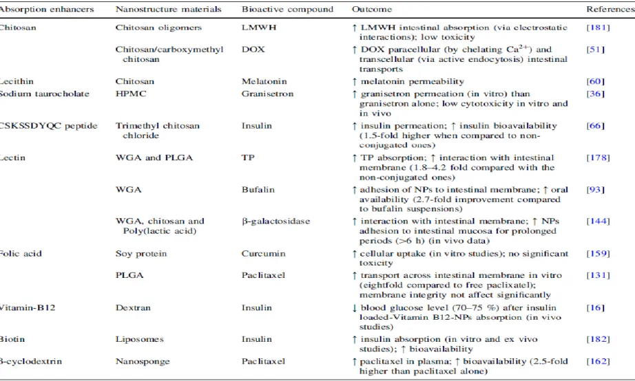
Strategies for Enhancing Nanostructures Delivery in GI—Absorption/Permeation Enhancers Various strategies have been applied to enhance permeation of bioactive compounds, such as (1) modification of their surface or (2) the alternative approach where the compounds are not chemically altered. Here the bioactive compounds are combined with another targeting functional agent or with a specific formulation (e.g., micro/nanoparticles), termed as absorption enhancers (AE) [8]. Therefore, this section will be focused on the second approach, the utilization of AE as a strategy to improve bioactive com- pounds’ absorption.
AE, also termed as bioavailability enhancers, are functional agents or formulations used to improve GI absorption of bioactive compounds by specifically facilitating their membrane permeation [8]. In the last years, several AE have been proposed; however their utility is often limited by their restricted biological effect and toxicity. Table 2 summarizes some approaches used to enhance absorption of bioactive compounds. Among all approaches, the use of chitosan as a AE has been extensively reviewed [20, 89]. Moreover, plant origin compounds have been stated as AE such as quercetin, curcumin, ginger and niaziridin [73, 157]. Also sodium caprate has being used as absorption-enhancing agent since it acts on TJs opening, modulating paracellular transport of bioactive compounds [184]. Krug et al. [80] demonstrated that caprate was able to open the paracellular pathway of molecules up to 10 kDa, supporting the application of caprate as an effective AE.
One of the main advantages of utilizing AE for poorly membrane-permeable compounds is the development of non-injection formulations (i.e., oral route), which are considered to be the most comfortable and convenient route of administration. Moreover, from an economic point of view, as AE will promote efficient systemic absorption, the waste of costly compounds is likely to be reduced [7].
Although several AE have been successful used, a part of them come with hurdles that limit their commercial use For example, some AE have been demonstrated to cause damage and irritation of intestinal mucosa [140]. Additionally, large-scale production is one of the main challenges for nanostructured AE production, mainly because it is harder to maintain size and composition, while avoiding agglomeration of these compounds during a scaling-up process [73]. Despite the presented disadvantages, several intestinal AE have registered progresses in terms of their application, being a few already in clinical trials [8]. The main barrier to the commercialization of AE seems to rely on the demonstration of safety and improved biological activity. Although this strategy may enable development of bioactive compounds with specific biological activity, all data indicate that AE will gradually become more broadly accepted in food and medical markets.
Nevertheless, scarce information has been found regarding characterization of biopolymeric nanostructures during the digestion process. Therefore, appropriate techniques to characterize these nanostructures during the passage through the GI tract in order to understand how they can resist to harsh conditions and their behavior during release of bioactive compounds to specific sites will be highlighted.
Separation, Purification and Characterization of Bio-Based Nanostructures Following Digestion and Absorption
Regulation for the use of nanostructures in the food industry led to a long and hard path in order to determine their safety for consumers. Despite the significant amount of work developed in the last years, it is not obvious how nanostructures behave after ingestion and digestion processes and how they will change their characteristics/ properties after the absorption.
Several techniques and methodologies have been presented in the last years for the characterization of nanostructures [e.g., dynamic light scattering, size exclusion chromatography, confocal scanning light microscopy (CLSM), scanning electron microscopy, transmission electron microscopy (TEM)] being unanimous used for virtually all types of nanostructures [90, 121]. While characterization methodologies are established among scientific community, separation and purification techniques of nanostructures from foods, after the passage through GI systems and in human fluids are not consensual. Recently, the existing methods for separation, purification and characterization of nanostructures in foods have been summarized [12, 79, 90, 121, 179]. Also, research projects were focused in development and validation of screening methods for the determination of nanostructures in food matrices [112].
It is known that nanostructures, besides food matrices, also interact with gastric fluids, leading to a change of their main characteristics. This change can happen due to the interaction with proteins (e.g., changing their charge and agglomeration state) [84] and pH values under GI conditions (e.g., leading to agglomeration) [122]. Several separation and purification methods have been explored in the last years for the characterization of nanostructures after ingestion, passage through GI systems and once incorporated in human fluids. Inorganic nanostructures (e.g., gold and silver) and others from synthetic materials have been the most studied [4, 111]. However, few works de- scribe methodologies and useful techniques for separation and purification of bio-based nanostructures. Their unique characteristics in terms of composition (e.g., polysaccharides, proteins and lipids), shape (e.g., spherical or planar), size (e.g., from 20 to 200 nm), charge (e.g., neutral, positive and negative) and degradability (e.g., acid or medium) make them a challenge for analytical and chemistry science. Thus, techniques and methodologies used should be chosen based on their characteristics and behavior.
M-M et al. [126] used an online flow field flow fractionation (FlFFF) with inductively coupled plasma mass spectrometry (ICP-MS) for the determination of particle size of selenium nanostructures (stabilized by pectin, mixed alginate/pectin, ovalbumin and -lactoglobulin) after GI tract passage. Results showed good agreement of particle size observed by FlFFF and the images obtained by TEM. The stability of different kinds of polymeric micelles (composed of polyethylene glycol (PEG), poly(lactic acid) and Tween 80) was evaluated in human serum, using fluorescence-based approach (Forster Resonance Energy Transfer). Asymmetrical Flow Field Flow Fractionation (AF4) was used for the separation of the micelles (further characterized by light scattering) [106]. In other interesting work, Lee et al. [84] evaluated chitosan nanostructures
integrity after GI tract passage and when in contact with a Caco-2 cell monolayer. The disintegration degree of chitosan nanostructures was determined by free chitosan quantification (using fluorescein isothiocyanate (FITC)-labeled chitosan) separated through ultracentrifugation. At the same time, nanostructure mean diameter and zeta potential were determined. CLSM was used to visualize chitosan nanostructures after their transport through Caco-2 cell monolayer allowing to understand the behavior of these nanostructures in cells. David-Birman et al. [26] evaluated the in vitro digestion of Lf-based nanostructures with anionic low-methoxy pectin (LMP), high-methoxy pectin (HMP), sodium alginate (ALG) and iota-carrageenan (CAR). The proteolysis products were characterized by sodium dodecyl sulfate polyacrylamide gel electrophoresis (SDS-PAGE). These authors observed the structures’ integrity during gastric and duodenal digestion.
They observed that these structures coated with HMP did not protect Lf from gastric proteolysis once intact Lf band was not detectable even after short digestion periods. The same authors concluded that ALG extends Lf resistance to proteolysis beyond that of LMP. Lf intact band was detected even after 40 min of gastric digestion. The digestion experiments revealed that Lf proteolysis leads to the formation of peptides, with specific SDS-PAGE bands. However, Lf-based nanostructures were relatively persistent and even reached the duodenal experiments.
It is expected that in the next few years, new studies exploring the detection and characterization of nanostructures under different environmental conditions will be achieved. This will allow improved traceability and evaluation exposure of these nanostructures to human body.
Nanotoxicity Assessment in GI Tract
The production of nanostructured systems presents an in- creasing risk of exposure to both nano-sized systems and associate products at concentration levels that could exceed those naturally present in the body. In some cases, nanostructure parts could be retained in the body for a long-term period due to incomplete excretion. Consequently, it may disturb normal functions of organs or tissues inducing metabolic toxicity, immunotoxicity or even genotoxicity [127]. Thus, in order to minimize and even prevent possible health risks, potential toxicity of these nanostructures has to be assessed in depth before their use in oral administration. The interactions of engineered nanostructures with biological systems and the consequent toxicity are due to materials themselves and also to their nano-size-related physicochemical characteristics (e.g., size, shape, aggregation, charge and surface properties). For instance, Loh et al. [94] reported that in vitro cytotoxicity of chitosan nanostructures against Caco-2 cells is less dependent on positive surface charges than on the particle size.
Several topics need to be addressed in studies of nanostructures toxicity, namely dose metric, standardized assays and reference materials, to draw general conclusions regarding toxicity of nanostructures [10]. Also, it is crucial to design a study by using a wide range of doses (e.g., doses resulting from biokinetic studies in vivo), so that a careful analysis of the dose–response correlations can be performed. An exposure and dose metric for engineered nanostructures, which have a range of either chemical compositions or structures, or even both, will depend on the mechanism of their pharmacokinetic and toxicological behavior [83].
Many toxicological studies (in vitro and in vivo) have been conducted on engineered nanostructures. Nevertheless, the absorption, distribution, metabolism and excretion processes of these nanostructures have not been completely addressed and understood to date. Additionally,
current testing and toxicity studies are mainly performed using a wide range of cell lines (e.g., Caco-2 cells), as well as in vivo models (e.g., mice). These circumstances led to an extensive number of tested nanostructures, experimental designs and model systems, which consequently result on several contradictory experimental data. Thus, standardized test protocols to permit risk assessment of nanostructures, especially dose thresholds, need to be regulated and established [10, 167]. The European Food Safety Authority (EFSA) provides a comprehensive document with key issues to be considered. Topics addressed include risk assessment, toxicological studies used for hazard identification and characterization of nanomaterials and a dose–response assessment [44].
Nanostructure small size confers a very large surface-to- volume ratio, which could lead to some undesired results after entering into the body. Possible nanostructure–cellular interactions may induce cytotoxicity and other physio- logical outcomes to GI tract. For example, oxidative stress, inflammation and immune responses (phagocytosis, complement activation and recruitment of inflammatory cells) [187], mitochondrial perturbation, ‘‘corona’’ formation (please see ‘‘Protein corona’’ section), protein denaturation [147], DNA damage and cell death [33, 81, 97]. Although some of these responses could be beneficial for some purposes, such as the activation of specific immune responses by nanostructures carrying antigens on vaccines [128, 168], some of those could have harmful effects. Despite the very scarce data on bio-based nanostructures’ immunological or cellular responsiveness of the GI tract, some of the potential biological mechanisms that could happen with these structures will be discussed.
Biological Mechanisms Induced by Nanostructures
Protein Corona
After the entry of nanostructures into the bloodstream, potential interaction with biological molecules (e.g., phospholipids, DNA and serum proteins) could affect the normal function of the body and consequently lead to toxicity effects. The binding of a mixture of cellular proteins to nanostructures forming the so-called protein corona could change the structure, stability and dynamic behavior of the nanostructures. This may provide different physio- logical responses of an organism [174]. For example, the protein corona can influence cell membrane endocytosis and exocytosis of nanostructures, as well as promote their uptake by phagocytes [119, 134].
The size, charge and type of coating on nanostructure surface have a strong influence on how they are going to be recognized by the cells [109]. Also, whether the particles are agglomerated or not on the biological fluids [33, 101] could strongly modify their uptake. Moreover, nanoparticles can induce conformational changes in proteins, affecting their physiological function [147]. Thus, in vivo fate of nanostructures and the biological responses could be altered when compared with in vitro studies.
The formation of a protein corona is believed to be the principal factor influencing its pattern of in vivo biodistribution, and thus its pharmacokinetic profile. There is still discussion on the advantages and disadvantages of adsorbed proteins [10, 30]. In some circumstances, it is useful to have these bound proteins as they can direct or target the nanoparticles to a specific area of the body [39]. Also, protein corona could play an important role in modulating toxicity of nanostructures [171]. For example, Peng et al.[119] reported that formation of a nanostructure-albumin complex is an effective and feasible means to prolong the nanostructure circulation time and reduce their toxicity. On the other hand, binding of proteins has also been shown to be
correlated with rapid uptake into the liver and spleen, and clearance of the particles by the reticuloendothelial system [174]. This can be a negative effect if one of the aims is to increase nanostructures circulation and the retention time in the body.
However, further studies are needed to understand interaction and potential impact of nanostructures with other cellular components (e.g., polysaccharides, proteins).
Oxidative Stress
The increased production of reactive oxygen species (ROS), revealing oxidative stress, has been proposed as a model for the assessment of toxicity induced by nanostructures [102]. High ROS levels could damage cells by peroxidizing lipids, inducing inflammation, changing DNA and other proteins and interfering with signaling and gene functions [143]. Oxidative stress induced by nanostructures is described to enhance inflammation through regulation of redox-sensitive transcription factors comprising nuclear factor kappa B (NFkB), activating protein 1 (AP-1), extracellular signal regulated kinases (ERK), c-Jun N-terminal kinases (JNK) and p38 mitogen-activated protein kinases pathways [3].
ROS can be originated from several sources such as in the phagocytic cell response to foreign material, scarce amounts of antioxidants, presence of transition metals, environmental factors and physicochemical properties (e.g., surface properties and size) of some nanostructures [6, 72]. Yan et al. [177] reported two pathways for the intracellular ROS production by nanostructures. ROS could indirectly be produced when nanostructures disturb the endogenous biochemical/physiological equilibrium of cells, damaging the structure of cellular organelles, such as mitochondria. One the other hand, ROS production de- pends on the direct contact of nanostructures with cellular components. Highly reactive nanoparticle surface dangling bonds can accept electrons from electron-donor groups, thus oxidizing cellular components.
Activation of the Immune System and Inflammation Response
The immune response includes both innate and adaptive defense mechanisms, which activate different cell populations. Monocytes, tissue macrophages, dendritic cells and neutrophils are some of the cellular components of the innate immune response. They carry out phagocytosis and produce inflammatory mediators, ROS and antimicrobial peptides [56]. Interaction between nanostructured systems and the innate immune system can induce undesirable effects such as cytotoxicity or inflammation. As previously stated, physicochemical characteristics of nanostructures could affect the way that immune system detects and reacts to them. The exposure of the body to ‘‘external’’ nanostructured systems could lead to harmful immune (activation or suppression) actions. When in contact with the blood, they can activate the complement system (which is a part of the innate humoral immune response), priming the surface of nanostructures with opsonic complement fragments (C3b, iC3b) for recognition and clearance by phagocytic cells [17, 187]. The adaptive immune response is mediated by antigen-specific lymphocytes (T and B cells). Both innate and adaptive immune responses can participate with their cells and secreted products. These humoral factors include inflammatory cytokines, e.g., in-terleukin (IL)-1, IL-6, interferon (INF)-, tumor necrosis factor- (TNF-a) and antibodies [45]. Semete et al. [141] studied the uptake of chitosan and PEG-coated PLGA nanostructures and the immunological response of Balb/C mice within 24 h of oral administration. An evaluation of the
subsequent immune reaction by analyzing secreted pro- and anti-inflammatory cytokines concentration profile was conducted. The expression of pro-inflammatory cytokines IL-2, IL-6, IL-12p70 and TNF- in plasma was found to remain at low concentration in PLGA nanostructures treated mice. The anti-inflammatory cytokines IL-10 and chemokines INF-, IL-4 and IL-5 remained at normal levels in the PLGA-treated mice. The authors stated that these results claim against an immunological contraindication for the oral administration of PLGA nanostructures in mice.
In Vitro and In Vivo Responses to Bio-Based Nanostructures
In vitro models can generate valuable data as a pre-stage for in vivo animal studies, therefore reducing the number of animal tests. The use of in vitro human cell lines of intestinal mucosa is very valuable to assess the behavior of orally delivered nanostructures [6].
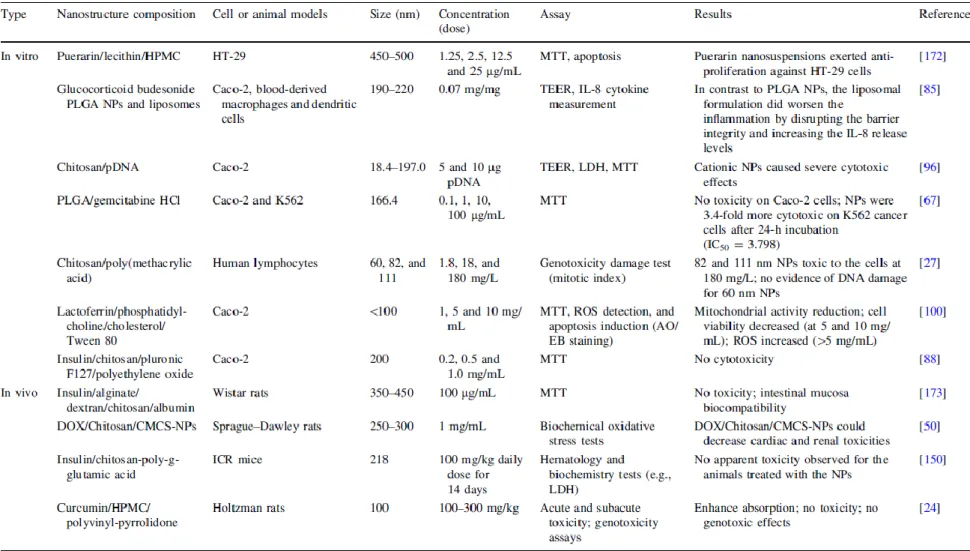
Wang et al. [172] studied in vitro the anti-colorectal cancer activity of puerarin nanosuspensions of lecithin and hydroxypropyl methylcellulose (HPMC) in the human colon cancer HT-29 cell line. Cytotoxicity assay, observation of morphological changes and early apoptosis revealed that puerarin nanosuspensions could significantly enhance the in vitro anti-proliferation effect against HT-29 cells compared to the puerarin-free solution (Table 3). In another study, Leonard et al. [85] developed a 3D inflamed intestinal mucosa equivalent, based on the co-culture of intestinal epithelial Caco-2 cells, blood-derived macro- phages and dendritic cells as components of the intestinal innate immune system (Table 3). The cells were all located on the porous membrane of a transwell insert. Inflammation was stimulated by the inflammatory cytokine IL-1.
The model was assayed with two different types of delivery vehicles (polymeric PLGA nanoparticles and liposomes) for glucocorticoid budesonide (a standard treatment for inflammatory bowel diseases). Glucocorticoid budesonide- loaded PLGA nanostructures showed low toxicity and good efficacy for recovery from inflammation, as indicated by transepithelial electrical resistance (TEER) value and pro- inflammatory protein release quantification. Additionally, PLGA nanoparticles only adhered to affected parts of the GI tract. Other examples of in vitro nanotoxicity studies are reported in Table 3.
The methods for the in vitro toxicity evaluation are capable of providing adequate data for many bulk materials. However, the in vivo interaction of nanostructures with the biological system is much more complicated and dynamic. Animal models (e.g., rats and mice) would be particularly useful to study aspects in vivo that cannot be obtained with in vitro systems, such as toxicokinetics in the body (i.e., absorption, distribution, metabolism and elimination) [52]. Toxicity of nanostructures can be investigated in greater detail in animal models and with minimal risk, time and cost compared to human clinical trials. Although there are some in vivo toxicity data available, most of them are related to acute or chronic exposure to nanomaterials used on inhalation, intravenously or by intraperitoneal administration, and very few after oral exposure [183]. Thus, the study of toxic effects induced by nanomaterials on animal models using this route is strongly needed. Feng et al. [50] investigated the ability of polyelectrolyte complex nanoparticles (CS/ CMCS-NPs), composed of chitosan (CS) and o-carboxymethyl chitosan (CMCS) as a pH responsive carrier, for the oral delivery of doxorubicin hydrochloride (DOX) (Table 3). The in vivo test using rats further confirmed the efficiency and safety of DOX:CS/CMCS-NPs as oral drug delivery system. Table 3 shows selected contributions to the in vivo studies of nanotoxicity of bio-based nanostructures in the GI tract.
and doubts persist, regarding dose selection, dose metrics, assay format, species of cells and matrices and lack of nano-relevant controls [35]. Moreover, a high number of in vivo explorations have been conducted in rodent models, which cannot fully mimic the complexity of the human physiology (e.g., intestine length) and human diseases [64].
Regulatory Considerations
The design of bio-based structures, through the use of nanotechnology, for oral delivery of bioactive compounds has been accepted by scientific community and several industrial organizations as a possible solution for some of the challenges faced by the food and pharmaceutical industry [15]. This technology is most commonly used to refer to the engineering (i.e., deliberate manipulation, manufacture, processing or selection) of materials, structures, devices and systems by controlling shape and size at the nanometer scale with modified or new functionalities, as compared to the same materials at macro- and microscale [15].
However, until now, there is not a globally recognized regulatory definition of nanotechnology or related terms (e.g., nanoscale or engineered nanomaterials). The National Nanotechnology Initiative Program defines nanotechnology as the understanding and control of matter at dimensions between approximately 1 and 100 nm, where unique phenomena enable novel applications [49], while Food and Drug Administration (FDA) has not established, to date, a regulatory definition [49]. Currently, FDA developed a framework based in two major points for considering whether products include nanomaterials or otherwise involved nanotechnology: (1) whether an engineered material or end product has at least one dimension in the nanoscale range (approximately 1–100 nm) or (2) whether an engineered material or end product exhibits properties or phenomena, including physical or chemical properties or biological effects, that are attributable to its dimension(s), even if these dimensions fall outside the nanoscale range, up to one micrometer [49]. The European Union (EU), through the Commission Regulation (EU) report No 1363/2013 that replaces the report No 1169/2011 of the European Parliament and of the Council, published the definition of engineered nanomaterials as any intentionally manufactured material containing particles in an unbound state, or as an aggregate or agglomerate and where, for 50 % or more of the particles in the number size distribution, one or more external dimensions is in the size range of 1–100 nm [42].
In terms of the current regulatory approaches in the EU, it is generally considered that the existing regulatory framework will cover potential uses of nanotechnologies in the food, either by the principles of the general food law (EC 178/2002) or by specific approval processes. Major gaps in existing regulations were not identified in a review undertaken by the UK Food Standards Agency [54], but there is uncertainty in some areas as to whether a number of specific applications of nanotechnologies would be picked up consistently, for example, the introduction of nanoscale preparations of existing food ingredients, or currently approved food additives. A review recently completed by the commission has indicated that the existing EU legislation is broadly adequate to cover potential risks of nanotechnology-based products. Although, in some areas, specific supporting instruments (e.g., guidelines and standardized protocols) may be needed. Moreover, some provisions should be clarified or adapted, in order to ensure the full effectiveness of the existing legislation in practice [40].
The Task Force on Novel Foods and Nanotechnology of the European Branch of the International Life Sciences Institute (ILSI Europe) set up an expert group to develop practical guidance on how to approach the safety assessment of products of nanotechnology. This guidance
is aimed at scientists involved in the research and development of such products specifically for food applications. Much of the debate on the safe use of nanotechnology for food applications has focused on the doubts, unknowns and lack of available information.
Engineered nanostructures, as with all materials added to foods, are a distinct group with individual chemical, biological, physiological, pharmacological and toxicological profiles. Several bodies and organizations have considered the terminology to be applied to this family of materials for the purposes of evaluation from different perspectives [43, 48, 65], and there is still an ongoing debate to achieve working definitions to ensure consistency [41].
Concluding Remarks
This review describes basic principles but also emerging trends and future challenges for designing the next generation of nanostructures. As previously stated, biopolymeric multilayer nanostructures assembled using LbL technique are promising candidates for more complex tasks of protection, encapsulation and release. Thus, these structures can be engineered and functionalized with de- sired characteristics. However, one of the major concerns of using engineered nanostructures in food is that there is insufficient knowledge on how physicochemical properties (at nanoscale) may change the biological fate of ingested bio-based materials (even if the bulk material is the same) and bioavailability of encapsulated bioactive compounds.
This could influence their toxicological properties and may lead to adverse effects on human health. Evidence that engineered nanostructures can cross natural barriers within the body is increasing. Although the health implications of this, if any, remain unclear and have to be linked with the background exposure to nanostructures (e.g., proteins, carbohydrates and fats) found naturally in food, and which are part of human daily diet, nanoscale proteins, carbohydrates and fats are unlikely to be a source of toxicity in their own right. However, scarce information is available about the possible interactions of engineered nanostructures with components of food. Moreover, potential toxic effects on GI tract or at systemic level, maintenance of their integrity following passage through the digestive system, or how they are absorbed, distributed and excreted from the body need to be assessed.
A great number of studies focused on the development of nanostructured systems for bioactive compounds delivery based in inorganic materials. However, few studies have focused on bio-based nanostructures behavior following oral ingestion. At the present stage, a better fundamental understanding of the mechanisms of action of these structures at the molecular level will provide a basis for their further optimization to ensure design of ideal nanocarriers. This will open more exciting opportunities for their use in the area of bioactive compounds delivery. While a range of in vitro screening tests have been developed, few in vivo studies in animals have been carried out, particularly via the oral route which is the only relevant route for prediction of risks in food. Therefore, it is critical to develop predictive and validated toxicological tests that can be used to screen potential human risks as well as new methodologies for measurement of engineered nanostructures in biological matrices.
The study of existing literature regarding biological effects following ingestion of bio-based nanostructures al- lows concluding that they can modulate biological responses. The exact determination of these effects still remains to be clearly identified, although intrinsic physicochemical characteristics of bio-based nanostructures such as size, surface nature and dispersion state (aggregation or agglomerated) seem to be of outmost importance.
![Fig. 3 Representative bio-based nanostructures for bioactive compounds gastrointestinal tract delivery, and their main characteristics [15, 68, 69, 71, 103, 110]](https://thumb-eu.123doks.com/thumbv2/123dok_br/15868685.1087736/32.892.138.768.113.496/representative-based-nanostructures-bioactive-compounds-gastrointestinal-delivery-characteristics.webp)