Corresponding author: Drª Lílian Maria Lapa Montenegro e-mail: [email protected]
Received 11 October 2017 Accepted 25 May 2018
doi: 10.1590/0037-8682-0372-2017
Major Article
Performance of the IS6110-TaqMan
®assay in the diagnosis
of extrapulmonary tuberculosis from
different biological samples
Fabiana Cristina Fulco Santos
[1], Laís Ariane de Siqueira Lira
[1],
Rosana de Albuquerque Montenegro
[1], Juliana Figueirêdo da Costa Lima
[1],
Andrea Santos Lima
[1]Haiana Charifker Schindler
[1]and Lílian Maria Lapa Montenegro
[1][1]. Departamento de Imunologia, Centro de Pesquisas Aggeu Magalhães, Fundação Oswaldo Cruz, Recife, PE, Brasil.
Abstract
Introduction: This study evaluated the performance of the IS6110-TaqMan® assay in different types of biological samples and tissues for laboratory diagnosis of extrapulmonary tuberculosis. Methods: 143 biological samples and tissues from patients
with suspected extrapulmonary tuberculosis from the health services of Recife/Pernambuco/Brazil were evaluated with the IS6110-TaqMan® assay. Results: The sensitivities of the IS6110-TaqMan® assay calculated for blood, urine, both blood and urine samples, tissue biopsies, extrapulmonary body fluid samples, and all samples from patients calculated together were 55.9%, 33.3%, 68.8%, 43.8%, 29.6%, and 73.7%, respectively, and the specificities were 80%, 100%, 78.6%, 100%, 100%, and 84.2%, respectively. Conclusions: The accuracy of qPCR was high in various clinical sample types. The analysis of more than one
type of clinical sample collected from the same patient with extrapulmonary tuberculosis enhances the diagnostic power of the IS6110-TaqMan® assay when compared with the use of only one clinical sample.
Keywords: Mycobacterium tuberculosis complex. IS6110-TaqMan® assay. Molecular diagnosis. Extrapulmonary tuberculosis.
INTRODUCTION
Tuberculosis (TB) is a major public health problem of global importance, and it is the second leading cause of death worldwide, with 1.5 million people in 20141. The World Health Organization (WHO) states that 80% of TB cases worldwide are concentrated in 22 countries, among which Brazil is in the 16th position2. In Brazil, 9,479 new cases of extrapulmonary TB
occurred in 2014, with 562 cases in the State of Pernambuco
alone3.
Although pulmonary TB is the most common manifestation of the disease, approximately one million people (~15% of the total) develop extrapulmonary TB. In Brazil, the most frequent clinical forms are pleural, ganglionar (lymph node), osteoarticular, and urogenital tuberculosis4.
Even though the detection of Mycobacterium tuberculosis
at the initial phase of the disease is vital for effective treatment initiation, the effective diagnosis of extrapulmonary TB is
challenging. This condition occurs mainly in cases where the disease is associated with other disorders. The extrapulmonary infections caused by the M. tuberculosis complex are frequently
paucibacillary (with a lower number of bacteria in biological specimens) compared to the pulmonary form5,6. Besides,
obtaining the patient’s extrapulmonary sample often requires invasive procedures and hospitalization, making it difficult to collect additional samples6,7.
Although classical signs and symptoms of TB facilitate diagnosis, extrapulmonary TB often manifests with symptoms that are nonspecific or related to the site of infection, rendering a confirmation of the disease diagnosis difficult. Therefore, complementary tools are of critical importance in diagnosing this form of the disease8. Conventional laboratory exams include
the direct microscopic detection of acid-fast bacilli (AFB) by Ziehl Nielsen staining and culture of clinical samples collected directly from the infection site, in the form of liquid and/or pleural biopsy, ganglion puncture, bone, urine, gastric wash, and others, in specific medium. The histopathological exam is also used for the detection of granulomatous lesions4,9. The examination of M.
tuberculosis by culture tests remains the gold standard test for
diagnostic confirmation4,10. However, the diagnostic sensitivity of
Nowadays, the most promising technique for the rapid diagnosis of TB is based on the polymerase chain reaction (PCR) method13-16. Real-time PCR (qPCR) has an advantage
over conventional PCR due to the ability to amplify and detect deoxyribonucleic acid (DNA) simultaneously through a fluorescence system. In addition, the quantification of nucleic acids is more precise and a highly reproducible using qPCR, as this method determines the values throughout the exponential phase of the reaction17. Studies have demonstrated the efficacy of
qPCR in detecting mycobacterial DNA in biological specimens, including pulmonary and extrapulmonary tissues18-20.
This study evaluated the performance of the IS6110-TaqMan® assay, sensitive and specific, in detecting DNA of the M. tuberculosis complex in different types of samples and biological
tissues for laboratory diagnosis of extrapulmonary tuberculosis.
METHODS
Study population and setting
A prospective double-blind study was conducted with 57 patients of both sexes with suspected extrapulmonary TB in different clinical forms (pleural, cutaneous, bone, lymph node, miliary, meningoencephalitis, renal, intestinal, hepatic). These patients were sourced from public health services specialized in TB in the metropolitan region of Recife, State of Pernambuco, northeastern Brazil, between December 2010 and January 2012.
A combination of clinical, epidemiological, and laboratory criteria was used to define the diagnosis of extrapulmonary TB according to recommendations from the American Thoracic Society10.
Specimens analyzed
Blood and urine samples were collected from each patient, as well as a third sample (a biopsy of tissues or another extrapulmonary body fluid), depending on the suspected clinical form of extrapulmonary TB. A total of 143 clinical samples were collected from 57 patients with initial suspected extrapulmonary TB: 49 blood samples, 46 urine samples, 27 biopsies of tissues, and 21 other extrapulmonary body fluids such as pleural, ascites, cerebrospinal and synovial fluid.
All clinical samples were collected before the initiation of specific anti-tuberculosis treatment. Culturing was performed on all samples, except for blood, followed by DNA extraction and IS6110-TaqMan® assay.
Collection and laboratory processing of biological samples
Blood: 4.5ml of blood was collected by venipuncture using
tubes (Vacutainer®, Becton and Dickson, England) containing ethylenediaminetetraacetic acid (EDTA). After collection, blood was processed for the separation of its components [plasma and peripheral blood mononuclear cells (PBMCs)] with Ficoll – PaqueTM PLUS (GE Healthcare Bio-Sciences, Uppsala, Sweden) to minimize inhibitory effects on the IS6110-TaqMan® assay.
Urine: 10ml of morning samples were collected in a sterile
tube on three consecutive days. Urine samples were initially decontaminated as per the Petroff protocol (NaOH 4%)21.
Biopsy (bone, lymph node, skin): fragments were collected
from the infection site and stored in a 0.9% saline solution. Extrapulmonary body fluids (cerebrospinal fluid, pleural,
ascetic, and synovial fluids):2-10ml were collected in a dry
and sterile tube.
Culture: cultures were established on Löwenstein-Jensen solid medium and incubated at 37ºC for 6-8 weeks in a level 3 biosafety laboratory. This protocol was followed for all biological samples except for blood. Mycobacterium species were identified
based on the growth rate and colony morphology. Tests of selective inhibition with para-nitrobenzoic acid (PNB) and thiophene-2-carboxylic acid hydrazide (TCH), niacin accumulation, and heat-stable catalase at 68°C were also performed21.
DNA extraction
DNA from clinical samples was extracted using the QIAamp DNA Mini Kit (Qiagen, Duesseldorf, Germany), as per the manufacturer’s instructions. The reference strain of M. tuberculosis (H37Rv)22 was grown on Löwenstein-Jensen (LJ)
solid medium, and the genomic DNA was extracted and purified using a Genomic PrepTM - Cells and Tissue DNA Isolation Kit (Amersham Biosciences, Piscataway, NJ, USA), as per the manufacturer's instructions.
qPCR conditions
The PCR reactions were performed in real time using an ABI Prism 7500 Sequence Detection System (Applied Biosystems, California, USA) with TaqMan®-specific probes (and ROX as a passive reference), TAQM3 (5'-AGGCGAACCCTGCCCAG-3') a n d TA Q M 4 ( 5 ' - G AT C G C T G AT C C G G C C A - 3 ' ) oligonucleotides, which amplify a target fragment of 122bp from IS611023. The cycling conditions followed the protocol
described by Broccolo et al.23. Milli-Q water negative controls
were included in all amplification reactions. All assays were performed in duplicate. The reactions included 1μl of DNA primers (300nm each), 1μl of probe (200 nM), 12.5μl of a
TaqMan® Universal PCR Master Mix kit, 9μl of the DNA template, and dd-water, for a final volume of 25µl.
Additionally, a qPCR assay targeting the housekeeping gene β-actin was performed. PCR reactions included 12.5µl
TaqMan®, 2.5µl of TaqMan® Endogenous Controls (Applied Biosystems, California, USA) (500nM probe and 300nM each primer) and 9µl of the DNA template plus Milli-Q water, for a final volume of 25µl. PCR conditions were: initial denaturation at 95°C for 10 min, followed by 40 cycles of denaturation at 95°C for 15s, and annealing and amplification at 60°C for 1
min20.
Dilution curve with a reference strain of Mycobacterium tuberculosis (H37Rv)
in urine and blood samples
Urine and blood samples were collected from one individual who was considered clinically healthy, in other words, without
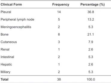
TABLE 1: Clinical forms of extrapulmonary tuberculosis.
Clinical Form Frequency Percentage (%)
Pleural 14 36.8
Peripheral lymph node 5 13.2
Meningoencephalitis 2 5.3
Bone 8 21.1
Cutaneous 3 7.9
Renal 1 2.6
Intestinal 2 5.3
Hepatic 1 2.6
Miliary 2 5.3
Total 38 100.0
concentration of 1 on the McFarland standard scale, then serial 10-fold dilutions were performed to achieve concentrations ranging from 3×10⁷ to 3×10¹⁰ bacilli/ml. The minimum detection
sensitivity of the assay was 15fg of DNA for both samples, corresponding to 3 bacilli/ml. This limit was defined by the amplification of the lowest dilution of the target DNA over the formation of primer dimers (the negative control of the reaction). To evaluate the positivity of biological samples, the threshold value of amplification (Ct) was established for positive and negative cases. The resulting Ct was 25 (25-33) and 34 (34-38) for the positive and negative tests, respectively (data not shown).
Statistical analysis
Culture and/or clinical responses to specific treatments were primarily considered the gold standard for performance
calculations. The IS6110-TaqMan® assay was also evaluated considering the results of the cultured samples as a gold standard (except for blood, for which no culturing was done). For the IS6110-TaqMan® assay, the sensitivity, specificity, and positive and negative predictive values were calculated, with a 95% confidence interval (CI) identified for each parameter. The database was constructed using Statistical Packages for the Social Sciences (SPSS) statistical software for Windows (Version 18, IBM Corporation, Armonk, NY, USA) which was also used to conduct statistical analyses, including cross tables and frequency calculations. For accuracy tests, the authors used the free software OpenEpi (Open Source Epidemiologic Statistics for Public Health, Version 3.01. Update 2013, available online at: www.OpenEpi.com).
For the blood samples, the IS6110-TaqMan® assay was positive when at least one blood phase, PBMC and/or plasma, was positive; negative results were obtained when both were negative24. The performance of the IS6110-TaqMan® assay was calculated in parallel for both blood phases.
In blood and urine samples, the performance of the
IS6110-TaqMan® assay was calculated alone and in parallel (blood and/ or urine positive or both negative).
The performance of the IS6110-TaqMan® assay was calculated for the combination of all biological sample types (blood and/or urine and/or biopsy or extrapulmonary liquid) from the same patient. In this case, a positive IS6110-TaqMan® assay was observed when at least one clinical sample had a positive result. Results were negative when all the patient's biological samples were negative.
Ethical considerations
This research was approved by the Ethics Committee of the Aggeu Magalhães Institute/FIOCRUZ (IAM/FIOCRUZ).
RESULTS
Clinical, epidemiological and laboratory findings
Out of the 57 participating patients with suspected extra-pulmonary TB, 38 (66.7%) had their final diagnosis defined as extrapulmonary TB (G1), and 19 (33.3%) patients were classified as non-TB (G2) with another kind of disease (different from TB) and
were considered a control group. Clinical forms of TB registered in the study are described in Table 1. Additionally, in the control group,
the following diagnoses were observed: lymphoma, bone cancer, sarcoidosis, and bacterial meningitis.
From the total number of patients included in the study, 32 (56.1%) were male. The age ranged from 1 to 89 years old, with an average of 34 years old. The majority (75%) of tested samples were negative for M. tuberculosis in culture.
Performance of the IS6110-TaqMan® assay in various clinical samples in patients with and
without extrapulmonary TB
The sensitivity, specificity, and positive/negative predictive values of the IS6110 TaqMan® assay on blood, urine, biopsy samples, and other body fluids, analyzed alone or in parallel, are described
in Table 2.
Considering the results of the cultured samples as a gold standard, the performance of TaqMan® ranged between 18-34% (sensitivity) and 75-100% (specificity). For urine samples, the sensitivity was 18.2% (CI = 4.6-41.0), and the specificity was 75% (CI = 60.9 -81.9). For biopsy tissues, these values were 34% (CI = 9.5-57.2) and 88.2% (CI = 65.7-96.7), respectively, and for extrapulmonary body fluids, 33.3% (CI = 9.7-70) and 100% (CI = 78.5 -100.0), respectively.
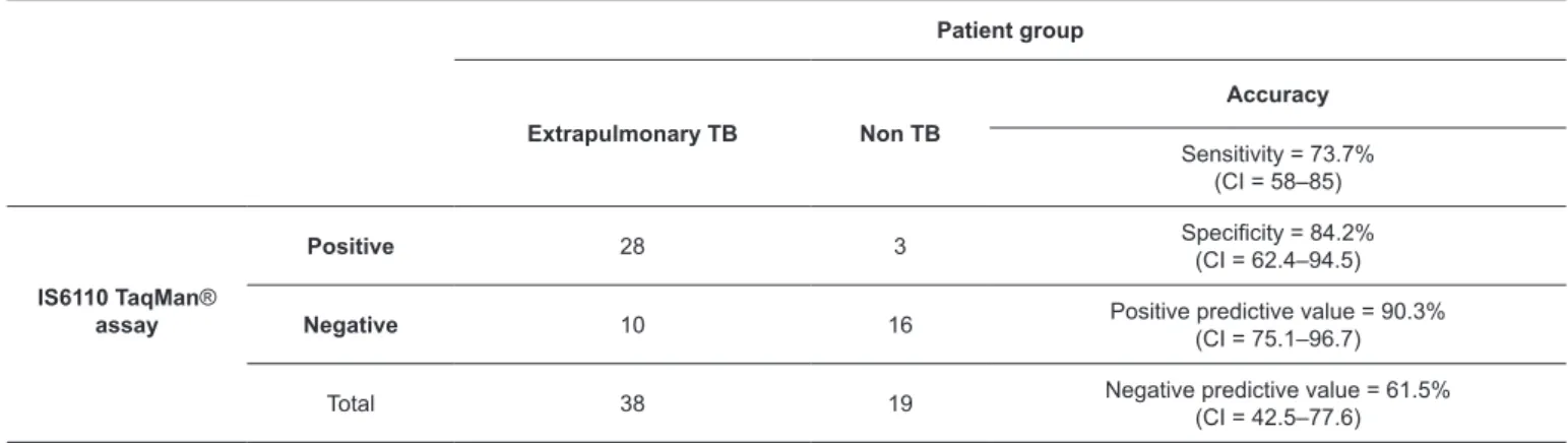
Considering all clinical samples from the same patients calculated in parallel, the sensitivity of the TaqMan® assay compared with culturing was 64.3% (CI = 39.2-89.4), and the specificity was 51% (CI = 36.2-66.1). These results are detailed in Table 3.
DISCUSSION
The development of sensitive and specific tools for the diagnosis of tuberculosis, mainly for paucibacillary forms such as extrapulmonary TB, remains a great challenge25. The
TABLE 2: Performance of the IS6110-TaqMan® assay in clinical sample between patients with extrapulmonary TB (G1) and No TB group (G2), using culture
and/or clinical diagnosis with the therapeutic response as gold-standard.
Clinical samples
Patients (n) IS6110-TaqMan
® assay
G1 G2 Sensitivity Specificity
Predictive value
positive negative
Blood 34 15 55.9% (19/34)
CI = 39.4–71.1
80% (12/15) CI = 54.8–93.0
86.4% (19/22) CI = 66.7–95.3
44.4% (12/27) CI = 27.6–62.7
Urine 33 13 33.3% (11/33)
CI = 19.8–50.4
100% (13/13) CI = 77.2–100.0
100% (11/11) CI = 74.1–100.0
37.1% (13/35) CI = 23.2–53.7
Blood + urine 32 14 68.8% (22/32)
CI = 51.4–82.1
78.6% (11/14) CI = 52.4–92.4
88% (22/25) CI = 70–95.8
52.4% (11/21) CI = 59.8–85.8
Biopsy 16 11 43.8% (7/16)
CI = 23.1–66,8
100% (11/11) CI = 74.1–100.0
100% (7/7) CI = 64.6–100.0
55% (11/20) CI = 34.2–74.2
Other
extrapulmonary body
fluids 15 7
29.6% (6/15) CI = 13.8–50.0
100% (7/7) CI = 64.6–100.0
100% (6/6) CI = 61–100.0
31.8% (7/16) CI = 16.4–52.7
TB: tuberculosis; G1 (Group 1): patients with extrapulmonary TB; G2 (Group 2): patients without TB (with a diagnosis other than TB); CI: confidence interval.
TABLE 3: Accuracy of the IS6110 TaqMan® assay in all biological sample types, analyzed together, perpatient.
Patient group
Extrapulmonary TB Non TB
Accuracy
Sensitivity = 73.7% (CI = 58–85)
IS6110 TaqMan® assay
Positive 28 3 Specificity = 84.2%
(CI = 62.4–94.5)
Negative 10 16 Positive predictive value = 90.3%
(CI = 75.1–96.7)
Total 38 19 Negative predictive value = 61.5%
(CI = 42.5–77.6)
TB: tuberculosis; CI: confidence interval.
fact that these are commonly patients with a low circulating bacterial load. In these cases, conventional exams of AFB and culture – the latter being considered a gold standard, usually yield negative results. In extrapulmonary TB, only one quarter of the cases can be diagnosed by bacteriological confirmation26.
From the 34 blood samples of patients with extrapulmonary TB, 15 were negative in the IS6110-TaqMan® assay. These false negative results might be related to the small quantities of targeted DNA present on the clinical samples or even the presence of PCR inhibitors27,28. On the other hand, three blood samples
were positive in the IS6110-TaqMan® assay, but these patients were finally diagnosed as non-TB, so the molecular results were
false positives. Therefore, it is possible that qPCR may detect the
presence of native genetic material from dead or latent organisms, generating discordant results with the gold standard used in the study. A study performed by Lima et al.29 analyzed blood samples
from patients under 15 years of age with extrapulmonary TB with nested PCR and showed similar results to the present study. Among the 33 urine samples from patients with extrapulmonary TB, 22 were negative in the IS6110-TaqMan® assay. These false negative results were probably due to the presence of components such as salts, enzymes, and other bacteria, which can compromise the purification process of DNA and therefore inhibit the amplification reaction in urine
15. In this study, there were no false positive urine results from
When the IS6110-TaqMan® assay for urine samples was compared with two different gold-standard criteria (1: culture
and/or response to antitubercular therapy; 2: culture alone),
the sensitivity increased by 15.1% when compared to the gold standard 1. These findings reinforce the limitation of culturing in cases of patients with paucibacillary forms30,31. Studies show
variability in positive results of the culture test (12% to 80%) in a range of clinical samples and biological tissues12,15,24.
Most biological samples analyzed in this study were blood and urine (66.4%), which are considered samples from ambulatory collection and represent a less invasive procedure for the patient. When blood and urine samples were evaluated in combination, the sensitivity of the molecular test (68.8%) increased by approximately 20% when compared with the results of the tests performed alone (55.9% and 33.3%, respectively).
Studies have shown that, despite the low sensitivity of molecular methods on urine samples, when these samples were analyzed in combination with blood samples, the sensitivity was increased by around 8-10%16,32. Lima et al.16 evaluated a
molecular diagnostic test for extrapulmonary TB in blood and urine samples. The sensitivity of the assay using combined samples (calculated in parallel) was 71.9%, but when using only urine samples, the sensitivity was 40.6%.
Rebollo et al.32 evaluated blood and urine samples from
patients diagnosed with TB using a simple PCR system, with the positive culture for each sample as the gold standard, and found a lower (42%) sensitivity. Despite their use of a conventional PCR system less sensitive than the IS6110-TaqMan® assay, the authors highlight the importance of using more than one type of sample from each patient with suspected TB, as this approach can increase the sensitivity. Cruz et al.11, when evaluating the usefulness of a nested-PCR method on samples of blood and urine of patients suspected to have extrapulmonary tuberculosis, also using IS6110 as target, found a sensitivity of 47.8% and 52% for blood and urine, respectively.
In this study, the sensitivity and specificity of the
IS6110-TaqMan® assay in extrapulmonary body fluid samples were 28.6% and 100%, respectively. Since the number of bacilli is lower in various extrapulmonary body fluids, it is known that false negative results arise mainly from the paucibacillary nature or even from the irregular distribution of bacilli in body fluids33, likely explaining the low sensitivity found in this study. Rosso et al.34 evaluated pleural fluid samples with the
IS6110-TaqMan® assay and obtained a better sensitivity (40%), probably because of the greater number of patients included in their study. Other research has also demonstrated a better performance of molecular tests in body fluids as compared with this study13,35-37.
We also evaluated the performance of the IS6110-TaqMan® assay in tissue samples. The sensitivity and specificity in this context were 43.8% and 100%, respectively. Out of the 16 tissue samples in group G1, the IS6110-TaqMan® assay was not able to detect Mycobacterium tuberculosis in 9 samples; these
results were considered false negatives. Possible causes were the quality and/or quantity of collected material, the presence of few bacilli on biopsied tissue, and collection outside the site
of infection, as well as the inhibitors present in the sample. No false positive results were found, showing that this test may aid in the confirmation of the disease.
Sun et al.35 evaluated the performance of qPCR on 90 kidney
biopsies and found a higher sensitivity than this study (83.3%), probably due to the inclusion of 30 patients with renal TB. Another work, which evaluated pleural fragments by qPCR using the same molecular target and extraction method, yielded similar results to this research (a sensitivity of 52.8% and specificity of 93.3%)34. In agreement with our study, Meghdadi
et al.38, using PCR targeting IS6110, demonstrated a sensitivity
and specificity of 48.6% and 100%, respectively.
In the different sample types evaluated, culture results demonstrated inferior performance in defining the final diagnosis of patients when compared with the gold standard 1. These results show the limitation of culture tests in the diagnosis of paucibacillary forms of TB, such as the extrapulmonary TB.
The main aim of this study was to evaluate the performance of detection of the IS6110 DNA sequence of M. tuberculosis
by qPCR in the laboratory diagnosis of extrapulmonary TB. Determination of sensitivity values for qPCR on each type of clinical sample yielded values ranging from 26.7% (in “other extrapulmonary body fluids”) to 55.9% (in blood samples), when analyzed alone. However, when the sensitivity of the
IS6110-TaqMan® assay was evaluated considering the combination of all clinical samples from a patient, it increased to 73.7%. A decrease in specificity (84.2%) was observed, however, compared to the analysis of clinical samples individually, particularly blood samples.
In regions where TB is endemic, negative results from the IS6110-TaqMan® assay must not be interpreted as excluding the possibility of the disease. We observed that the positive predictive values obtained were equal to or higher than 86% in all analyses, reinforcing the confirmatory power of the assay in the detection of M. tuberculosis in patients with suspected
extrapulmonary TB, despite the small sample sizes. However, the predictive values for the various tests analyzed (Table 2 and Table 3) cannot be inferred for the general population. These
values are not comparable because the prevalence of TB is not the same in all situations.
Our results demonstrate the importance of investigating more than one type of clinical sample collected from the same patient with suspected extrapulmonary tuberculosis, which might improve the predictive power of the IS6110-TaqMan assay®.
The IS6110-TaqMan® assay can be an important tool to facilitate the laboratory diagnosis of extrapulmonary TB, mainly in the most severe cases of the disease, to initiate specific therapy. However, more research is needed to clarify the value of this new technique as a diagnostic test in the clinical setting.
Acknowledgements
Conflict of interest
The authors declare that there is no conflict of interest.
Financial support
This study was funded by Programa de Excelência em Pesquisa/Clinical
Research (Grant number: 402058/2012-7), Research Program for the Single Health Service: Shared Health Management 2012 – Pernambuco and Instituto Aggeu Magalhães/Fundação Oswaldo Cruz.
REFERENCES
1. Gulland A. Tuberculosis killed 1.5 million people in 2014. BMJ. 2015;351:h5798.
2. World Health Organization (WHO). Global Tuberculosis Report 2016. Geneva: WHO; 2016. 214p. Available from: http://www.who. int/tb/publications/global_report/en/
3. Ministério da Saúde (MS). Sistema de Informação de Agravos de Notificação. (SINAN). Brasília: MS; 2014. Acessado em 12 de abril de 2016. Disponível em: http://dtr2004.saude.gov.br/sinanweb/ tabnet/dh?sinannet/tuberculose/bases/tubercbrnet.def.
4. Ministério da Saúde (MS). Secretaria de Vigilância em Saúde. Departamento de Vigilância Epidemiológica. Manual de recomendações para o controle da tuberculose no Brasil. Brasília: MS; 2011. 284p.
5. Marouane C, Smaoui S, Kammoun S, Slim L, Messadi-Akrout F. Evaluation of molecular detection of extrapulmonary tuberculosis and resistance to rifampicin with GeneXpert®MTB/RIF. Med Mal Infect. 2016;46(1):20-4.
6. Lange C, Mori T. Advances in the diagnosis of tuberculosis. Respirology. 2010;15(2):220-40.
7. Hillemann D, Rüsch-Gerdes S, Boehme C, Richter E. Rapid molecular detection of extrapulmonary tuberculosis by automated GeneXpert®MTB/RIF system. J Clin Microbiol. 2011;49(4):1202-5. 8. Bento J, Silva AS, Rodrigues F, Duarte R. Diagnostic tools in
tuberculosis. Acta Med Port. 2011;24(1):145-54.
9. McGrath EE, Anderson PB. Diagnostic tests for tuberculous pleural effusion. Eur J Clin Microbiol Infect Dis. 2010;29(10):1187-93. 10. Lewinsohn DM, Leonard MK, LoBue PA, Cohn DL, Daley CL,
Desmond E, et al. Official American Thoracic Society/Infectious Diseases Society of America/Centers for Disease Control and Prevention Clinical Practice Guidelines: Diagnosis of Tuberculosis in Adults and Children. Clin Infect Dis. 2017;64(2):111-5.
11. Lawn SD, Zumla AI. Diagnosis of extrapulmonary tuberculosis using the Xpert(®) MTB/RIF assay. Expert Rev Anti Infect Ther. 2012;10(6):631-5.
12. Furini AAC, Pedro HSP, Rodrigues JF, Montenegro LML, Machado RLD, Franco C, et al. Detection of Mycobacterium tuberculosis
complex by nested polymerase chain reaction in pulmonary and extrapulmonary specimens. J Bras Pneumol. 2013;39(6):711-8. 13. Amin I, Muhammad I, Awan Z, Shahid M, Afzal S, Hussain A. PCR
could be a method of choice for identification of both pulmonary and extra-pulmonary tuberculosis. BMC Res Notes. 2011;4:332. 14. Maurya AK, Kant S, Nag VL, Kushwaha RA, Kumar M, Dhole
TN. Comparative evaluation of IS6110 PCR via conventional methods in rapid diagnosis of new and previously treated cases of extrapulmonary tuberculosis. Tuberk Toraks. 2011;59(3):213-20.
15. Cruz HL, Montenegro RA, Lima JF, Poroca DR, Lima JF, Montenegro LM, et al. Evaluation of a nested-PCR for
Mycobacterium tuberculosis detection in blood and urine samples.
Braz J Microbiol. 2011;42(1):321-9.
16. Lima JFC, Guedes GMR, Lima JFA, Lira LAS, Santos FCF, Arruda ME, et al. Single-tube nested PCR assay with in-house DNA extraction for Mycobacterium tuberculosis detection in blood and
urine. Rev Soc Bras Med Trop. 2015;48(6):731-8.
17. Novais CM, Pires-Alves M, Silva FF. PCR em tempo real: uma inovação tecnológica da reação em cadeia da polimerase. Biotecnologia. 2004;33:10-3.
18. Armand S, Vanhuls P, Delcroix G, Courcol R, Lemaître N. Comparison of the Xpert MTB/RIF test with an IS6110-TaqMan real-time PCR assay for direct detection of Mycobacterium tuberculosis in respiratory and nonrespiratory specimens. J Clin
Microbiol. 2011;49(5):1772-6.
19. Yang YC, Lu PL, Huang SC, Jenh YS, Jou R, Chang TC. Detection of Mycobacterium tuberculosis complex in respiratory specimens. J
Clin Microbiol. 2011;49(3):797-801.
20. Lira LAS, Santos FCF, Carvalho MSZ, Montenegro RA, Lima JFC, Schindler HC, et al. Evaluation of a IS6110-Taqman real-time PCR assay to detect Mycobacterium tuberculosis in sputum samples of
patients with pulmonary TB. J Appl Microbiol. 2013;114(4):1103-8. 21. Ministério da Saúde (MS). Secretaria de Vigilância em Saúde.
Departamento de Vigilância Epidemiológica. Manual nacional de vigilância laboratorial da tuberculose e outras micobacterioses. Brasília: MS; 2008. 406p.
22. Cole ST, Brosch R, Parkhill J, Garnier T, Churcher C, Harris D, et al. Deciphering the biology of Mycobacterium tuberculosis from
the complete genome sequence. Nature. 1998;393(6685):537-44. 23. Broccolo F, Scarpellini P, Locatelli G, Zingale A, Brambilla AM,
Cichero P, et al. Rapid diagnosis of Mycobacterial infections and quantification of Mycobacterium tuberculosis load by two
Real time calibrated PCR assays. J Clin Microbiol. 2003;41(10): 4565-72.
24. Wu SH, Ho CM, Lu JJ. Diagnosis of tuberculosis by PCR-based amplification of mpt64 gene from peripheral blood. Int J Bio Lab Sci.2013;2(1):25-30.
25. Purohit M, Mustafa T. Laboratory diagnosis of extra-pulmonary tuberculosis (EPTB) in resource-constrained setting: state of the art, challenges and the need. J Clin Diagn Res. 2015;9(4):EE01-6. 26. Lee JY. Diagnosis and treatment of extrapulmonary tuberculosis.
Tuberc Respir Dis. 2015;78(2):47-55.
27. Al-Soud WA, Râdström P. Purification and Characterization of PCR-inhibitory components in blood cells. J Clin Microbiol. 2001;39(2):485-93.
28. Valentine-Thon E. Quality control in nucleic acid testing-where do we stand? J Clin Virol. 2002;25(3):13-21.
29. Lima JF, Montenegro LM, Montenegro RA, Cabral MM, Lima AS, Abath FG, et al. Performance of nested PCR in the specific detection of Mycobacterium tuberculosis complex in blood samples
of pediatric patients. J Bras Pneumol. 2009;35(7):690-7.
30. Pai M, Ling DI. Rapid diagnosis of extrapulmonary tuberculosis using nucleic acid amplification tests: What is the evidence? Future Microbiol. 2008;3(1):1-4.
31. Ajantha GS, Shetty PC, Kulkarni RD, Biradar U. PCR as a diagnostic tool for extra-pulmonary tuberculosis. J Clin Diagn Res.
2013;7(6):1012-5.
33. Green C, Huggett JF, Talbot E, Mwaba P, Reither K, Zumla AI. Rapid diagnosis of tuberculosis through the detection of mycobacterial DNA in urine by nucleic acid amplification methods. Lancet Infect Dis. 2009;9(8):505-11.
34. Rosso F, Michelon CT, Sperhacke RD, Verza M, Olival L, Conde MB, et al. Evaluation of real-time PCR of patient pleural effusion for diagnosis of tuberculosis. BMC Res Notes. 2011;4:279.
35. Sun L, Yuan Q, Feng JM, Yang CM, Yao L, Fan QL et al. Rapid diagnosis in early stage renal tuberculosis by real-time polymerase chain reaction on renal biopsy specimens. Int J Tuber Lung Dis.
2010;14(3):341-6.
36. Mehta PK, Raj A, Singh N, Khuller GK. Diagnosis of extrapulmonary tuberculosis by PCR. FEMS Immunol Med Microbiol. 2012;66(1):20-36.
37. Khosravi AD, Alami A, Meghdadi H, Hosseini AA. Identification of Mycobacterium tuberculosis in clinical specimens of patients
suspected of having extrapulmonary tuberculosis by application of nested PCR on five different genes. Front Cell Infect Microbiol. 2017;7:3. doi: 10.3389/fcimb.2017.00003.
38. Meghdadi H, Khosravi AD, Ghadiri AA, Sina AH, Alami A. Detection of Mycobacterium tuberculosis in extrapulmonary
biopsy samples using PCR targeting IS6110, rpoB, and nested-rpoB