Mariana Santos
Moreda Graça
Caracterização de novos complexos da LAP1 e a
sua relevância na distonia DYT1
Characterization of novel LAP1 complexes and their
relevance in DYT1 dystonia
Universidade de Aveiro 2014
Secção Autónoma de Ciências da Saúde
Mariana Santos
Moreda Graça
Caracterização de novos complexos da LAP1 e a sua
relevância na distonia DYT1
Characterization of novel LAP1 complexes and their
relevance in DYT1 dystonia
Tese apresentada à Universidade de Aveiro para cumprimento dos requisitos necessários à obtenção do grau de Doutor em Ciências Biomédicas, realizada sob a orientação científica da Professora Doutora Odete Abreu Beirão da Cruz e Silva, Professora Auxiliar com Agregação da Secção Autónoma de Ciências da Saúde da Universidade de Aveiro, e coorientação científica da Professora Doutora Sandra Maria Tavares da Costa Rebelo, Professora Auxiliar Convidada da Secção Autónoma de Ciências da Saúde da Universidade de Aveiro.
Apoio financeiro da FCT e do FSE no âmbito do III Quadro Comunitário de Apoio (Bolsa de Doutoramento SFRH/ BD/65353/2009).
o júri
presidente Professor Doutor António Carlos Mendes de Sousa
Professor Catedrático do Departamento de Engenharia Mecânica da Universidade de Aveiro
Professora Doutora Maria João Gameiro de Mascarenhas Saraiva
Professora Catedrática do Instituto de Ciências Biomédicas Abel Salazar da Universidade do Porto
Professor Doutor Carlos Jorge Alves Miranda Bandeira Duarte
Professor Associado com Agregação da Faculdade de Ciências e Tecnologia da Universidade de Coimbra
Professor Doutor João Carlos Cruz de Sousa
Professor Auxiliar da Escola de Ciências da Saúde da Universidade do Minho
Doutora Sónia Cristina das Neves Ferreira
Investigadora de Pós-Doutoramento da Universidade de Coimbra
Professora Doutora Ana Gabriela Henriques
Professora Auxiliar Convidada da Secção Autónoma de Ciências da Saúde da Universidade de Aveiro
Professora Doutora Odete Abreu Beirão da Cruz e Silva
Professora Auxiliar com Agregação da Secção Autónoma de Ciências da Saúde da Universidade de Aveiro
Professora Doutora Sandra Maria Tavares da Costa Rebelo
Professora Auxiliar Convidada da Secção Autónoma de Ciências da Saúde da Universidade de Aveiro
agradecimentos Ao Prof. Edgar da Cruz e Silva pela orientação na fase inicial do meu doutoramento, por todo o conhecimento partilhado e pela oportunidade de trabalhar neste projeto.
À Prof. Odete pela orientação e oportunidade de trabalhar no Laboratório de Neurociências. À Sandra Rebelo pela orientação, e por me encorajar e acompanhar durante este trabalho.
A todos os colegas do CBC pela ajuda e apoio. Em particular, às minhas “companheiras de bancada” Filipa e Pati, e à Joana Rocha, à Joana Oliveira, à Lili, à Regina, ao Roberto e à Sara Soares por todo o apoio, companheirismo e por animarem os meus dias no laboratório. À Patrícia Costa por me ter acompanhado neste trabalho. Ao Luís Godinho pela ajuda e conhecimentos partilhados. À Sara Domingues, à Sara Esteves, ao Korrodi, à Sandra Vieira, à Margarida Fardilha, à Fátima e ao Miguel por estarem sempre disponíveis a ajudar.
À Mónica Ferreira, que me acompanha desde que cheguei ao CBC, e à Mónica Almeida. Obrigado pelos bons momentos partilhados ao longo destes anos e pela vossa amizade.
E ainda aos nossos colaboradores Dr. William Dauer e Dr. Thorsten Muller por terem contribuído para a realização de alguns objetivos deste trabalho.
Aos amigos de sempre pela vossa amizade. À Tatiana, ao Daniel, ao Avelino, ao Pedro, ao Álvaro, à Ângela, à Inês e ao Chico.
Ao Eurico por todo o apoio, carinho e paciência.
palavras-chave Proteína 1B associada com a lâmina (LAP1B), invólucro nuclear, fosforilação de proteínas, proteína fosfatase 1 (PP1), torsinaA, distonia.
resumo A distonia é a terceira doença de movimento mais comum depois do tremor essencial e da doença de Parkinson e abrange um variado número de síndromes clínicos. A distonia DYT1 é a forma mais comum de distonia isolada de início precoce e é causada por uma mutação no gene DYT1, a qual resulta na perda de um ácido glutâmico na proteína torsinaA (ΔE-torsinaA). A torsinaA localiza-se no retículo endoplasmático e no invólucro nuclear (IN), enquanto que a ΔE-torsinaA é anormalmente distribuída para o IN. No IN, a torsinaA interage com a proteína 1 associada com a lâmina (LAP1), cuja função ainda não é totalmente conhecida. A LAP1 é uma proteína da membrana interna do núcleo e três isoformas (LAP1A, B e C) desta proteína já foram descritas na literatura.
As interações proteína-proteína têm vindo a ser determinantes no estudo de doenças humanas e de sinalização celular. Neste trabalho, foi identificada uma nova interação entre a LAP1B humana e a proteína fosfatase 1 (PP1). A PP1 é uma fosfatase serina/treonina que é responsável pela desfosforilação de cerca de um terço de todas as proteínas em células eucarióticas. A versatilidade da PP1 é possível através da sua ligação a diferentes proteínas reguladoras responsáveis pela sua especificidade e atividade. A LAP1B é desfosforilada pela PP1 e liga-se a esta através de um motivo semelhante ao RVxF que se encontra no seu domínio nucleoplasmático. No decorrer deste trabalho, identificamos também uma nova isoforma da LAP1 humana – LAP1C, que nunca tinha sido descrita em células humanas. A expressão da LAP1B e LAP1C humanas parece ser regulada durante a diferenciação celular e neuronal e são ambas desfosforiladas pela PP1. Além disso, estas proteínas são provavelmente crucias para a manutenção do IN e regulação da mitose. Dado que a PP1 interage com a LAP1 no nucleoplasma e esta interage com a torsinaA no espaço perinuclear, a existência do tricomplexo PP1/LAP1/torsinaA foi validada em cérebro de rato e linhas celulares. A torsinA é desfosforilada pela PP1 mas liga-se a esta, provavelmente, através da LAP1. Dada a localização aberrante da ΔE-torsinaA no IN, é possível que o papel dos complexos formados pela LAP1 no IN e a sua regulação por fosforilação esteja na base da patologia da distonia DYT1. Além disso, a distonia DYT1 tem sido relacionada com uma disfunção nos circuitos dos gânglios de base e da sinalização dopaminérgica. Curiosamente as proteínas PP1 e DARPP-32 desempenham um papel fundamental na sinalização dopaminérgica. Assim, os nossos resultados podem ajudar a compreender os mecanismos moleculares e celulares inerentes à distonia DYT1, onde a fosforilação de proteínas pode desempenhar um papel crucial.
keywords Lamina associated polypeptide 1 (LAP1), nuclear envelope, protein phosphorylation, protein phosphatase 1 (PP1), torsinA, dystonia.
abstract Dystonia is the third most common movement disorder, following essential tremor and Parkinson’s disease, and comprises a large number of clinical syndromes. DYT1 dystonia is recognized as the most common form of early onset isolated dystonia and is caused by a mutation in the DYT1 gene, resulting in the loss of a single glutamic acid within the torsinA protein (ΔE-torsinA). TorsinA resides in the endoplasmic reticulum and nuclear envelope (NE), but the mutant form is abnormally relocated to the NE. At the NE, torsinA interacts with lamina associated polypeptide 1 (LAP1), whose function is poorly understood. LAP1 is a transmembrane protein of the inner nuclear membrane that was described to exist as three alternatively spliced isoforms (LAP1A, B and C) in rat.
Protein-protein interactions are becoming increasingly important in the study of human diseases and signaling pathways. In this work, human LAP1B was identified as a novel protein phosphatase 1 (PP1) binding protein. PP1 is a major Ser/Thr phosphatase that is estimated to dephosphorylate about one third of all proteins in eukaryotic cells. The versatility of PP1 is only possible by complexing with different binding proteins that play regulatory and targeting roles. We found that LAP1B is a substrate of PP1 and binds to the latter through an RVxF-like motif located in the nucleoplasmic domain. In the course of this work, we also identified a novel human LAP1 isoform - LAP1C that is N-terminally truncated. Human LAP1B and LAP1C isoforms seem to be developmentally regulated and are both dephosphorylated by PP1. Moreover, LAP1 proteins seem to be important for NE integrity and mitosis regulation. Given that PP1 interacts with LAP1 in the nucleoplasm, which in turn interacts with torsinA in the perinuclear space, the existence of the tricomplex PP1/LAP1/torsinA was validated in rat brain and cultured cells. Moreover, torsinA was dephosphorylated by PP1 but probably binds to the latter via LAP1. The aberrant behavior of ΔE-torsinA in the NE implicates nuclear dysfunction in DYT1 dystonia pathogenesis. Thereby, the role of LAP1 complexes in the NE and its regulation by protein phosphorylation may underlie the pathology of DYT1 dystonia and other NE-related diseases. Moreover, DYT1 dystonia has been related with a dysfunction in the basal ganglia circuit, including dopamine signaling disturbance. Interestingly, PP1/DARPP-32 cascade plays a key role in mediating the actions of dopamine and modulating the phosphorylation and activity of effectors in dopaminoceptors neurons. Thus, our results may lead to novel insights into the molecular and cellular mechanisms of DYT1 dystonia, where protein phosphorylation cascades represent a regulatory mechanism.
Characterization of novel LAP1 complexes and their relevance in DYT1 dystonia 7
CONTENTS
Abbreviations ... 13
Chapter I – Introduction ... 17
I.1. Protein phosphorylation ... 19
I.1.1. Protein phosphatases ... 21
I.1.2. Protein phosphatase 1 ... 22
I.2. The nuclear envelope ... 39
I.2.1. Lamins, the main components of the nuclear lamina ... 40
I.2.2. Properties and targeting of INM proteins ... 43
I.2.3. NE disassembly and reassembly during mitosis ... 46
I.2.4. Lamina associated polypeptide 1 (LAP1) ... 47
I.2.5. Relevance of nuclear envelope proteins in disease ... 56
I.3. TorsinA ... 58
I.3.1. TorsinA interactors and related functions ... 60
I.4. Dystonia ... 67
I.4.1. Early-onset generalized isolated dystonia ... 69
I.5. Objectives ... 76
Chapter II - LAP1B is a novel PP1 binding protein ... 77
II.1. Introduction ... 81
II.2. Materials and Methods ... 83
II.2.1. Antibodies ... 83
II.2.2. Expression vectors and DNA constructs ... 83
II.2.3. Yeast co-transformation analysis ... 84
II.2.4. Expression of recombinant proteins in Escherichia coli ... 85
II.2.5. Blot overlay assays ... 85
II.2.6. Cell culture and transfection ... 86
II.2.7. Brain dissection ... 86
II.2.8. Co-immunoprecipitation ... 87
II.2.9. Immunocytochemistry ... 87
II.2.10. In vitro dephosphorylation of LAP1B ... 88
8
II.3. Results ... 89
II.3.1. Identification of LAP1B as a novel putative PP1 regulatory protein ... 89
II.3.2. LAP1B and PP1 interact in vitro ... 92
II.3.3. The novel complex LAP1B:PP1 is also formed in vivo ... 94
II.3.4. Both LAP1B isoforms bind to PP1 ... 97
II.3.5. Localization of the LAP1B:PP1 complex ... 97
II.3.6. PP1 specifically binds to LAP1B ... 100
II.3.7. LAP1B is a novel substrate for PP1 ... 100
II.4. Discussion ... 102
Chapter III - Characterization of human LAP1 isoforms ... 107
III.1. Introduction ... 111
III.2. Materials and Methods ... 113
III.2.1. Antibodies ... 113
III.2.2. Expression vectors and DNA constructs ... 113
III.2.3. Brain dissection ... 113
III.2.4. Cell culture and transfection ... 114
III.2.5. LAP1B knockdown ... 115
III.2.6. RT-PCR and sequencing ... 115
III.2.7. RNA isolation ... 116
III.2.8. Northern blot analysis... 116
III.2.9. Co-immunoprecipitation ... 117
III.2.10. LAP1 solubilization assay ... 117
III.2.11. Nano-HPLC and Mass spectrometry ... 118
III.2.12. In vitro translation (IVT) ... 119
III.2.13. SDS-PAGE and immunoblotting ... 119
III.2.14. Bioinformatics analysis ... 119
III.2.15. Quantification and Statistical Analysis ... 120
III.3. Results ... 121
III.3.1. Knockdown of human LAP1 ... 121
III.3.2. In silico analysis of the TOR1AIP1 gene ... 123
III.3.3. Analysis of LAP1 transcripts ... 128
Characterization of novel LAP1 complexes and their relevance in DYT1 dystonia 9
III.3.5. Identification of a putative promoter in LAP1C sequence ... 135
III.3.6. Functional characterization of LAP1 isoforms ... 136
III.3.7. Regulation of both isoforms by pos-translational modifications ... 140
III.4. Discussion ... 143
Chapter IV - Potential role of LAP1 in nuclear envelope dynamics... 149
IV.1. Introduction ... 153
IV.2. Materials and Methods ... 155
IV.2.1. Antibodies ... 155
IV.2.2. Cell culture and cell cycle arrest ... 155
IV.2.3. Immunocytochemistry ... 155
IV.2.4. LAP1 knockdown ... 156
IV.2.5. Protein phosphorylation analysis ... 156
IV.2.6. SDS-PAGE and immunoblotting ... 156
IV.2.7. Quantification and Statistical Analysis ... 157
IV.3. Results ... 158
IV.3.1. Intracellular levels and phosphorylation state of LAP1 during mitosis .... 158
IV.3.2. Localization of LAP1 during mitosis ... 159
IV.3.3. Effects of LAP1 knockdown ... 161
IV.4. Discussion ... 164
Chapter V - Novel insights into DYT1 dystonia pathophysiology… ... 167
Chapter V.A – Characterization of DYT1 dystonia cellular models ... 170
V.A.1. Introduction ... 171
V.A.2. Material and methods ... 172
V.A.2.1. Antibodies ... 172
V.A.2.2. Expression vectors and DNA constructs ... 172
V.A.2.3. Cell culture and transfection ... 172
V.A.2.4. Immunocytochemistry ... 173
V.A.2.5. Statistical Analysis ... 173
V.A.3. Results ... 174
V.A.4. Discussion ... 180
Chapter V.B – DYT1 dystonia-associated mutant affects cytoskeletal… ... 182
10
V.B.2. Material and methods ... 185
V.B.2.1. Antibodies and drugs ... 185
V.B.2.2. Expression vectors and DNA constructs ... 185
V.B.2.3. Cell culture and transfection ... 185
V.B.2.4. Immunocytochemistry ... 185
V.B.2.5. Statistical Analysis ... 186
V.B.3. Results ... 187
V.B.4. Discussion ... 190
Chapter V.C – Identification of the novel LAP1/PP1/TorsinA complex ... 192
V.C.1. Introduction ... 194
V.C.2. Materials and Methods ... 196
V.C.2.1. Antibodies ... 196
V.C.2.2. Expression vectors and DNA constructs ... 196
V.C.2.3. Cell culture and transfection ... 196
V.C.2.4. Brain dissection ... 197
V.C.2.5. Co-immunoprecipitation ... 197
V.C.2.6. Blot overlay assay ... 197
V.C.2.7. Immunocytochemistry ... 198
V.C.2.8. Detection of protein phosphorylation ... 198
V.C.2.9. LAP1 knockdown ... 199
V.C.2.10. SDS-PAGE and immunoblotting ... 199
V.C.2.11. Quantification and Statistical Analysis ... 199
V.C.3. Results ... 200
V.C.3.1. Identification of the novel LAP1/PP1/TorsinA complex ... 200
V.C.3.2. Localization of the trimeric complex ... 202
V.C.3.3. LAP1 as a bridging protein between torsinA and PP1 ... 204
V.C.3.4. TorsinA is dephosphorylated by PP1 ... 205
V.C.3.5. Effects of LAP1 knockdown ... 207
V.C.4. Discussion ... 209
Chapter VI - Discussion and concluding remarks ... 213
VI.1. LAP1B, a novel PP1 binding protein ... 215
Characterization of novel LAP1 complexes and their relevance in DYT1 dystonia 11
VI.3. Potential role of LAP1 proteins during cell cycle ... 217
VI.4. Relevance of PP1/LAP1/torsinA complex in DYT1 dystonia ... 218
VI.4.1. DYT1 dystonia, a nuclear envelope-related disease ... 219
VI.4.2. Potential role of the PP1/LAP1/TorsinA complex in NE dynamics ... 219
VI.4.3. Abnormal dopamine signaling in DYT1 dystonia ... 222
VI.5. Concluding remarks ... 223
References ... 225
Appendix I – Culture media and solutions ... 259
Appendix II – Primers ... 270
Appendix III – Bacteria and yeast strains ... 271
Characterization of novel LAP1 complexes and their relevance in DYT1 dystonia 13
ABBREVIATIONS
AAA+ ATPases associated with a variety of cellular activities
ADLD Autosomal dominant leukodystrophy
AKAP A-kinase anchoring protein
AMPA α-amino-3-hydroxy-5-methyl-4-isoxazolepropionic acid
ATM Ataxia-telangiectasia mutated
ATR Ataxia-telangiectasia and Rad 3-related
BAF Barrier autointegration factor
BM Binding motif
CaMKII Ca2+/calmodulin-dependent kinase II
CDK Cyclin-dependent kinase
cDNA Complementary deoxyribonucleic acid
CENP-E Centromere-associated protein E
CMS Control missense
CNS Central nervous system
Co-IP Co-immunoprecipitation
CREB cAMP-responsive element-binding protein
DARPP-32 Dopamine and cAMP-regulated phosphoprotein, Mr 32 kDa
DIV Days in vitro
DNA Deoxyribonucleic acid
DOPAC 3-,4-dihydroxyphenylacetic acid
ECL Enhanced chemiluminescence
EDMD Emery-Dreifuss muscular dystrophy
ER Endoplasmic reticulum
EST Expressed sequence tag
FI Fluorescence intensity
FITC Fluorescein isothiocyanate
GABA γ-aminobutyric acid
GFP Green fluorescente protein
GSK3β Glycogen synthase kinase 3β
HP1 Heterochromatin protein 1
14
HVA Homovanilic acid
INM Inner nuclear membrane
IP Immunoprecipitation
IVT In vitro translation
KASH Klarsicht/ANC-1/Syne-1 homology
KLC Kinesin light chain
LAP Lamina associated polypeptide
LBR Lamin B receptor
LINC Linker of nucleoskeleton and cytoskeleton
LTD Long-term depression
LTP Long-term potentiation
LULL1 Lumenal domain- like LAP1
MAD2 Mitotic arrest deficient 2
MEF2 Myocyte enhancer factor-2
mRNA Messenger ribonucleic acid
MS Mass spectrometry
Mypt1 Myosin phosphatase targeting subunit 1
NE Nuclear envelope
NIPP1 Nuclear inhibitor of PP1
NLS Nuclear localization signal
NMDA N-methyl-D-aspartate
NPC Nuclear pore complex
OA Okadaic acid
ONM Outer nuclear membrane
ORF Open reading frame
PK Protein kinase
PLK1 Polo-like kinase 1
PNUTS Phosphatase 1 nuclear targeting subunit
PP Protein phosphatase
PP1c Protein phosphatase 1 catalytic subunit
PPPs Phosphoproteins phosphatases
PPMs Metal-dependent protein phosphatases
QDO Quadruple dropout medium
Characterization of novel LAP1 complexes and their relevance in DYT1 dystonia 15
RNA Ribonucleic acid
RNAi RNA interference
RT-PCR Reverse transcriptase- polymerase chain reaction
SAC Spindle assembly checkpoint
SAP155 Spliceosome-associated protein 155
SD Synthetic dropout medium
Ser Serine
shRNA Short/Small hairpin RNA
SUN Sad1-UNC84 homology
Thr Threonine
TM Transmembrane
TRF2 Telomeric repeat binding factor 2
Tyr Tyrosine
UTR Untranslated region
VAMP Vesicle-associated membrane protein
VMAT2 Vesicular monoamine transporter 2
Wt Wild-type
Characterization of novel LAP1 complexes and their relevance in DYT1 dystonia 17
CHAPTER I - INTRODUCTION
Characterization of novel LAP1 complexes and their relevance in DYT1 dystonia 19
I.1. PROTEIN PHOSPHORYLATION
Reversible protein phosphorylation is the most common type of pos-translational modification in eukaryotic organisms. It involves either the addition of phosphate groups via the transference of the terminal phosphate from ATP to an amino acid residue by protein kinases or its removal by protein phosphatases (Fig. I.1). Protein phosphorylation is a crucial mechanism for signal transduction that regulates the biological activity of diverse proteins. For example, protein phosphorylation allows for the regulation of enzymatic activities and is important for modulation of protein-protein interactions (Barford et al., 1998). About one third of all eukaryotic proteins can be phosphorylated, mainly at serine (Ser), threonine (Thr) and tyrosine (Tyr) amino acids. Proteomic analysis of more than 2000 human proteins revealed that phosphorylation at Ser, Thr and Tyr accounts for 86.4%, 11.8% and 1.8%, respectively, of the phosphorylated amino acids (Olsen et al., 2006). Analysis of the human genome revealed that it contains about 518 kinases that are classified as Tyr kinases or Ser/thr kinases (Johnson and Hunter, 2005). Whereas the number of Tyr kinases is approximately the same as that for Tyr phosphatases, the number of Ser/Thr kinases is much higher when compared to Ser/Thr phosphatases. The discrepancy of this number can be explained by the unique manner by which Ser/Thr phosphatases are regulated. These enzymes have additional interacting and regulatory proteins that bind to the phosphatase catalytic subunits and control their activity and/or subcellular location (Bollen et al., 2010; Cohen, 2002b; Moorhead et al., 2007).
Figure I.1. Schematic representation of reversible protein phosphorylation. Protein kinases transfer a phosphate group from ATP to the target protein (protein phosphorylation), while protein phosphatases catalyze the hydrolysis of the phosphate group from the target protein (protein dephosphorylation).
20
Protein phosphorylation is a major mechanism for controlling several intracellular events in eukaryotic cells, such as, metabolism, contractility, membrane transport and secretion, transcription and translation, cell division, fertilization and memory (reviewed in Cohen, 1989). Additionally, aberrant protein phosphorylation of key proteins has been linked to many diseases and dysfunctional states, such as, cancer, metabolic disorders and inflammatory and neurological diseases. In neurodegenerative diseases, such as Alzheimer’s disease, there is evidence for abnormal regulation of protein phosphorylation. Alzheimer’s disease is primarily characterized by the presence of neuritic plaques and neurofibrillary tangles in the brains of affected individuals. Neurofibrillary tangles result from the aggregation of hyperphosphorylated tau protein into paired helical filaments. Glycogen synthase kinase 3β (GSK3β) and protein kinase A (PKA) may be key kinases involved in tau phosphorylation (Delobel et al., 2002a), while inhibition of protein phosphatase 2A (PP2A) and protein phosphatase 1 (PP1) lead to tau hyperphosphorylation (Bennecib et al., 2000; Planel et al., 2001). Moreover, the inactivation of PP1 leads to a relative increase in Alzheimer’s amyloid precursor protein processing by the non-amyloidogenic pathway (da Cruz e Silva et al., 1995a). Other neurodegenerative diseases associated with abnormal protein phosphorylation are Parkinson’s disease and Huntington’s disease. Parkinson’s disease is characterized by the presence of Lewy bodies (proteinaceous inclusions) that contain phosphorylated and aggregated α-synuclein. It was demonstrated that α-synuclein is constitutively phosphorylated, predominantly on serine residues (Okochi et al., 2000), and the levels of phosphorylated α-synuclein were found to be higher in Parkinson’s disease patients compared to the control cases (Foulds et al., 2011). Synapsin I, a major phosphoprotein important in regulating neurotransmitter release, is abnormally phosphorylated in mice expressing the Huntington’s disease mutation. It has been suggested that an imbalance between kinase and phosphatase activities also occurs in Huntington’s disease (Lievens et al., 2002). Therefore, protein phosphorylation systems represent attractive targets for diagnostics and therapeutics in several diseases. Indeed, kinases are the second most important group of drug targets in the pharmaceutical industry’s (Cohen, 2002a). In contrast, protein phosphatases were recognized later as potential therapeutic targets. Further, the therapeutic value of protein phosphatases interacting proteins as targets is only currently being addressed (da Cruz e Silva et al., 2004; Fardilha et al., 2010).
Characterization of novel LAP1 complexes and their relevance in DYT1 dystonia 21
I.1.1. Protein phosphatases
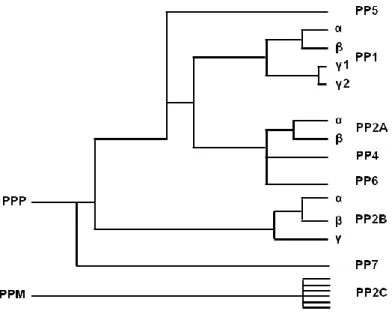
Based on substrate specificity, eukaryotic protein phosphatases can be classified into two families: ser/thr protein phosphatases, that catalyze the dephosphorylation of ser and thr amino acids, and tyr phosphatases, which catalyze the dephosphorylation of tyr amino acids. Ser/thr protein phosphatases can be categorized into three families: phosphoproteins phosphatases (PPPs), metal-dependent protein phosphatases (PPMs) and the aspartate-based phosphatases represented by FCP/SCP (TFIIF-associating component of RNA polymerase II CTD phosphatase/small CTD phosphatase). Members of the PPPs family include: PP1, PP2A, PP2B (also known as calcineurin), PP4, PP5, PP6 and PP7. PP1 and PP2A are some of the most well conserved enzymes with a variety of cellular functions attributed to the interaction with a large number of regulatory subunits (reviewed in Honkanen and Golden, 2002; Moorhead et al., 2009; Shi, 2009). PP2B alone is inactive, acquiring phosphatase activity after binding with Ca2+-calmodulin. In contrast, PP1 and PP2A are mainly active in the absence of divalent cations, despite dephosphorylation of some substrates being strongly stimulated by Mn2+ (Cohen, 1989; Wang et al., 2008a). PP4, PP5 and PP6, like PP1, PP2A and PP2B, were identified in all mammalian tissues examined. At the structural level, PP4 and PP6 are closely related to the catalytic subunit of PP2A. PP5 contains an N-terminus tetratricopeptide repeat domain that is a protein-protein interaction motif (Cohen, 1997; Shi, 2009). In contrast with other PPPs, human PP7 is not ubiquitous and was primarily found in retina. PP7 contains multiple Ca2+ binding sites and its activity is dependent of Mg2+ (Kutuzov et al., 2002). These PPPs contain a common catalytic core domain that is conserved among species. The remarkable degree of evolutionary conservation of these enzymes (Fig. I.2) is related to their essential role in the regulation of fundamental cellular processes (Honkanen and Golden, 2002). PP1, PP2A, and PP2B together with PP2C of the PPM family, account for the majority of the protein ser/thr phosphatase activity in vivo (Barford et al., 1998). The PPM family includes PP2C and pyruvate dehydrogenase phosphatase, which are Mn2+/Mg2+ dependent enzymes. Unlike most PPP family members, PPMs do not have additional regulatory subunits but contain additional domains and conserved motifs that may determine substrate specify. Members of the FCP/SCP family use an aspartate-based catalysis mechanism (reviewed in Moorhead et al., 2009; Shi, 2009).
22
Figure I.2. Phylogenic tree representing the homology between members of the phosphoprotein phosphatase (PPP) family based on their primary amino acid sequence. PP1-PP7 belongs to the PPP family that is structurally different from PP2C (metal-dependent protein phosphatase family, PPM) (adapted from Honkanen and Golden, 2002).
I.1.2. Protein phosphatase 1
PP1 and PP2A together are responsible for more than 90% of the protein phosphatase activity in eukaryotic cells. However, in terms of substrate diversity, PP1 is predicted to catalyze the majority of protein dephosphorylation events (Bollen et al., 2010; Heroes et al., 2013). As a major phosphatase, PP1 is present in various eukaryotic organisms. Eukaryotic genomes contain multiple genes encoding PP1 isoforms with the exception of yeast Saccharomyces cerevisiae which only contains one PP1 gene. PP1 isoforms are about 70% identical in the central region and mainly differ at the N- and C-terminal sequences. Moreover, PP1 was shown to be one of the most conserved eukaryotic proteins. PP1 sequences are highly conserved between different species, as is the case for Giardia lamblia which expresses a PP1 isoform very similar to the mammalian form. This suggests that PP1 may have similar functions in different organisms (reviewed in Ceulemans and Bollen, 2004; Lin et al., 1999). In mammals, PP1 isoforms are encoded by three genes: PPP1CA, PPP1CB and PPP1CC that encode for PP1alpha (PP1α), PP1beta/delta (PP1β/δ) and PP1gamma (PP1γ), respectively (Barker et al., 1994; Barker et al., 1993; Sasaki et al., 1990). These isoforms are about
Characterization of novel LAP1 complexes and their relevance in DYT1 dystonia 23
90% identical in amino acid sequence; most of the differences are located at the N- and C-terminals (Fig. I.3). The gene encoding PP1γ undergoes alternative splicing to originate an ubiquitous PP1gamma1 (PP1γ1) variant and a PP1gamma2 (PP1γ2) variant enriched in testis (da Cruz e Silva et al., 1995b).
PP1gamma1 MADLDKLNIDSIIQRLLEVRGSKPGKNVQLQENEIRGLCLKSREIFLSQPILLELEAPLK 60 PP1gamma2 MADLDKLNIDSIIQRLLEVRGSKPGKNVQLQENEIRGLCLKSREIFLSQPILLELEAPLK 60 PP1alpha MSDSEKLNLDSIIGRLLEVQGSRPGKNVQLTENEIRGLCLKSREIFLSQPILLELEAPLK 60 PP1beta MADG-ELNVDSLITRLLEVRGCRPGKIVQMTEAEVRGLCIKSREIFLSQPILLELEAPLK 59 *:* :**:**:* *****:*.:*** **: * *:****:******************** PP1gamma1 ICGDIHGQYYDLLRLFEYGGFPPESNYLFLGDYVDRGKQSLETICLLLAYKIKYPENFFL 120 PP1gamma2 ICGDIHGQYYDLLRLFEYGGFPPESNYLFLGDYVDRGKQSLETICLLLAYKIKYPENFFL 120 PP1alpha ICGDIHGQYYDLLRLFEYGGFPPESNYLFLGDYVDRGKQSLETICLLLAYKIKYPENFFL 120 PP1beta ICGDIHGQYTDLLRLFEYGGFPPEANYLFLGDYVDRGKQSLETICLLLAYKIKYPENFFL 119 ********* **************:*********************************** PP1gamma1 LRGNHECASINRIYGFYDECKRRYNIKLWKTFTDCFNCLPIAAIVDEKIFCCHGGLSPDL 180 PP1gamma2 LRGNHECASINRIYGFYDECKRRYNIKLWKTFTDCFNCLPIAAIVDEKIFCCHGGLSPDL 180 PP1alpha LRGNHECASINRIYGFYDECKRRYNIKLWKTFTDCFNCLPIAAIVDEKIFCCHGGLSPDL 180 PP1beta LRGNHECASINRIYGFYDECKRRFNIKLWKTFTDCFNCLPIAAIVDEKIFCCHGGLSPDL 179 ***********************:************************************ PP1gamma1 QSMEQIRRIMRPTDVPDQGLLCDLLWSDPDKDVLGWGENDRGVSFTFGAEVVAKFLHKHD 240 PP1gamma2 QSMEQIRRIMRPTDVPDQGLLCDLLWSDPDKDVLGWGENDRGVSFTFGAEVVAKFLHKHD 240 PP1alpha QSMEQIRRIMRPTDVPDQGLLCDLLWSDPDKDVQGWGENDRGVSFTFGAEVVAKFLHKHD 240 PP1beta QSMEQIRRIMRPTDVPDTGLLCDLLWSDPDKDVQGWGENDRGVSFTFGADVVSKFLNRHD 239 ***************** *************** ***************:**:***::** PP1gamma1 LDLICRAHQVVEDGYEFFAKRQLVTLFSAPNYCGEFDNAGAMMSVDETLMCSFQILKPAE 300 PP1gamma2 LDLICRAHQVVEDGYEFFAKRQLVTLFSAPNYCGEFDNAGAMMSVDETLMCSFQILKPAE 300 PP1alpha LDLICRAHQVVEDGYEFFAKRQLVTLFSAPNYCGEFDNAGAMMSVDETLMCSFQILKPAD 300 PP1beta LDLICRAHQVVEDGYEFFAKRQLVTLFSAPNYCGEFDNAGGMMSVDETLMCSFQILKPSE 299 ****************************************.*****************:: PP1gamma1 KKK---PNATRPVTPPRG---MITKQAKK--- 323 PP1gamma2 KKK---PNATRPVTPPRVGSGLNPSIQKASNYRNNTVLYE 337 PP1alpha KNKGKYGQFSGLNPGGRPITPPRN---SAKAKK--- 330 PP1beta KKAKYQYG---GLNSGRPVTPPRT---ANPPKKR--- 327 *: . **:**** : :
Figure I.3. Analysis of homology of PP1 isoforms using CLUSTALW algorithm. Identical (*); conservative (:); similar (.).
PP1 holoenzymes are composed by a highly conserved catalytic subunit called protein phosphatase 1 catalytic subunit (PP1c) complexed with one or two variable regulatory subunits. The crystal structure of mammalian PP1c revealed that PP1 is a metalloenzyme with two divalent metal ions (Mn2+ and Fe2+) at the center of the catalytic site (Egloff et al., 1995; Goldberg et al., 1995). The catalytic site of PP1 is located at the intersection of three potential substrate-binding grooves (Fig. I.4): the hydrophobic, the acidic and the C-terminal grooves (Peti et al., 2013). Most PP1 regulatory subunits interact with the PP1 catalytic subunit through a conserved PP1 binding motif termed the RVxF motif. The RVxF motif binds to a hydrophobic groove of PP1c that is distant from the catalytic site (Egloff et al., 1997). The residues of PP1c
24
responsible for binding of the RVxF motif are conserved in all isoforms of different species (Barford et al., 1998; Egloff et al., 1997). Initially, the RVxF motif was defined as a five-residue motif with the consensus sequence [R/K] XA(0-1) [V/I] XB [F/W], where
XA is any amino acid and XB is any amino acid except proline (Wakula et al., 2003).
Later on, a more specific consensus sequence for the RVxF motif was proposed: [HKR]-[ACHKMNQRSTV]-V-[CHKNQRST]-[FW] (Meiselbach et al., 2006). The latter definition has only 40% of sensitivity but is more specific compared to the first one (Ceulemans and Bollen, 2006; Meiselbach et al., 2006). A remarkable aspect is that the RVxF motif is often N-terminally flanked by basic residues and C-terminal flanked by acidic residues and this affects the binding affinity for the RVxF motif (Meiselbach et al., 2006; Wakula et al., 2003). Based on the two proposed definitions for the RVxF motif, Hendrickx and colleagues (Hendrickx et al., 2009) performed an in silico screening for novel PP1 interactors in combination with biochemical validation, and proposed a novel consensus sequence: [KRL][KRSTAMVHNQ][VI]{FIMYDP}[FW]. The binding of PP1 to regulatory proteins through the RVxF motif does not cause major effects on the conformation and activity of PP1 (Egloff et al., 1997; Meiselbach et al., 2006; Wakula et al., 2003). However, the RVxF motif mediates the initial anchoring of regulatory subunits to PP1 and thereby promotes the occupation of secondary binding sites, and this often does affect the activity and/or substrate specificity of PP1 (Bollen, 2001). Additional PP1 binding motifs were identified, the SILK and MyPhone motifs. The SILK motif has the consensus sequence [GS]IL[RK] (Hendrickx et al., 2009) and it was first described for inhibitor-2. The SILK motif was shown to be essential for PP1 inhibiton by inhibitor-2 (Huang et al., 1999) and can functionally replace the RVxF motif in nuclear inhibitor of PP1 (NIPP1) (Wakula et al., 2003). The SILK motif is always positioned N-terminal to the RVxF sequence and binds in a hydrophobic groove on the opposite face of the PP1 active site (Bollen et al., 2010). The myosin phosphatase targeting subunit 1 (Mypt1) has a N-terminal PP1 binding motif (MyPhone motif) with the consensus sequence RxxQ[VIL][KR]x[YW], where x can be any residue (Terrak et al., 2004). The MyPhone motif is present in other PP1 regulatory proteins and is also N-terminal to the RVxF sequence (Bollen et al., 2010). Some members of the anti-apoptotic Bcl-2 family have, in addition to the RVxF motif, another PP1 binding motif with the consensus sequence F-X-X-[KR]-X-[KR] (Ayllon et al., 2001; Godet et al., 2010). This motif, termed apoptotic signature, was also found in other PP1 binding proteins (Esteves et al., 2012b).
Characterization of novel LAP1 complexes and their relevance in DYT1 dystonia 25
Figure I.4. Representation of the PP1α structure. A- PP1α contains two divalent metal ions (pink spheres) at the center of the catalytic site (green), which is located at the intersection of three potential substrate-binding grooves: the hydrophobic (blue), the acidic (orange) and the C-terminal (red). B- 130º rotation of A to show the binding sites for the RVxF (purple), SILK (cyan) and MyPhone (wheat) motifs (Bollen et al., 2010).
Close to 200 PP1 interacting proteins have been identified and many more are expected to be found (Esteves et al., 2012a; Esteves et al., 2012b; Fardilha et al., 2010; Fardilha et al., 2011; Hendrickx et al., 2009; Heroes et al., 2013). As explained, the versatility of PP1 is largely determined by the binding of its catalytic subunit to different specific regulatory subunits. These PP1 binding proteins can function as inhibitors of the catalytic activity, substrate-specifying subunits, targeting subunits or substrates (Fig. I.5) (Bollen et al., 2009; Bollen et al., 2010). Many substrates that directly associate with PP1c are enzymes that are activated by dephosphorylation, as is the case for focal adhesion kinase, E3 ubiquitin ligase and caspase 2 (Bollen et al., 2010). In contrast, PP1α dephosphorylates NEK2, Aurora-A and C-Nap1 and keeps these proteins in an inactive state (Mi et al., 2007). Some substrates are dephosphorylated specifically on a single residue, whereas others are dephosphorylated on multiple residues (Bollen et al., 2010). Many PP1 binding proteins mediate the targeting of PP1 to specific subcellular compartments or protein complexes. This brings PP1 in close proximity to specific substrates (Bollen et al., 2010; Ceulemans et al., 2002a). For example, spinophilin directs PP1 to dendritic spines in brain, near to potential substrates, which mediate the regulation of PP1 synaptic function (Allen et al., 1997). NIPP1 was initially identified as a nuclear inhibitor of PP1c but it also targets PP1 to dephosphorylate spliceosome-associated protein 155 (SAP155), not as an inhibitor of PP1 (Tanuma et al., 2008). Some PP1 binding proteins selectively inhibit
26
PP1 dephosphorylation of only a subset of substrates such as glycogen phosphorylase. Thus, these proteins are defined as substrate specifiers rather than as inhibitors. In addition, some substrate specifiers enhance PP1 activity toward PP1 substrates, as is the case of the MYPT1. Interaction of MYPT1 with PP1 not only promotes the dephosphorylation of the myosin regulatory light chain but also decreases PP1 activity towards other substrates. PP1 true inhibitors are capable of blocking the PP1 active site and inhibit the dephosphorylation of all substrates. Innibitor-1 and DARPP-32 (dopamine and cAMP-regulated phosphoprotein, Mr 32 kDa) potently inhibit PP1c when phosphorylated on a thr residue, while inhibitor-2 and -3 activity does not require prior phosphorylation (Bollen, 2001; Bollen et al., 2010; Ceulemans and Bollen, 2004). Targeting and inhibitor proteins were also found to associate simultaneously with PP1 forming a trimeric complex (Lesage et al., 2007; Terry-Lorenzo et al., 2002).
Figure I.5. Schematic representation of the PP1 holoenzyme structure. The protein phosphatase 1 catalytic subunit (PP1c) interacts with regulatory subunits that can be substrates, targeting proteins, inhibitors of the catalytic activity or substrate-specifiers.
PP1 is the most widely expressed ser/thr phosphatase and regulates a variety of cellular functions. It is involved in glycogen metabolism, transcription, protein synthesis, cellular division and meiosis, and apoptosis. When nutrients are abundant PP1 stimulates the synthesis of glycogen and also enables the return to the basal state of protein synthesis and the recycling of transcription and splicing factors. PP1 is required for anaphase progression, exit from mitosis and is also responsible for maintenance of the cells in G1 or G2 cell cycle phases. In addition, PP1 can also promote apoptosis
Characterization of novel LAP1 complexes and their relevance in DYT1 dystonia 27
involved in neurotransmission, neurite outgrowth and synapse formation (reviewed in Ceulemans and Bollen, 2004; Cohen, 2002b). Most PP1 binding proteins identified so far have an annotated function. In accordance with the broad action of PP1, its binding proteins are also linked to diverse cellular functions. However, they function predominantly in signal transduction events, including regulation of nucleic acid, cell cycle, protein synthesis, stress response, metabolism and transport (Esteves et al., 2012b; Heroes et al., 2013). Some PP1 binding proteins do not show cell or tissue-specific expression, while others are selectively expressed in brain, testis or white blood cells (Heroes et al., 2013), accordingly with the high expression levels of PP1 in those tissues (da Cruz e Silva et al., 1995b; Fardilha et al., 2011; Heroes et al., 2013). Regarding the subcellular localization, PP1 binding proteins are mainly found in the nucleus, cytoplasm and plasma membrane (Esteves et al., 2012b; Heroes et al., 2013). This is consistent with the fact that all PP1 isoforms can be found in the nucleus and cytoplasm (Andreassen et al., 1998).
I.1.2.1. PP1 functions in the nucleus
The nucleus is a highly dynamic subcellular compartment where reversible protein phosphorylation is a crucial regulatory mechanism (Moorhead et al., 2007; Olsen et al., 2006). The activity of protein phosphatases that regulate nuclear events are often enriched in the nucleus, as is the case for PP1 (Kuret et al., 1986). All PP1 isoforms can be found in the nucleus and cytoplasm with PP1γ and PP1β/δ showing additional accumulation in the nucleoli. The localization of PP1 isoforms is dynamic and changes throughout mitosis. Moreover, each PP1 isoform shows different localization patterns during cell cycle, suggesting isoform-specific roles. During mitosis PP1α was found in centrosomes, PP1β/δ was located in chromosomes, and PP1γ was differentially found at kinetochores, chromosomes, cleavage furrow and midbody throughout mitosis progression (Andreassen et al., 1998; Trinkle-Mulcahy et al., 2003; Trinkle-Mulcahy et al., 2001). The specific locations of PP1 isoforms can be due to different affinities for regulatory subunits which have distinct subcellular distribution themselves (Moorhead et al., 2007; Trinkle-Mulcahy and Lamond, 2006).
28
I.1.2.1.1. mRNA processing and transcription
The two most abundant nuclear PP1 binding proteins are phosphatase 1 nuclear targeting subunit (PNUTS) and NIPP1 (Tran et al., 2004), both bind to RNA and are thought to play a role in pre-mRNA splicing. PNUTS may anchor PP1 to specific RNA complexes in the nucleus (Kim et al., 2003). Moreover PNUTS can inhibit the phosphatase activity of PP1γ and PP1α towards exogenous substrates in vitro (Allen et al., 1998; Kreivi et al., 1997). NIPP1 is a potent inhibitor of PP1 that has a nucleoplasmic distribution, but also accumulates in nuclear speckles where it binds to pre-mRNA splicing factors (Trinkle-Mulcahy et al., 1999). When overexpressed in cells, NIPP1 is capable of redirecting PP1γ and PP1α to nuclear speckles (Trinkle-Mulcahy et al., 2001). NIPP1 was also found to mediate the interaction of PP1 with a regulator of pre-mRNA splicing (CDC5L). It was suggested that CDC5L and PP1-NIPP1 complex may be involved in the splicing reaction and in the spliceosome disassembly (Boudrez et al., 2000). NIPP1 also associates with the spliceosomal component SAP155 and recruits PP1 to dephosphorylate the latter (Tanuma et al., 2008). Indeed, previous reports showed that activity of ser/thr phosphatases, including PP1, is required for pre-mRNA splicing (Mermoud et al., 1992; Mermoud et al., 1994). PP1 may also be involved in alternative 5’ splice site selection, possibly by dephosphorylating splicing factors of the SR family (Cardinali et al., 1994). Furthermore, PP1 was found to associate with splicing factors regulating its activity, including PTB-associated RNA splicing factor and non-POU-domain-containing, octamer binding protein (Liu et al., 2011) and transformer2-beta1 (Novoyatleva et al., 2008).
In the nucleus, PP1 has been linked to other processes, such as, regulation of transcription. The transcription by RNA polymerase II relies on the reversible phosphorylation of the C-terminal domain of the largest subunit of the polymerase. PP1 was found to associate with RNA polymerase II in nuclear extracts and to dephosphorylate, the C-terminal domain of the polymerase, at least in vitro (Ceulemans and Bollen, 2004; Washington et al., 2002). The transcription factor cAMP-responsive element-binding protein (CREB) was initially identified as a mediator of cAMP-induced gene expression. CREB has several phosphorylation sites that differentially regulate its activity (reviewed in Carlezon et al., 2005). PP1 dephosphorylates CREB
Characterization of novel LAP1 complexes and their relevance in DYT1 dystonia 29
and inhibits c-AMP dependent transcription, thus regulating CREB activity (Hagiwara et al., 1992). It was also reported that histone deacetylase associates with CREB and PP1, and promotes CREB Ser133 dephosphorylation via interaction with PP1 (Canettieri et al., 2003). Furthermore, PP1 was found in association with several transcription factors in the nucleus, namely Hox11 (Kawabe et al., 1997; Riz and Hawley, 2005), human factor C1 (Ajuh et al., 2000) and myocyte enhancer factor-2 (MEF2)(Perry et al., 2009). Hox11 is a homeobox proto-oncogene that may function as a transcription factor for G1/S cell cycle progression. The interaction with PP1 and
PP2A are important effectors of Hox11 transcriptional activity (Riz and Hawley, 2005). Human factor C1 is involved in regulation of G0/G1 phase of the cell cycle and can inhibit the phosphatase activity of PP1 toward phosphorylase a (Ajuh et al., 2000). MEF2 interaction with PP1α blocks MEF2-dependent transcription and MEF2 mediated neuronal survival (Perry et al., 2009).
I.1.2.1.2. Cell Cycle
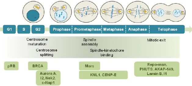
PP1 has different localization patterns during the cell cycle suggesting an association with different regulatory subunits. The PP1 complexes are capable of controlling cell cycle progression in diverse aspects (Fig. I.6). Protein phosphatases are important regulators of G1/S transition by maintaining retinoblastoma proteins dephosphorylated, which enables the latter proteins to recruit stimulators of G1/S transition (e.g. E2F transcription factor) (Bollen and Beullens, 2002). The retinoblastoma protein pRB is dephosphorylated at the end of mitosis by PP1, but at the G1/S transition PP1 is inactivated by a cyclin-dependent kinase (CDK) phosphorylation, allowing S phase entry (Liu et al., 1999; Rubin et al., 2001). PP1, like other protein phosphatases, controls the entry into mitosis by regulating the activity of mitotic kinases. Activation of CDK1 in association with cyclins is crucial for entry into mitosis and requires dephosphorylation by Cdc25B and Cdc25C phosphatases at the centrosomes and nucleus. Dephosphorylation of Cdc25C on Ser287 partially activates Cdc25C and it is mediated by PP1, resulting in the activation of a pool of CDK1 at the G2/M transition (reviewed in Bollen et al., 2009).
30
Figure I.6. Activity of PP1 and its regulatory proteins during cell cycle progression. The upper panel shows a timeline of cell cycle stages, the middle panel underlines the major events known to be regulated by PP1 and the lower panel shows the activity stage of PP1 regulatory proteins. PP1 regulates G1/S transition by dephosphorylation of the retinoblastoma protein pRB. BRCA prevents centrosome maturation in a PP1-dependent manner. PP1 keeps Aurora A, Nek2 and c-Nap1 dephosphorylated to prevent centrosome splitting. The inhibitor-2 (I2) restrain PP1 activity and promotes Nek2 activation and centrosome splitting. PP1-Mars complex stabilize microtubules. PP1 dehosphorylates KNL1 and CENP-E and stabilizes microtubule-kinetochore attachments. PP1 and its binding proteins Repo-man and PNUTS are required for chromatin decondensation at the end of mitosis. AKAP-149 recruits PP1 to the nuclear envelope to dephosphorylate lamin B, promoting nuclear envelope reassembly. Inhibitor 1 (I1) is inactivated by PP1 allowing PP1 to dephosphorylate proteins required for mitotic exit.
When cells enter mitosis, one of the first steps for mitotic spindle formation is the separation of duplicated centrosomes (duplication occurs at S phase), the main microtubule organizing centers that form the two spindle poles. Formation of a stable mitotic spindle is crucial for accurate separation of chromosomes during mitosis. PP1 may prevent the premature splitting of the duplicated centrosomes at the beginning of mitosis by inactivating the kinases involved in this process (Fig. I.6). One of these kinases is Aurora A, which is required for centrosome separation. Aurora A and PP1 antagonize each other and inhibitor-2 additionally regulates this complex by inhibiting PP1 and activating Aurora A. Centrosome separation also depends on Nek2a activity and its substrates C-Nap1 and Rootletin. PP1, Nek2 and C-Nap1 form a trimeric complex. During interphase, PP1 dephosphorylates Nek2 leading to its inhibition and also dephosphorylates c-Nap1 maintaining centrosome cohesion. Additionally, inhibitor-2 associates with the Nek2/PP1 complex, where it inhibits PP1, and thereby promotes Nek2 activation and centrosome splitting. In turns, Nek2 phosphorylates PP1, reducing its phosphatase activity (Bollen et al., 2009; Ceulemans and Bollen, 2004).
Characterization of novel LAP1 complexes and their relevance in DYT1 dystonia 31
Recently, it was reported that a leucine rich repeat protein that binds to PP1 termed PPP1R42 also regulates centrosome separation. PPP1R42 is a positive regulator of PP1 and its depletion reduces PP1 activity and consequently leads to Nek2 activation (Devaul et al., 2013). It seems that separation of centrosomes at early mitosis requires inactivation of PP1 and that PP1 maintains centrosomes together during interphase (reviewed in Bollen et al., 2009; Ceulemans and Bollen, 2004). Indeed, PP1 was shown to be involved in centrosome maturation (G2 phase), a process characterized by the accumulation of γ-tubulin and other proteins. The BRCA protein promotes ubiquitination and further degradation of γ-tubulin, and thus inhibits centrosome maturation. During interphase PP1α activates BRCA by dephosphorylation, suggesting an additional role for PP1 in centrosome maturation (reviewed in Bollen et al., 2009). Furthermore, PP1 also binds to the centrosomal proteins SFI1, CEP192, CEP170 and inhibitor-3 (Esteves et al., 2012a; Hendrickx et al., 2009; Huang et al., 2005).
Furthermore, mitotic spindle assembly that begins with microtubule outgrowth at the centrosomes, is regulated by different phosphatases, including PP1 (Fig. I.6) (Bollen et al., 2009). It was proposed that the PP1 binding protein Mars targets the centrosomal microtubule stabilizer dTACC to be dephosphorylated by PP1, thus contributing to mitotic spindle stability (Tan et al., 2008). Others functions that have been attributed to PP1 are the maintenance of microtubules-kinetochores attachment and spindle assembly checkpoint (SAC) silencing. Stable attachment of kinetochores to spindle microtubules is crucial for accurate chromosome separation. Aurora B phosphorylates diverse proteins to destabilize the binding of microtubules to kinetochores, thus preventing erroneous attachments. Conversely, PP1 stabilizes correct microtubule-kinetochore attachments during metaphase by opposing Aurora B-mediated phosphorylation (reviewed in Bollen et al., 2009; Funabiki and Wynne, 2013). It was demonstrated that the kinetochore protein KNL1 targets PP1γ to kinetochores and this recruitment is required for PP1γ dephosphorylation of other Aurora B substrates at kinetochores (Liu et al., 2010). PP1γ is also directed to kinetochores through binding to the centromere-associated protein E (CENP-E). Binding and dephosphorylation of CENP-E by PP1 is required for stable attachment of kinetochores to microtubules (Kim et al., 2010b). On the other hand, Aurora B inhibits targeting of PP1 to kinetochores by phosphorylation of the PP1 binding motif in KNL1 and CENP-E (Kim et al., 2010b; Liu
32
et al., 2010). In addition, polo-like kinase 1 (PLK1) was reported to promote kinetochore-microtubule attachment and SAC silecing. The SAC signaling pathway is activated on unattached kinetochores to block the metaphase-anaphase transition, until chromosomes are properly attached to the mitotic spindle. When the chromosomes align at methaphase, the levels of the PLK1 at kinetochore decrease and this seems to be dependent on PP1 recruitment. Like Aurora B, PLK1 substrates are likely to be dephosphorylated in a PP1-dependent manner (Liu et al., 2012). Moreover, PLK1 directly binds to the PP1 interactor MYPT1. Depletion of MYPT1 increases PLK1 phosphorylation on Thr210 and thus increases its kinase activity, suggesting that MYPT1/PP1 antagonizes PLK1 activity (Yamashiro et al., 2008). It was also shown that, in yeast, PP1 localization at the kinetochores is necessary for SAC silencing, indicating that dephosphorylation of kinetochore-associated proteins is required for SAC silencing. In yeast the association of PP1 with Spc7 (KLN1 in human) and kinesins Klp5/6 is needed for SAC silencing and kinetochore-microtubule attachment. IF18A, the vertebrate orthologue of Klp5/6, also interacts with PP1 (Meadows et al., 2011).
Protein phosphatases, including PP1, are required for mitotic exit (Bollen et al., 2009). Mitotic exit is characterized by mitotic spindle breakdown, chromosome decondensation and reassembly of interphase structures, particularly the NE. PP1 was shown to be required for kinetochore disassembly, possibly by dephosphorylation of a chromatin or kinetochore-bound substrate (Emanuele et al., 2008). Moreover, it was demonstrated that inhibitor-2 is required for accurate chromosome segregation and cytokinesis by regulating the Aurora B and PP1 activity (Wang et al., 2008b). PP1 and its regulatory subunits Repo-man (recruits PP1 onto mitotic chromatin at anaphase) and PNUTS are required for chromatin decondensation. Repo-man was initially found to recruit PP1γ to chromatin at anaphase and when overexpressed also recruits PP1α to chromatin (Trinkle-Mulcahy et al., 2006). The PP1γ/Repo-man complex, in particular, mediates the dephosphorylation of histone H3 at the end of mitosis and regulates chromosomal targeting of Aurora B (Qian et al., 2011). Dephosphorylation of histone H3 by PP1 seems to be correlated with chromosome decondensation in budding yeast and nematodes (Hsu et al., 2000). PNUTS is targeted to the reforming nucleus in telophase concomitantly with chromatin decondensation and promotes chromatin decondensation in a PP1-dependent manner (Landsverk et al., 2005). Furthermore, it
Characterization of novel LAP1 complexes and their relevance in DYT1 dystonia 33
was reported that PP1 is involved in the first step of nuclear envelope (NE) reassembly by stimulating the targeting of membrane vesicles to chromatin in Xenopus egg extracts (Ito et al., 2007). Moreover, the reassembly of the nuclear lamina is mediated in part by dephosphorylation of lamin B (Thompson et al., 1997). The PP1 regulatory subunit A-kinase anchoring protein (AKAP)-149 recruits PP1 to the NE upon NE assembly in vitro and promotes lamin B dephosphorylation and polymerization (Steen et al., 2000). Mitotic exit also requires CDK1 inactivation by cyclin B degradation and dephosphorylation of CDK1 and other kinases substrates. PP1 activity is repressed at early-mid mitosis by CDK1 phosphorylation on PP1 residue Thr320 (Kwon et al., 1997) and by binding to inhibitor-1. CDK1 inactivation at the end of mitosis allows PP1 auto-dephosphorylation promoting partial PP1 activation. PP1 is then able to dephosphorylate and inactivate inhibitor-1, allowing for the complete activation of PP1. Active PP1 dephosphorylates mitotic phosphoproteins required for mitotic exit (Wu et al., 2009).
A role for PP1 in the regulation of cytokinesis has also been suggested (Cheng et al., 2000; Fernandez et al., 1992), consistent with the co-localization of PP1γ and F-actin at the cleavage furrow and spindle midzone (Trinkle-Mulcahy et al., 2003). PP1γ was also found in the center of the midbody during cytokinesis (Zeitlin et al., 2001).
I.1.2.2. PP1 signaling in the brain
PP1 isoforms α, β/δ and γ1 are ubiquitously expressed in mammalian tissues but have higher abundance in brain. Within the brain, the mRNAs for these isoforms were widely distributed with particular incidence in the hippocampus and the cerebellum (da Cruz e Silva et al., 1995b). At the protein level PP1α and PP1γ1 have highest levels of expression in the striatum, where they are relatively enriched in the medium-sized spiny neurons (da Cruz e Silva et al., 1995b; Ouimet et al., 1995). At the ultrastructural level, PP1α and PP1γ1 are highly and specifically concentrated in dendritic spines in the striatum. At spines, PP1α and γ1 are concentrated at the postsynaptic density. The localization of PP1α and PP1γ1 at dendritic spines, the principal sites for excitatory synapses in the central nervous system, suggests that these isoforms are involved more extensively in postsynaptic mechanisms of neurotransmission rather than PP1β/δ. In
34
addition, PP1α, γ1 and β/δ are also located at the cell body, dendritic shafts and axons (Bordelon et al., 2005; Ouimet et al., 1995). Additionally, PP1β/δ and PP1γ1 were detected in all cytoskeletal fractions (neurofilaments, microtubules and actin cytoskeleton) from hindbrain proteins. However, PP1β/δ was found enriched in the microtubules whereas PP1γ1 was found mostly in the actin cytoskeleton fraction (Strack et al., 1999). Indeed, PP1, alongside with PP2A, were reported to have a role in the neurite structure. Inhibition of PP1 and PP2A in cultured hippocampal neurons leads to decrease number and length of neurites, synapse loss and also hyperphosphorylation of the microtubule-stabilizing protein tau (Malchiodi-Albedi et al., 1997). Hyperphosphorylation of tau leads to tau aggregation and formation of neurofibrillary tangles, a feature of Alzheimer’s disease.
I.1.2.2.1. PP1/DARPP-32 signaling pathway
Medium-sized spiny neurons are highly enriched for DARPP-32 (Ouimet et al., 1984), which has a crucial role in the biology of dopamineceptive neurons. Dopamine and others neurotransmitters in the striatum alter the (de)phosphorylation state of 32 (Greengard et al., 1999). When phosphorylated by PKA on Thr34, DARPP-32 is a potent inhibitor of PP1 (Hemmings et al., 1984). Conversely, phosphorylation of DARPP-32 on Thr75 by CDK5 turns DARPP-32 into an inhibitor of PKA and blocks phosphorylation on Thr34 (Bibb et al., 1999). Thus, DARPP-32 modulates PP1 and PKA activities in the striatum, which in turn regulate the expression of neuropeptides and ion channels and pumps (Fig. I.7). Dopamine has opposite effects on PKA signalling when bound to D1 or D2 dopamine receptors of medium-sized spiny neurons. These neurons contain both D1 class (D1, D5) and D2 class (D2, D3, D4) dopamine receptors. However, D1 receptors are predominantly expressed in striatonigral neurons, while D2 receptors are mainly found in striatopallidal neurons. Dopamine has an excitatory effect on striatonigral neurons expressing D1 receptors and causes increased activity of adenylyl cyclase and cAMP-mediated activation of PKA, which phosphorylates DARPP-32 on Thr34 converting DARPP-32 into a PP1 inhibitor. Ligands that stimulate PKA also promote the dephosphorylation of Thr75 by PP2A. In contrast, striatopallidal neurons expressing D2 receptors are inhibited by dopamine by two mechanisms: inhibition of adenylyl cyclase and thus PKA; and Ca2+ and
PP2B-Characterization of novel LAP1 complexes and their relevance in DYT1 dystonia 35
mediated dephosphorylation of DARPP-32 and restore of PP1 activity (reviewed in Greengard et al., 1999).
Figure I.7. Regulation of DARPP-32/PP1 signaling pathway. Dopamine acts on D1 receptors to increase cAMP formation and activation of PKA, which phosphorylates DARPP-32 on Thr34 leading to PP1 inhibition. Conversely, activation of D2 receptors leads to an increase in Ca2+ levels and activity of PP2B, which dephosphorylates DARPP-32. D2 receptors can also act to decrease cAMP formation. Cdk5 phosphorylates DARPP-32 on Thr75 converting DARPP-32 into a PKA inhibitor. Further, PP2A dephosphorylates DARPP-32 on Thr75. Both adenosin and serotonin act on A2A and 5-HT4/6,
respectively, and promote cAMP formation, PKA activation and thus DARPP-32 Thr34 phosphorylation. Glutamate act on NMDA and AMPA receptors leading to increase Ca2+ levels and PP2B activity, whereas GABA action on GABAA receptors has an opposite effect. Stimulatory effects are represented as solid
arrows and inhibitory effects as dashed arrows.
DARPP-32/PP1 pathway in medium-sized spiny neurons is also affected by other neurotransmitters (Fig, I.7). Adenosine via A2A adenosine receptor stimulates adenylyl
cyclase and activates PKA, leading to the phosphorylation of DARPP-32 at Thr34 (Svenningsson et al., 1998). Serotonin binding to 5-HT4/6 receptor also promotes the
activity of adenylyl cyclase and PKA and thus phosphorylation of DARPP-32 at Thr34 (Svenningsson et al., 2002). Glutamate acting on N-methyl-D-aspartate (NMDA) and α-amino-3-hydroxy-5-methyl-4-isoxazolepropionic acid (AMPA) receptors stimulates DARPP-32 dephosphorylation by PP2B by influx of Ca2+. In contrast, γ-aminobutyric
36
acid (GABA) acting on GABAA receptors stimulates DARPP-32 phosphorylation by
hyperpolarization of the neuron and decreases influx of Ca2+, resulting in the inactivation of PP2B. Inhibition of PP1 by phospho-DARPP-32, in concert with PKA and other kinase activities, results in an increased phosphorylation of various downstream effector proteins. Higher levels of phosphorylation are associated with decreased activity of GABAA receptors, Na+ channels, and the Na+/K+-ATPase and
increased activity of NMDA and AMPA glutamate receptors, L-, N-, and P-type Ca2+ channels and CREB (Greengard et al., 1999).
I.1.2.2.2. PP1 regulation of synaptic plasticity
PP1 has been associated with changes in glutamatergic transmission. Glutamate is the major excitatory neurotransmitter in the mammalian nervous system and its effects are mediated in part by NMDA and AMPA receptors. NMDA and AMPA glutamate receptor subunits are pivotal in synaptic plasticity and its activity is regulated by protein phosphorylation (Munton et al., 2004; Soderling and Derkach, 2000). The efficiency of transmission at glutamatergic synapses can be strengthened (long-term potentiation, LTP) by brief, high-frequency stimulation or weakened (long-term depression, LTD) by prolonged, low-frequency stimulation (Malenka, 1994). It was reported that inhibition of PP1 is required for LTP induction in hippocampal neurons. Stimulation that induces LTP leads to cAMP-dependent phosphorylation of inhibitor-1 resulting in decreased PP1 activity. Moreover, this also results in increased phosphorylation of Ca2+/calmodulin-dependent kinase II (CaMKII) at Thr286 and thus increased activity of CaMKII (Blitzer et al., 1998), which phosphorylates the AMPA receptor subunits and potentiates synaptic current (Soderling and Derkach, 2000). Moreover, at the postsynaptic density, PP1 dephosphorylates CaMKII on Thr286 and inactivates it (Strack et al., 1997).
Conversely, LTD was associated with activation of PP1 after dephosphorylation and inactivation of inhibitor-1 by PP2B in hippocampus (Mulkey et al., 1994). Dephosphorylation of AMPA receptors is correlated with LTD. Introduction of peptides that disrupt the binding of PP1 to its regulatory subunits inhibited NMDA receptor-dependent LTD and addition of active PP1 enhanced LTD. It was also reported that synaptic activation of NMDA receptor in cultured hippocampal neurons caused a
Characterization of novel LAP1 complexes and their relevance in DYT1 dystonia 37
redistribution of PP1 to synapses (Morishita et al., 2001). Recently, it was reported that PP1α regulates the NR2B subunit of the NMDA receptor by dephosphorylating a specific residue on N2RB (Farinelli et al., 2012).
I.1.2.2.3. Localization of PP1 to dendritic spines
Regulation of dendritic spine motility is mediated by signaling pathways that involve PP1 and some of its regulatory subunits. PP1α and PP1γ are specifically concentrated at the postsynaptic density of dendritic spines (Bordelon et al., 2005; Ouimet et al., 1995). Among the known neuronal PP1 regulators that are also postsynaptically localized are spinophilin (Allen et al., 1997), neurabin I (McAvoy et al., 1999), yotiao (Westphal et al., 1999), neurofilament light chain (Terry-Lorenzo et al., 2000) and AKAP79 (Le et al., 2011).
Spinophilin (also named neurabin II) and neurabin I can both target PP1 to dendritic spines. Spinophilin was identified as a PP1 binding protein enriched in spines heads (Allen et al., 1997). Spinophilin binds to F-actin and it was demonstrated that the F-actin binding domain is necessary and sufficient for targeting PP1 to dendritic spines (Grossman et al., 2002). Spinophilin can also target PP1 to the postsynaptic density in close proximity with the AMPA receptors (Yan et al., 1999). Thus, PP1 is important for AMPA receptor activity and synaptic plasticity not only through DARPP-32/PP1 pathway, but also by association with spinophilin. Spinophilin is also important for PP1-mediated regulation of NMDA receptors. Consistent with altered glutamatergic transmission, spinophilin-knockout mice showed reduced LTD and altered spine density and filopodia formation (Feng et al., 2000). Recently, it was reported that spinophilin can target CaMKII to F-actin as well as targets PP1 to CaMKII (Baucum et al., 2012).
Neurabin I is a F-actin binding protein mainly expressed in neural tissues, that binds to PP1α and PP1γ. Neurabin I is phosphorylated on Ser461 by PKA disrupting the interaction with PP1 (McAvoy et al., 1999). PP1 also binds yotiao, a protein member of the AKAP family. It seems that yotiao attaches PP1 and PKA to NMDA receptors regulating synaptic transmission mediated by the NMDA receptor (Westphal et al., 1999). At the postsynaptic density PP1 also binds to neurofilament light chain, a component of the intermediate filament network in neurons, which can be
38
phosphorylated by PKA and by protein kinase N (PKN) (Terry-Lorenzo et al., 2000). AKAP79 was recently identified as a PP1 regulatory protein (Le et al., 2011) but it was previously described to anchor PKA, protein kinase C (PKC) and PP2B at the postsynaptic density regulating AMPA receptors phosphorylation in these pathways (Bauman et al., 2004).
Characterization of novel LAP1 complexes and their relevance in DYT1 dystonia 39
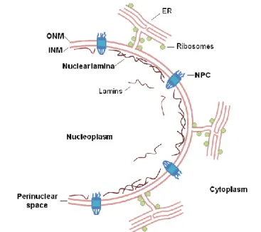
I.2. THE NUCLEAR ENVELOPE
The eukaryotic nucleus is a complex organelle enclosed by a double membrane, the NE. The NE separates the cytoplasm from de the nucleus in eukaryotic cells and is structurally composed by the inner nuclear membrane (INM), the outer nuclear membrane (ONM), the nuclear lamina and the nuclear pore complexes (NPCs) (Fig. I.8) (reviewed in Gerace and Burke, 1988; Worman and Courvalin, 2005).The INM and the ONM are separated by a perinuclear space (Stewart et al., 2007) that is 40-50 nm wide in mammalian cells. However these membranes are joined in some regions at the NPCs, structures that regulate molecular transport between the cytoplasm and the nucleoplasm. NPCs are large macromolecular complexes composed of 30 different proteins, termed nucleoporins (reviewed in Fahrenkrog and Aebi, 2003). The ONM is continuous with the endoplasmic reticulum (ER) and contains various proteins found in the ER and associated ribosomes (Pathak et al., 1986). Nonetheless, the ONM also contains specific proteins that are involved in nuclear positioning by linking the nucleus to the cytoskeleton (Crisp et al., 2006; Starr and Han, 2002; Wilhelmsen et al., 2005). In contrast, the INM contains a set of distinctive integral membrane proteins. Using proteomic approaches nearly 70 transmembrane proteins were found to associate with the INM (Malik et al., 2010; Schirmer et al., 2003) but, so far, few have been characterized in detail. The major components of the nuclear lamina are A-type and B-type lamins (Aebi et al., 1986), which are found in association with proteins of the INM and also in the nucleoplasm.