Journal of Movement Disorders 2012;5:5-8 pISSN 2005-940X / eISSN 2093-4939
Reorganization of the Human Somatosensory Cortex
in Hand Dystonia
ORIGINAL ARTICLE
Maria Jose Catalan Kenji Ishii
William Bara-Jimenez Mark Hallett
Human Motor Control Section, Medical Neurology Branch, National Institute of Neurological Disorders and Stroke,
National Institutes of Health, Bethesda, Maryland, 20892 USA
Received January 22, 2012 Revised April 9, 2012 Accepted April 9, 2012 Corresponding author Mark Hallett, MD
Human Motor Control Section, Medical Neurology Branch, National Institute of Neurological Disorders and Stroke,
National Institutes of Health, Building 10, Room 7D37 10 Center Drive MSC 1428, Bethesda, MD 20892-1428, USA Tel +1-301-496-9526 Fax +1-301-480-2286 E-mail [email protected]
•- The authors have no financial conflicts
of interest.
Copyright © 2012 The Korean Movement Disorder Society 5 Background and Purpose: Abnormalities of finger representations in the somatosensory cortex have been identified in patients with focal hand dystonia. Measuring blood flow with positron emission tomography (PET) can be use to demonstrate functional localization of re-ceptive fields. Methods: A vibratory stimulus was applied to the right thumb and little finger of six healthy volunteers and six patients with focal hand dystonia to map their receptive fields using H215O PET. Results: The cortical finger representations in the primary somatosensory cortex were closer to each other in patients than in normal subjects. No abnormalities were found in secondary somatosensory cortex, but the somatotopy there is less well distinguished. Conclusions: These data confirm prior electrophysiological and functional neuroimaging observations showing abnormalities of finger representations in somatosensory cortex of pa-tients with focal hand dystonia. Journal of Movement Disorders 2012;5:5-8 Key Words: Dystonia, Somatosensory cortex, Receptive field, Cortical representation,
Positron emission tomography.
Writer’s cramp is a task-specific focal hand dystonia characterized by agonist and antago-nist muscle co-contraction causing abnormal posturing. Previous studies have shown abnor-mal activation of the primary sensorimotor cortex during writing1 and structural primary somatosensory cortex (SI) involvement.2 There is considerable evidence of sensory involve-ment in writer’s cramp.3-7 In the present study we mapped the finger representations in so-matosensory cortices [SI and secondary soso-matosensory cortex (SII)] in writer’s cramp pa-tients using H215O positron emission tomography (PET).
Methods
We studied 6 patients (2 males and 4 females) with writer’s cramp of their right hands. Mean age was 46.5 (range from 26 to 59). The diagnosis of idiopathic hand dystonia was based on the medical history, physical and neurological examinations, and after excluding other diseases by laboratory tests and magnetic resonance imaging (MRI) scans. All patients were studied after at least 1 year without any botulinum toxin injections. Control subjects consisted of 6 normal volunteers, 2 males and 4 females, mean age 47.5, range 28 to 60. They had no history of neurological disease and no abnormalities on physical and neuro-logical examinations. All subjects were right-handed by the Edinburgh Inventory.8 The pro-tocol was approved by the Institutional Review Board, and all the subjects gave written in-formed consent for the study.
For each subject, PET scans of regional cerebral blood flow (rCBF) were performed using H215O as a tracer. The experimental paradigm consisted of three conditions: vibratory stimula-tion on right thumb (D1), vibratory stimulastimula-tion on right little finger (D5), and rest. Each con-dition was repeated five times.
Subjects lay in a supine position. The right arm was lying on a support to maintain a con-stant arm position. Each subject underwent 15 consecutive scans at 10-minute intervals. Vi-bratory stimulation on each individual finger was started 20 seconds before injection and
6
Journal of Movement Disorders ▐ 2012;5:5-8
tinued during the whole scan. The subjects were instructed to pay attention to the stimulation site. Surface electromyogra-phy from right flexor and extensor carpi radialis was recorded during the study to see whether dystonia was induced. For the rest scan, subjects lay quietly. The vibrator (Brüel and Kjaer, Mini-Shaker type 4810) had two different frequencies, fast (100 Hz) and slow (25 Hz). The stimulus was delivered using these two different frequencies alternating in a random order.
The PET scans were performed using a GE Advance sys-tem (General Electric, Schenectady, NY, USA). Data were ac-quired in 3D mode and reconstructed into 35 contiguous tr-ansaxial planes separated by 4.25 mm (center-to-center). In-plane and axial resolution were 5.2 mm and 4.6 mm full-width half-maximum (FWHM) respectively, after reconstruction. Emission scans were attenuation corrected with a transmis-sion scan collected before each sestransmis-sion during exposure of a Germanium-68/Gallium-68 external rotating source. Recon-structed images were obtained by summing the activity dur-ing the 60 second period followdur-ing the first detection of an increase in cerebral radioactivity after the intravenous bolus injection of 10 mCi of 15O-water. Magnetic resonance images were obtained using a General Electric scanner (1.5 T). For each subject a Sagittal T1-weighted, matrix 256 × 256, 124 contiguous sagittal slices with 1.5 mm thickness were collect-ed. After reconstruction, the MR images were aligned paral-lel with the intercommissural line, and interpolated to yield a cubic voxel size of 0.938 mm3, which permitted coregistra-tion with the PET.
Data analysis was performed with statistical parametric mapping 96 (SPM96 from the Wellcome Dept. of Cognitive Neurology, London, UK) implemented in Matlab (Math-works Inc., Natick, MA, USA). The scans from each subject were realigned and normalized to Montreal Neurological In-stitute template. Following realignment, the mean image was used to coregister PET data onto the same individual’s MRI scan. Afterwards the MRI image was normalized using the same parameters. Each image was smoothed using a Gauss-ian filter (FWHM = 8 mm for all direction). In the stereotaxic standard space, each voxel was 2 × 2 × 2 mm in size.
After the appropriate design matrix was specified, the con-dition effects were estimated according to the general linear model at each and every voxel. Differences in global cerebral blood flow between scans were removed by proportional scal-ing with global flow as a confoundscal-ing variable.9 To test the hypothesis about the specific regional effects of the condition, the estimates were compared using linear contrasts. Analysis of data was performed as single subject analysis. Because the hypothesis was somatotopic organization in the somato-sensory cortices, we restricted the cortical areas for the anal-ysis to the somatosensory cortices. Although the images were normalized to standard space, the exact anatomic locations of the local maxima were identified for each individual with
reference to the gyral anatomy identified on each individual MRI. The location of each finger in SI was determined by the highest change in rCBF posterior to the central sulcus. The location of each finger in SII was determined by looking at the highest change in rCBF in insula and operculum.
The Mann-Whitney-Wilcoxon test was used for statistical comparison of the distances between the finger representa-tions, and the level of significance was p < 0.05.
Results
Vibratory stimulation of each finger evoked increase in rCBF in the contralateral postcentral sulcus. The location of D1 in relation to D5 in normal subjects was located in a ho-muncular fashion in SI: inferior (6 of 6 subjects), anterior (5/6) and lateral (5/6). Similarly, for hand dystonia patients, the lo-cation of D1 in relation to D5 was inferior (5/6), anterior (6/6), and lateral (4/6). Only one patient showed inferior-to-superi-or topography inverted (D1 superiinferior-to-superi-or to D5).
Patients with hand dystonia showed some degradation of the homuncular S1 organization, with changes in the relative positions of D1 and D5. The unidirectional distances between D1 and D5 in the control subjects and patients were, respec-tively, 15.3 mm and 8.0 mm in the x axis; 7.3 mm and 5.0 mm in the y axis; and 15.7 mm and 8.7 mm in the z axis, showing that the distance between fingers 1 and 5 was decreased. There were no significant difference in the horizontal (x, y) plane (17.6 mm vs. 10.5 mm, p = 0.3), and sagittal (y, z) plane (18.0 mm vs. 10.1 mm, p = 0.1). However, there was significant difference in the coronal (x, z) plane (24.0 mm vs. 12.9 mm, p = 0.009). The mean three-dimensional distance between fingers 1 to 5 representations was significantly decreased, 25.3 mm for controls vs. 14.1 mm for dystonia (p = 0.02).
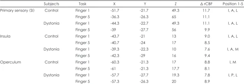
Individual finger representations could be identified in SII in all control subjects and dystonia patients, for both insula and operculum. In the insula, the location of D1 in relation to D5 was inferior (4/6), anterior (4/6) and lateral (3/6) for con-trol subjects, and inferior (5/6), anterior (6/6) and lateral (1/6) for dystonia patients. Location in the operculum showed more variability in both groups. The mean coordinates for each region are in Table 1. There were no significant differ-ences in distance between fingers 1 and 5 for the two groups (Table 2).
Discussion
corti-www.e-jmd.org 7 Reorganization of Somatosensory Cortex in Dystonia ▐ Catalan MJ, et al.
ces is modulated based on learning and experience.13,14 Our study shows degradation of the normal organization of SI in patients with hand dystonia. The cortical finger rep-resentations in SI were closer to each other in patients than in normal subjects, mainly in the coronal plane, but also in the tridimensional distance. Only one patient showed inversion of the finger representation in the superior-inferior topogra-phy. The results confirm the previous findings using electroen-cephalography, magnetoenelectroen-cephalography, functional MRI.3-5,7 There appears to be an abnormal clustering of digit represen-tations and a disruption of the normal homuncular arrange-ment in the SI. The explanation for this may be due to expan-sions of the representations and overlapping, similar to what has been shown in studies of learning-induced cortical remod-eling in monkeys.10,14 This finding appears to be primary be-cause representation of the symptomatic fingers are more af-fected than asymptomatic ones.7
Activation of each individual finger was identified in each subject in both insula and operculum, but there was more top-ographical variability than in SI. We did not find differences between control and dystonia patients. Our findings in
nor-mal subjects are consistent with previous reports in non-hu-man primates and hunon-hu-mans that have shown somatotopic ar-rangement with large inter-individual differences. The differ-entiation was not as clear as that seen in the homunculus in the SI.15-18 It is uncertain whether the failure to find a differ-ence between groups in our work is because of the intrinsic variability or the relatively small number of subjects that we have studied. Current knowledge of SII function is predomi-nantly from animal studies, and role of SII in humans remain unclear. A functional role for SII areas in processing somato-sensory stimuli has been implicated as being important for tactile processing, learning and memory.18-22
REFERENCES
1. Ibáñez V, Sadato N, Karp B, Deiber MP, Hallett M. Deficient activa-tion of the motor cortical network in patients with writer’s cramp. Neu-rology 1999;53:96-105.
2. Garraux G, Bauer A, Hanakawa T, Wu T, Kansaku K, Hallett M. Ch-anges in brain anatomy in focal hand dystonia. Ann Neurol 2004;55: 736-739.
3. Bara-Jimenez W, Catalan MJ, Hallett M, Gerloff C. Abnormal somato-sensory homunculus in dystonia of the hand. Ann Neurol 1998;44:
Table 1. Mean coordinates for peak delta (Δ) rCBF in each somatosensory cortical region (SI and SII), for control subjects and dystonia pa-tients
Subjects Task X Y Z Δ rCBF Position 1-5
Primary sensory (SI) Control Finger 1 -51.7 -21.7 49.3 11.7 I, A, L
Finger 5 -36.3 -26.3 65 11.1
Dystonia Finger 1 -44.3 -22.7 49.3 11.1 I, A, L
Finger 5 -39 -27.7 56 9.9
Insula Control Finger 1 -43.7 -21 13 9.0 I, A, L
Finger 5 -40.7 -24 17 8.5
Dystonia Finger 1 -39.3 -22.3 10 7.6 I, A, M
Finger 5 -42.3 -29 16 9.4
Operculum Control Finger 1 -60.3 -21.3 17 8.8 I, M
Finger 5 -61 -21.3 17.7 8.1
Dystonia Finger 1 -57.7 -27.7 19.3 7.8 I, P, L
Finger 5 -57.3 -26.3 20 8.9
Mean Δ rCBF (mL/min/100 mL). Relative position of finger 1 relative to finger 5. rCBF: regional cerebral blood flow, SI: primary somato-sensory cortex, SII: secondary somatosomato-sensory cortex, I: inferior, A: anterior, P: posterior, L: lateral, M: medial.
Table 2. Mean distance (SD) between fingers 1 and 5 in control subjects and dystonia patients, in unidimensional (X, Y, Z) bidimensional (X-Y, X-Z, Y-Z) and tridimensional measurements; in SI and SII cortices
Primary sensory cortex (SI) Operculum (SII) Insula (SII)
Control subjects
Dystonia patients
Control subjects
Dystonia patients
Control subjects
8
Journal of Movement Disorders ▐ 2012;5:5-8
828-831.
4. Elbert T, Candia V, Altenmüller E, Rau H, Sterr A, Rockstroh B, et al. Alteration of digital representations in somatosensory cortex in focal hand dystonia. Neuroreport 1998;9:3571-3575.
5. Butterworth S, Francis S, Kelly E, McGlone F, Bowtell R, Sawle GV. Abnormal cortical sensory activation in dystonia: an fMRI study. Mov Disord 2003;18:673-682.
6. Tinazzi M, Rosso T, Fiaschi A. Role of the somatosensory system in primary dystonia. Mov Disord 2003;18:605-622.
7. Nelson AJ, Blake DT, Chen R. Digit-specific aberrations in the prima-ry somatosensoprima-ry cortex in Writer’s cramp. Ann Neurol 2009;66:146-154.
8. Oldfield RC. The assessment and analysis of handedness: the Edin-burgh inventory. Neuropsychologia 1971;9:97-113.
9. Friston KJ, Frith CD, Liddle PF, Dolan RJ, Lammertsma AA, Frack-owiak RS. The relationship between global and local changes in PET scans. J Cereb Blood Flow Metab 1990;10:458-466.
10. Byl NN, Merzenich MM, Jenkins WM. A primate genesis model of focal dystonia and repetitive strain injury: I. Learning-induced dedif-ferentiation of the representation of the hand in the primary somato-sensory cortex in adult monkeys. Neurology 1996;47:508-520. 11. Xerri C, Merzenich MM, Jenkins W, Santucci S. Representational
plasticity in cortical area 3b paralleling tactual-motor skill acquisition in adult monkeys. Cereb Cortex 1999;9:264-276.
12. Braun C, Schweizer R, Heinz U, Wiech K, Birbaumer N, Topka H. Task-specific plasticity of somatosensory cortex in patients with writ-er’s cramp. Neuroimage 2003;20:1329-1338.
13. Hallett M. Is dystonia a sensory disorder? Ann Neurol 1995;38:139-140.
14. Wang X, Merzenich MM, Sameshima K, Jenkins WM. Remodelling of hand representation in adult cortex determined by timing of tactile stimulation. Nature 1995;378:71-75.
15. Hari R, Karhu J, Hämäläinen M, Knuutila J, Salonen O, Sams M, et al. Functional organization of the human first and second somatosensory cortices: a neuromagnetic study. Eur J Neurosci 1993;5:724-734. 16. Krubitzer L, Clarey J, Tweedale R, Elston G, Calford M. A redefinition
of somatosensory areas in the lateral sulcus of macaque monkeys. J Neurosci 1995;15(5 Pt 2):3821-3839.
17. Burton H, Fabri M, Alloway K. Cortical areas within the lateral sulcus connected to cutaneous representations in areas 3b and 1: a revised in-terpretation of the second somatosensory area in macaque monkeys. J Comp Neurol 1995;355:539-562.
18. Maeda K, Kakigi R, Hoshiyama M, Koyama S. Topography of the se-condary somatosensory cortex in humans: a magnetoencephalo-graph-ic study. Neuroreport 1999;10:301-306.
19. Hari R, Forss N. Magnetoencephalography in the study of human so-matosensory cortical processing. Philos Trans R Soc Lond B Biol Sci 1999;354:1145-1154.
20. Rowe MJ, Turman AB, Murray GM, Zhang HQ. Parallel organiza-tion of somatosensory cortical areas I and II for tactile processing. Clin Exp Pharmacol Physiol 1996;23:931-938.
21. Huttunen J, Wikström H, Korvenoja A, Seppäläinen AM, Aronen H, Ilmoniemi RJ. Significance of the second somatosensory cortex in sensorimotor integration: enhancement of sensory responses during finger movements. Neuroreport 1996;7:1009-1012.