Margarida Sâncio da
Cruz Fardilha
Caracterização do Interactoma da PP1 do Testículo
Humano
Characterization of the PP1 Interactome from Human
Testis
Margarida Sâncio da
Cruz Fardilha
Caracterização do Interactoma da PP1 do Testículo
Humano
Characterization of the PP1 Interactome from Human
Testis
Dissertação apresentada à Universidade de Aveiro para cumprimento dos requisitos necessários à obtenção do grau de Doutor em Biologia, realizada sob a orientação científica do Prof. Doutor Edgar F. da Cruz e Silva, Professor Associado do Departamento de Biologia da Universidade de Aveiro.
o júri
presidente Prof. Dr. António Manuel Melo de Sousa Pereira
Professor Catedrático da Universidade de Aveiro
Prof. Dr. Gustavo Cardoso Nunes Caldeira
Professor Catedrático da Universidade de Aveiro
Prof. Dr. Mário Manuel da Silva Leite de Sousa
Professor Associado com Agregação do Instituto de Ciências Biomédicas de Abel Salazar da Universidade do Porto
Prof. Dr. Edgar Figueiredo da Cruz e Silva
Professor Associado da Universidade de Aveiro
Prof. Dr. António José de Brito Fonseca Mendes Calado
Professor Auxiliar da Universidade de Aveiro
Prof. Dra. Odete Abreu Beirão da Cruz e Silva
Professora Auxiliar da Universidade de Aveiro
Prof. Dr. Ricardo Enrique Pérez Tomás
agradecimentos
À FCT (EU V Framework Program, BD/13658/97), ao Centro de Biologia Celular, à Universidade de Aveiro.
Al Prof. Dr. Ricardo Pérez, por su hospitalidad y por haberme hecho sentir como en casa en su laboratorio de Barcelona.
A todos os colegas do laboratório pela amizade que construímos ao longo destes anos e, em especial, ao Miguel, à Catarina, à Wenjuan, à Ana Paula e à Cristina por estarem sempre disponíveis para me ajudar.
Quero também agradecer aos estudantes de licenciatura que deram os primeiros passos na ciência comigo, e que trazem para o laboratório muita dedicação e alegria; à Lígia, ao Krespim, à Catarina, à Rita, à Sara, à Rosália e ao Filipe.
À Dra. Odete A. B. da Cruz e Silva pelas opiniões pertinentes que me deu ao longo deste percurso mas principalmente por ser uma grande amiga. Ao Prof. Dr. Edgar da Cruz e Silva por me ensinar a ser uma melhor cientista partilhando comigo todos os seus conhecimentos; por ter sempre respostas para as minhas dúvidas; por me atribuir responsabilidades e por me ensinar a ser paciente.
À minha família, pais, sogros, irmãs, cunhada e Graça pela ajuda incondicional na educação dos meus filhos.
Aos meus filhos e ao meu marido por todas as alegrias que partilhamos e por me fazerem sentir a pessoa mais feliz do mundo.
resumo A fosforilação de proteínas é um dos principais mecanismos reguladores de cascatas de transdução de sinais em eucariotas. A fosforilação é catalizada por proteínas cinases e é revertida pela acção de proteínas fosfatases. Embora as proteínas fosfatases tenham sido descobertas à mais de sessenta anos, a sua importância central em múltiplos mecanismos celulares só muito recentemente foi reconhecida.
PP1, uma fosfatase específica para serina/treonina, está envolvida em importantes mecanismos celulares, como o ciclo celular, a contracção muscular e a apoptose, entre outros. O seu papel em tão variados processos celulares depende das suas interacções com subunidades reguladoras. Até à data, foram descritas mais de 50 subunidades reguladoras que se ligam à subunidade catalítica da PP1, sendo determinantes para a sua função num local específico da célula.
Das três isoformas da PP1 conhecidas, PP1 , PP1 e PP1 , a isoforma gama sofre splicing alternativo, originando a PP1 1, ubíqua e, a PP1 2 específica de testículo.
Incubação de espermatozóides imaturos com inibidores de fosfatases induz a sua motilidade, sendo a PP1 2 a fosfatase envolvida. Este facto levou-nos a procurar proteínas de testículo humano capazes de interagir especificamente com a PP1 2 que possam ser alvos terapêuticos no tratamento da infertilidade, ou na contracepção masculina. Para atingir este objectivo utilizou-se o sistema Dois Híbrido de Levedura no rastreio de uma biblioteca de testículo humano, na busca de novas proteínas que se ligam à PP1 usando como isco a PP1 1 (no YTH1) ou a PP1 2 (no YTH2).
Obtivemos 120 clones positivos no YTH1 e 155 no YTH2. Entre eles encontravam-se clones que codificam reguladores da PP1 previamente descritos, como a Nek2 e a NIPP1, assim como novas proteínas. Efectuámos um estudo detalhado de um novo gene, codificando uma nova proteína, que denominamos SEARP-T. Esta proteína de 93kDa é expressa maioritariamente em testículo e, a sua localização intracelular foi determinada por
imunocitoquímica de fluorescência. Ambas as proteínas, PP1 2 e SEARP-T, estão presentes na cauda e no segmento equatorial da cabeça do
espermatozóide. Estes resultados clarificam as funções da PP1 no testículo humano e na motilidade do esperma e, indicam que o sistema Dois Híbrido de Levedura é um bom método para compreender o papel da PP1 em múltiplos eventos de regulação celular.
abstract Protein phosphorylation is a major regulatory mechanism notably of signal transduction cascades in eukaryotic cells. Protein phosphorylation is catalysed by protein kinases and can be reversed by the action of protein phosphatases. Although phosphatases were discovered more than sixty years ago, their importance as central players in multiple cellular mechanisms was only recently recognized.
PP1, a Serine/Threonine specific phosphatase, is involved in important cellular mechanisms such as the cell cycle, muscle contraction and apoptosis, among others. Its role in such diverse cellular processes depends on its interactions with targeting/regulatory subunits. To date, more than 50 regulatory subunits have been identified that bind the catalytic subunit of PP1 determining its function in a specific cellular location.
Several isoforms of PP1 are known, termed PP1 , PP1 and PP1 . The gamma isoform undergoes alternative splicing to yield a ubiquitously expressed PP1 1 and a testis-specific PP1 2 isoform.
Incubation of non-motile immature sperm with phosphatase inhibitors induces sperm motility, and PP1 2 was implicated in this process. This led us to search for PP1 2-specific interactors in human sperm that could be targeted for infertility therapeutics or in male contraception. To achieve this goal the Yeast Two Hybrid system was used to screen a human testis library for new PP1 binding proteins using both PP1 1 (YTH1) and PP1 2 (YTH2) as baits. We recovered 120 positive clones in YTH1 and 155 positive clones in YTH2. Among these were clones encoding “bona fide” PP1 interactors such as Nek2 and NIPP1, and also previously uncharacterized proteins. We undertook a more detailed study of a novel gene encoding a novel protein that we termed SEARP-T. This protein of 93KDa is expressed mainly in testis and fluorescence immunocytochemistry was used to determine its intra sperm localization. Both PP1 2 and SEARP-T proteins are present in the tail and in the equatorial segment of the head.These results provide new insights into PP1 function in human testis and sperm motility, and indicates that the Yeast Two Hybrid System provides a mean to understand the roles PP1 plays in diverse cellular regulatory events.
INDEX
ABBREVIATIONS ...11
I
INTRODUCTION ...15
I.1 PROTEIN PHOSPHORYLATION AS A DYNAMIC PROCESS...15
I.2 ABNORMAL PHOSPHORYLATION IN DISEASE...16
I.3 PHOSPHATASES AND THEIR CLASSIFICATION ...17
I.3.1 The PPP Family...18
I.3.2 The PPM Family...31
I.4 PHOSPHATASE INHIBITORS...33
I.4.1 Toxins as natural inhibitors of PPs...34
I.4.2 Protein inhibitors ...40
I.5 DISTRIBUTION AND EXPRESSION OF PROTEIN PHOSPHATASES .43 I.5.1 PP1...43
I.5.2 PP2A ...43
I.5.3 PP2B ...44
I.6 PP1 STRUCTURE AND FUNCTION...45
I.6.1 Historical background...46
I.6.2 Recombinant PP1c...47
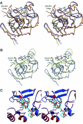
I.6.3 PP1c crystal structure and catalytic mechanism...49
I.7 PP1 TARGETING/BINDING PROTEINS ...55
I.7.1 “RVxF motif”...58
I.8 PP1 IN TESTES AND SPERM...61
I.8.1 PP1γ knockout mice...62
I.9 SIGNAL TRANSDUCTION THERAPEUTICS...63
I.10 AIMS...64
II
YEAST TWO HYBRID SCREENS ...67
II.1 INTRODUCTION ...67
II.2 MATERIALS AND METHODS...69
II.2.1 Isolation of plasmids from bacteria ...69
II.2.2 DNA digestion with restriction enzymes ...71
II.2.3 DNA purification...71
II.2.4 DNA ligation ...72
II.2.5 Preparation of competent cells ...73
II.2.6 Bacteria transformation with plasmid DNA...73
II.2.7 Electrophoretic analysis of DNA...74
II.2.8 DNA purification from low melting point agarose gels ...74
II.2.9 DNA sequencing...75
II.2.10 Yeast transformation with plasmid DNA...76
II.2.12 SDS-PAGE...77
II.2.13 Staining gels with Coomassie Blue ...78
II.2.14 Immunoblotting ...79
II.2.15 Immunodetection by enhanced chemiluminescence (ECL)...79
II.2.16 cDNA library screening by yeast mating...80
II.2.17 Library titering...81
II.3 RESULTS ...82
II.3.1 Construction of the bait plasmids ...83
II.3.2 Expression of the bait proteins ...87
II.3.3 Identification of PP1 interacting clones...88
II.4 DISCUSSION ...92
III
CHARACTERIZATION OF THE POSITIVE CLONES ...95
III.1 INTRODUCTION ...95
III.2 MATERIALS AND METHODS...95
III.2.1 Plasmid isolation from yeast and transformation into bacteria ...95
III.2.2 Analysis of the positive plasmids by restriction digestion, sequencing and database searching...96
III.2.3 Yeast colony hybridization...96
III.2.4 Isolation of total RNA from mammalian tissues...98
III.2.5 Electrophoretic analysis of RNA...98
III.2.6 Northern blot analysis ...99
III.2.7 PCR (polymerase chain reaction)...100
III.2.8 cDNA synthesis and RT-PCR ...101
III.2.9 Overlay Blot ...102
III.3 RESULTS ...102
III.3.1 Preliminary analysis of the positive clones...102
III.3.2 Yeast Colony hybridization analysis ...105
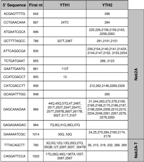
III.3.3 Identification of the positive clones...106
III.3.4 Nek2 (NIMA- related kinase 2)...108
III.3.5 NIPP1-T (Nuclear Inhibitor of Protein Phosphatase 1 in Testis)...117
III.3.6 PPPR15B (Phosphoprotein Phosphatase Regulatory Subunit 15B)...123
III.3.7 KIAA1949...129
III.3.8 I2-L (Protein Phosphatase 1 Inhibitor 2 Like)...132
III.4 DISCUSSION ...135
IV
SEARP – a novel PP1 interacting protein ...141
IV.1 INTRODUCTION ...141
IV.2 MATERIALS AND METHODS...153
IV.2.1 Tissue preparation...153
IV.2.2 Immunoprecipitation...154
IV.2.7 Immunocytochemistry ...158
IV.2.8 Immunohistochemistry ...158
IV.3 RESULTS ...161
IV.3.1 SEARP sequence analysis...161
IV.3.2 Tissue distribution of SEARP ...165
IV.3.3 Confirmation of SEARP/PP1 2 interaction ...169
IV.3.4 Intracellular localization of SEARP and PP1 2 in COS-7 cells ...173
IV.3.5 Sperm maturation and SEARP expression...176
IV.3.6 Colocalization of SEARP and PP1 2 in human spermatozoa...183
IV.4 DISCUSSION ...185
V
DISCUSSION AND CONCLUSION ...189
V.1 THE YEAST TWO HYBRID SYSTEM ...189
V.2 MECHANISMS INVOLVED IN SPERM MOTILITY...192
V.2.1 PP1 2/I2-L/GSK-3 ...193
V.2.2 PP1 2/sds22...194
V.2.3 PP1 2/AKAP220/PKARII ...195
V.2.4 PP1 2/AKAP110/PKA ...196
V.3 SPLICING AS A FEASIBLE THERAPEUTICAL TARGET...197
V.4 CONCLUDING REMARKS...200
REFERENCES...203
Appendix I - Culture media and solutions...233
SDS-PAGE Solutions ...234
Yeast Two-Hybrid Solutions...236
Solutions for yeast colony hybridization experiments ...238
Solutions for Northern Analysis ...240
Solutions for the 2D gel electrophoresis ...240
Immnunoprecipitation Solutions...241
Appendix II - Primers ...242
Appendix III - Bacteria and yeast strains ...243
ABBREVIATIONS
aa AD Ade Amp APS BD BLAST cAMP cDNA cds Chr CK2 Cys dATP dCTP DEPC dGTP DMSO DNA dNTP dsDNA dTTP EDTA EST GAL4 GSK3 His Amino acid Activation domain Adenine Ampicillin Ammonium persulfate Binding domainBasic Local Alignment Search Tool
Cyclic AMP (Adenosine 3’,5’-monophosphate) Complementary deoxynucleic acid
Protein coding sequence Chromosome Casein kinase 2 Cystein 2’-deoxyadenosine-5’-triphosphate 2’-deoxycytidine-5’-triphosphate Diethylpyrocarbonate 2’-deoxyguanosine-5’-triphosphate Dimethylsulfoxide Deoxynucleic acid Deoxynucleotide triphosphate Double strand deoxynucleic acid 2’-deoxythymidine-5’-triphosphate Ethylenodiaminotetraacetic acid Expressed sequence tag
Gal4 transcription factor Glycogen synthase kinase-3 Histidine
Leu LiAc NMDA nt OD ORF PCR PEG PKA PMSF PP1 PPM PPP QDO RNA RT-PCR SAP SD SDS SDS-PAGE Ser TBS TDO TEMED Thr Tris Trp Tyr UAS UV X- -gal YPD Leucine Lithium acetate N-methyl-D-aspartic acid Nucleotide Optical density Open Reading Frame Polymerase Chain Reaction Polyethylene glycol
Protein kinase A Phenyl methylsulfoxide Protein phosphatase 1
Mg2+-dependent protein phosphatase
Phosphoprotein phosphatase Quadruple dropout
Ribonucleic acid
Reverse transcriptase - polymerase chain reaction Shrimp alkaline phosphatase
Supplement dropout Sodium dodecyl sulfate
Sodium dodecyl sulphate – polyacrylamide gel electrophoresis Serine
Tris-buffered saline solution Triple dropout
N,N,N’,N'-tetramethyletylendiamine Threonine
Tryptophan Tyrosine
Tris (hydroxymethyl)-aminoethane chloride Upstream activating sequence
Ultraviolet
5-bromo-4-chloro-3-indolyl-alpha-D-galactopyranoside Yeast extract, Peptone and Dextrose medium for S. cerevisiae
YPDA YTH
YPD with adenine Yeast Two-hybrid system
I
INTRODUCTION
I.1
PROTEIN PHOSPHORYLATION AS A DYNAMIC PROCESS
The reversible phosphorylation of structural and regulatory proteins is a major intracellular control mechanism in eukaryotes. It is involved in almost all cellular functions, from metabolism to signal transduction, cell division and memory. The phosphorylation state of a protein is a dynamic process controlled by both protein kinases and protein phosphatases (Fig. I.1).
Figure I.1: The dynamic process of protein phosphorylation.
The protein kinases and protein phosphatases, the key controlling elements, are regulated by a myriad of extracellular and intracellular signals. Unlike the protein kinases that all belong to a single gene family, the protein phosphatases are divided into several distinct and unrelated protein/gene families. The Ser/Thr-specific protein phosphatases (Ser/Thr-PPs) comprise two distinct families, the PP1/PP2A/PP2B gene family and the PP2C gene family. The specific phosphatase family, as well as including the Tyr-specific phosphatases, also comprises the so-called dual Tyr-specificity phosphatases (capable of dephosphorylating Ser, Thr and Tyr residues). Besides these intracellular phosphatases involved in signal transduction, there are also unrelated non-specific alkaline and acid phosphatases that are usually found either in specialized intracellular compartments or in the extracellular milieu.
The sequencing of entire genomes has revealed that approximately 3% of all eukaryotic genes encode protein kinases or protein phosphatases (Plowman et al., 1999). Surprisingly, there appear to be 2-5 times fewer protein phosphatases than protein kinases. This imbalance is even more pronounced when the analysis is limited to Ser/Thr-PP and kinases, particularly in vertebrates. The human genome, for instance, encodes approximately 20 times fewer Ser/Thr-PP than Ser/Thr-kinases. Thus, whereas the diversity of the Ser/Thr-protein kinases has kept pace with the increasing complexity of evolving organisms, that of Ser/Thr-PP has not. In the past decade it has became apparent that the diversity of Ser/Thr-PP is achieved not only by the evolution of new catalytic subunits, but also by the ability of a single catalytic subunit to interact with multiple regulatory (R) subunits.
I.2
ABNORMAL PHOSPHORYLATION IN DISEASE
Reversible protein phosphorylation is the major metabolic control mechanism of eukaryotic cells. Indeed, cellular health and vitality are dependent on the fine equilibrium of protein phosphorylation systems. Not surprisingly many diseases and dysfunctional states are associated with the abnormal phosphorylation of key proteins (e.g. cancer, diabetes, etc.).
In neurodegenerative diseases such as Alzheimer’s Disease there is evidence for abnormal regulation of protein kinases. Altered activities and protein levels of several specific kinases suggest that abnormal phosphorylation contributes to the pathogenesis of these diseases. In Alzheimer’s Disease, neurofibrillary degeneration results from the aggregation of abnormally phosphorylated Tau protein into paired helical filaments. Protein kinase A (PKA) and glycogen synthase kinase 3 (GSK3 ) are likely to be key kinases involved (Delobel et al., 2002), and PP2A is thought to be the main tau phosphatase (Planel et al., 2001). Furthermore, activation of protein kinase C (PKC), or inactivation of protein phosphatase 1 (PP1) leads to a relative increase in the utilization of the non-amyloidogenic pathway for Alzheimer’s amyloid precursor protein (APP) processing (Gandy and Greengard, 1994; da Cruz e Silva et al., 1995a).
major anatomical hallmark of PD). Alpha-synuclein was demonstrated to be constitutively phosphorylated indicating that its function is regulated by phosphorylation/dephosphorylation mechanisms (Okochi et al., 2000). Motor and cognitive deficits in HD are likely caused by progressive neuronal dysfunction preceding neuronal cell death. Synapsin I is one of the major phosphoproteins regulating neurotransmitter release. In mice expressing the HD mutation, synapsin I is abnormally phosphorylated suggesting that an early impairment in its phosphorylation may alter synaptic vesicle trafficking and lead to defective neurotransmission in HD (Lievens et al., 2002).
Altered phosphorylation has also been implicated in the etiology and/or symptoms of heart failure (Neumann, 2002) as well as in Diabetes (Sridhar et al., 2000).
Therefore, protein phosphorylation systems represent attractive targets for diagnostics and therapeutics of several neurodegenerative and non-neurodegenerative diseases.
I.3
PHOSPHATASES AND THEIR CLASSIFICATION
The Ser/Thr-PPs, based on biochemical parameters, were initially divided into two classes: the type-1 phosphatases (PP1) that were inhibited by two heat-stable proteins, inhibitor-1 (I-1) and inhibitor-2 (I-2), and preferentially dephosphorylated the -subunit of phosphorylase kinase; and the type-2 phosphatases, insensitive to the heat-stable inhibitors and that preferentially dephosphorylated the -subunit of phosphorylase kinase (Ingebritsen and Cohen, 1983; Cohen, 1989). Type-2 phosphatases were further subdivided into cation independent (PP2A), Ca2+-dependent (PP2B) and Mg2+-dependent (PP2C)
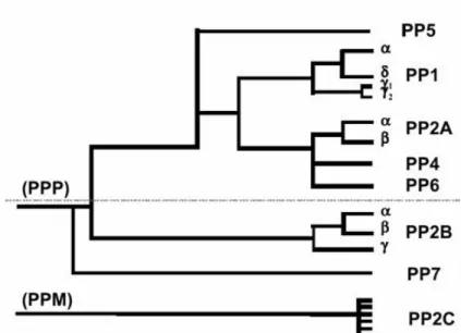
classes. The use of okadaic acid, a specific phosphatase inhibitor, further facilitated the discrimination between different classes (Cohen et al., 1989). Although widely in use, this classification does not reflect the actual phylogenetic relationship between the different Ser/Thr-PP. Molecular cloning revealed that PP2A was in fact much more related to PP1 than to PP2C (Berndt et al., 1987; da Cruz e Silva et al., 1987). From a phylogenetic point of view it is more reasonable to group PP1, PP2A and PP2B in family I or PPP [that also includes the bacteriophage λ, orf221 phosphatase (Cohen et al., 1988)] and PP2C in family II or PPM (Fig. I.2).
Figure I.2: Phylogenic tree depicting the degree of similarity between the known phosphatases
based on their primary amino acid sequence. PP1-PP7 belong to a single gene family (PPP) that is structurally distinct from the PP2C family (PPM). The phosphatases above the dashed line are highly sensitive to inhibition by the naturally occurring toxins, okadaic acid, mycrocystin and calyculin A (Honkanen and Golden, 2002).
I.3.1
The PPP Family
The application of recombinant DNA techniques to the field yielded not only the primary structure of all four phosphatase types, but also documented the existence of isoforms for each type and revealed the existence of previously undetected phosphatases in a variety of eukaryotic cells (Berndt et al., 1987; da Cruz e Silva et al., 1987; da Cruz e Silva and Cohen, 1987; Cohen et al., 1988; da Cruz e Silva et al., 1988). Three genes are known to encode type 1 phosphatase catalytic subunits, termed PP1α, PP1β and PP1γ. At least PP1γ is known to undergo tissue-specific processing to yield an ubiquitously expressed PP1γ1 isoform and a testis-specific PP1γ2 isoform (da Cruz e Silva et al., 1995b).
Two genes are known encoding PP2A catalytic subunits, termed PP2Aα and PP2Aβ, and the three known PP2B catalytic subunit genes (Aα, Aβ and Aγ) are also subject to complex regulation to yield several alternatively spliced isoforms from each.
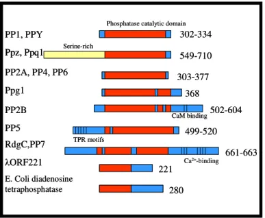
novel phosphatases (da Cruz e Silva et al., 1988). PP4, PP5 and PP6 (Cohen, 1997) are present in all mammalian tissues examined. In contrast, human PP7 (Huang and Honkanen, 1998), also found in Arabidopsis thaliana by Andreeva et al. (1998) and two Drosophila phosphatases are tissue specific (PPY is testis-specific and RdgC is restricted to photoreceptor organs and a small region in the brain). PP7 has been detected in the human retina and also in specialized sensory cells in plants (Fig. I.3).
PP1, PPY PP2A, PP4, PP6 Ppg1 PP2B PP5 RdgC,PP7 E. Coli diadenosine tetraphosphatase ORF221 Ppz, Ppq1 Serine-rich
Phosphatase catalytic domain
CaM binding TPR motifs Ca2+-binding 302-334 549-710 303-377 368 502-604 499-520 661-663 221 280 PP1, PPY PP2A, PP4, PP6 Ppg1 PP2B PP5 RdgC,PP7 E. Coli diadenosine tetraphosphatase ORF221 Ppz, Ppq1 Ppz, Ppq1 Serine-rich
Phosphatase catalytic domain
CaM binding TPR motifs Ca2+-binding 302-334 549-710 303-377 368 502-604 499-520 661-663 221 280
Figure I.3: Domain organization of PPP family members. The numbers of amino acids in each are
indicated on the right.
I.3.1.1
PP1 - Protein Phosphatase 1
Eukaryotic genomes contain between one (Saccharomyces cerevisiae) and eight (Arabidopsis thaliana) genes that encode isoforms of PP1 catalytic subunit. These isoenzymes typically show an overall sequence identity of approximately 90% and cannot be distinguished by either their substrate specificity or their ability to interact with R subunits in vitro (Zhang et al., 1993; Schillace and Scott, 1999). The sequence of the catalytic core of PP1 (residues 41-269 of the mammalian PP1 isoform) is almost identical in all isoforms, showing a high degree of similarity with the corresponding fragment of the catalytic subunits of PP2A and PP2B (Egloff et al., 1995; Goldberg et al., 1995). A detailed description of PP1, its properties and binding proteins will be given below.
I.3.1.2
PP2A - Protein Phosphatase 2A
PP2A has one of the most highly expressed catalytic subunits. It has been detected in various cell types and can comprise 0.3-1% of cellular proteins (Ruediger et al., 1991; Lin et al., 1998). The first descriptions of PP2A holoenzymes in the late 1970s and early 1980s demonstrated that the prevalent holoenzymes in rabbit skeletal muscle were heterotrimers composed of a catalytic subunit (C), a structural A subunit and a regulatory B subunit. cDNAs have been identified for two A subunits (α,β), two C subunits (α,β) and over twenty B subunits. For example, PR55 (B subunit), a 54 kDa protein (B’ subunit), PR72, a 74 kDa protein (B’’ subunit) and PR130. It has becoming increasingly clear that a major function of these regulatory subunits is to target the PP2A holoenzyme to distinct intracellular locations, signaling complexes and substrates. The A subunit is composed almost entirely of 15 imperfect repeats, each of 39 amino acids. These HEAT repeats (so named because they were found in Huntingtin, Elongation factor, PP2A subunit and the TOR kinase) are found in a variety of proteins. It seems that the repeats consist of two α helices connected by an intra-repeat loop, and that these loops are the binding sites for the B and C subunits (Ruediger et al., 1992; Ruediger et al., 1994; Ruediger et al., 1999) (Fig. I.4). The crystal structure of the mammalian Aα subunit (PR65α) has been solved: the subunit is a hook-shaped protein with the C subunit binding to the inner face of the hook; the various B subunits attach to the medial surface of the hook (Fig. I.5) (Cegielska et al., 1994).
A
C
B
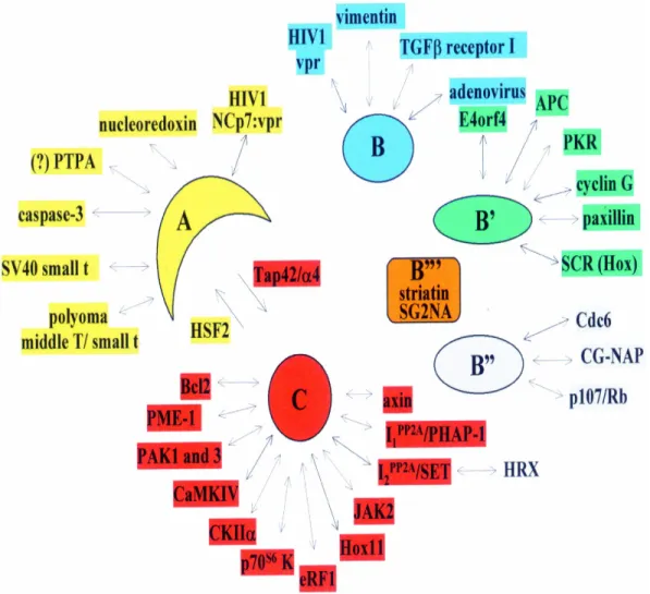
C subunit regulators and interactors
Natural products (okadaic acid, calyculin, cantharidin, etc.) Polypeptide inhibitors (I1/pp32, I2/SET, Hox11) Methylesterase PME-1
Tap42/α4, CK2α, axin, Bcl-2
B subunit and their targets
B/PR55 Vimentin, CamKIV, E4orf4, others B’/B56/PR61 B’’/PR72 Cyclin G, APC P107, CDC6 Polyoma T antigens A C B
C subunit regulators and interactors
Natural products (okadaic acid, calyculin, cantharidin, etc.) Polypeptide inhibitors (I1/pp32, I2/SET, Hox11) Methylesterase PME-1
Tap42/α4, CK2α, axin, Bcl-2
B subunit and their targets
B/PR55 Vimentin, CamKIV, E4orf4, others B’/B56/PR61 B’’/PR72 Cyclin G, APC P107, CDC6 Polyoma T antigens
Figure I.4: The heterotrimeric form of
PP2A includes a catalytic C subunit, a structural A subunit and a regulatory B subunit. Several cellular and viral proteins that interact with PP2A components are indicated.
I(1)PP2A and I(2)PP2A (Li et al., 1995) and stimulation by ceramide. The C subunit of PP2A can be phosphorylated in vitro by the tyrosine kinases p60v-src, p56lck, epidermal growth factor receptor and insulin receptor (Chen et al., 1992). Phosphorylation occurs at Tyr-307 at the extreme C-terminus of the protein and leads to 90% loss of activity. Dephosphorylation reactivates the phosphatase. This reactivation is prevented by okadaic acid, suggesting an auto-dephosphorylation reaction. A carboxymethyltransferase (Lee and Stock, 1993; Xie and Clarke, 1994a, b) has PP2A C subunit as the major substrate. The methylation of the α-carboxyl group of the C-terminal Leu-309 of PP2A in vitro has only moderate stimulatory effects on phosphatase activity and impairs binding of peptide-specific antibodies to the C-terminus of PP2A C subunit (Favre et al., 1994). Interestingly, the C-terminal sequence of PP2A C subunit is conserved from yeast to man suggesting that modifications of the C-terminus may be a conserved regulatory mechanism (Zolnierowicz et al., 1994). This modification may influence the interaction of the C subunit with its regulatory subunits. In vitro ceramide activates the trimeric forms of PP2A that contain either the PR55a or the 54kDa (B’) subunit. In contrast, PP2A dimmeric form and the isolated C subunit are insensitive to ceramide (Dobrowsky et al., 1993).
Figure I.5: Crystal structure of the mammalian Aα subunit of PP2A.
PP2A plays a central role in processes as varied as cell cycle regulation, cell signaling and neuronal function.
Cell Cycle Control and PP2A
PP2A was first implicated in the control of DNA replication when PP2A C subunit was purified as replication protein C on the basis of its ability to stimulate the efficient initiation of simian virus 40 (SV40) DNA replication (Virshup and Kelly, 1989). PP2A C subunit restored SV40 DNA replication activity to extracts from G1 phase cells suggesting that specific PP2A activity was regulated in the cell cycle as well (Virshup et al., 1989). More recently, strong evidence has been obtained for the involvement of PP2A in chromosomal DNA replication (Lin et al., 1998). The immunodepletion of A-subunit-containing forms of PP2A from Xenopus egg extracts inhibit subsequent DNA replication. This inhibition was relieved by replenishing the extracts with purified free PP2A C subunit. It was shown that PP2A activity was required for initiation of chromosomal replication rather than elongation. Immunodepletion of A/C dimers alone did not inhibit DNA replication, suggesting that a specific heterotrimeric form of PP2A is required (Lin et al., 1998). The finding of a novel PR72-related B subunit that recruits PP2A to the human replication initiator protein CDC6 suggests that this family of B subunits (the PR72/130/B’’ family) may be involved in initiation of both chromosomal and viral DNA replication (Yan et al., 2000). Another novel PR72 family member, PR59, has been isolated in a two hybrid screen using p107, the Rb-related cell-cycle regulatory protein (Voorhoeve et al., 1999a). PR59 may target PP2A to dephosphorylate p107 in response to stimuli such as UV irradiation that causes G1 arrest (Voorhoeve et al., 1999b). So, the PR72-related proteins may be the PP2A regulatory subunit family involved in regulation of the G1/S transition.
Interestingly, a body of data suggests that a subset of PP2A holoenzymes function as tumor suppressors. The PP1/PP2A inhibitor okadaic acid can promote tumor formation suggesting that phosphatase inhibition can function similarly to kinase activation. The most compelling evidence that PP2A is a tumor suppressor remains the finding that DNA tumor virus small and middle T antigens replace B subunits in the PP2A heterotrimer and modulate substrate specificity (Pallas et al., 1990; Walter et al., 1990). Other potentially oncogenic human proteins that inhibit PP2A function have been reported, including HOX11 and SET oncogenes (Li et al., 1996b; Kawabe et al., 1997). Set expression and
induced transformation (Al-Murrani et al., 1999; Baharians and Schonthal, 1999). Although some studies indicate that PP2A might exert tumor suppressive functions, other findings demonstrate the requirement for PP2A in cell growth and survival, which is not a characteristic of a tumor suppressor. This discrepancy might be due to the fact that PP2A is a multitask enzyme system, rather then a single enzyme. The puzzling observation that PP2A exerts inhibitory as well as stimulatory effects on cell growth could be due to the activity of different PP2A complexes with distinct subcellular location and diverse substrate specificity.
PP2A and neuronal function
The roles of PP2A in dephosphorylation of brain proteins are obscure. PP2A is a major activity that dephosphorylates voltage-sensitive sodium channels (Chen et al., 1995), and is also a major brain phosphatase that dephosphorylates autophosphorylated CaMKII. PP2A selectively dephosphorylates soluble CaMKII rather then CaMKII associated with postsynaptic density, where it becomes a substrate for PP1 (Strack et al., 1997a). The recent demonstrations that PP2A associates with and regulates CaMKIV (Westphal et al., 1998), p70S6 kinase and p21-activated kinase (Westphal et al., 1999a), support the proposal that PP2A might be a general regulator of protein kinases (Barnes et al., 1995). PP2A is also known to be associated with neurofilaments, dephosphorylating NF-M and NF-L (Saito et al., 1995; Strack et al., 1997b). It probably regulates either the stability of neurofilaments or their interactions with other components of the neuronal cytoskeleton. Another potential function of PP2A is the regulation of the state of phosphorylation of microtubulule-associated proteins (MAPs). Neuronal-specific MAPs, including Tau and MAP2, bind to microtubules and regulate their stability. Neural MAPs are phosphorylated at multiple sites by a variety of protein kinases. Phosphorylation causes disassociation of MAPs from microtubules and their loss of stability. Accumulation of hyperphosphorylated Tau in neurofibrillary tangles is a pathological mark of Alzheimer’s disease (T. H. Lee, 1995; Mandelkow et al., 1996; Billingsley and Kincaid, 1997). Disruption of the microtubule cytoskeleton as a result of the loss of Tau-mediated stabilization may be one mechanism of neuronal cell death.
Signaling Cascades and PP2A
PP2A also plays an important role in the regulation of specific signal transduction cascades, as witnessed by its presence in a number of macromolecular signaling modules,
where it is often found in association with other phosphatases and kinases. Viral proteins target specific PP2A enzymes in order to deregulate chosen cellular pathways in the host and promote viral progeny. The observation that a variety of viruses utilize PP2A to alienate cellular behavior emphasizes the fundamental importance of PP2A in signal transduction. Other examples of the importance of PP2A in signaling cascades include the Wnt signaling pathway and the TOR (Target Of Rapamycin) signaling pathway.
Wnt signaling regulates vertebrate axis formation in early embryogenesis, and deregulation of the Wnt pathway is found in multiple epithelial cancers, including those of colon, skin and liver. One major effect of Wnt signaling is increased transcription driven by heterodimeric β-catenin/TCF transcription factor (Brown and Moon, 1998). In the absence of Wnt signaling β-catenin is maintained at low levels by phosphorylation-regulated proteolysis. Phosphorylation is mediated by a large multiprotein complex containing the adenomatous polyposis coli protein (APC), the serine/threonine kinase GSK3β and axin. PP2A has also been placed in this complex. It was shown by a two hybrid screen (Seeling et al., 1999) that an amino-terminal domain of APC interacted with all members of the B’/B56 family of PP2A regulatory subunits. It was also found (Hsu et al., 1999) that the carboxyl terminus of axin binds to PP2A via the catalytic subunit. Thus, the role of B56-containing PP2A heterotrimers in Wnt signaling may be to regulate the activity of GSK3β, keeping it dephosphorylated and thus active, leading to lower β-catenin levels in vivo. It was shown that PP2A C subunit dephosphorylated and activated GSK3β in vitro.
TOR signaling activates a response to nutrients, leading to TOR kinase activation with subsequent phosphorylation and activation of the ribosomal S6 kinase and phosphorylation and inactivation of eukaryotic initiation factor 4E-BP1, an inhibitor of translation initiation factor eIF4E (Gingras et al., 1999). The result is stimulation of mRNA translation. Rapamycin inhibits TOR leading to growth arrest. TOR signaling may be due largely to its effect on PP2A and related phosphatases (Di Como and Arndt, 1996). TOR directly phosphorylates the Tap42 protein of S. cerevisiae, which then binds to either the C subunit of PP2A or the related yeast phosphatase Sit4 (Jiang and Broach, 1999). The Tap42-PP2A C dimer does not contain PP2A A or B, subunits representing a novel form of
yeast Tap42, α4, has been shown to bind PP2A C (Murata et al., 1997) and also the related phosphatases PP4 and PP6 (Jiang and Broach, 1999).
In summary, many proteins have been identified that interact with PP2A. Some of them affect PP2A activity [such as I(1)PP2A, I(2)PP2A, PTPA, Tap42/α4, SV40 small t, polyoma middle T/small t, HIV-1 NCp-7Vpr, adenovirus E4orf4CKIIα, Hox11 and PKR], some are PP2A substrates (such as Bcl2, p70s6 kinase, CaMKIV, vimentin, paxillin and SCR), for some PP2A it self is a substrate (such as caspase-3, JAK2, PME-1 and PKR) and some act as targeting proteins (such as eRF1, axin, APC and CG-NAP). An overview is given in Figure I.6.
I.3.1.3
PP2B - Protein Phosphatase 2B
PP2B, or calcineurin, is a Ca2+/calmodulin-dependent protein phosphatase. The
enzyme consists of two subunits, the catalytic A subunit of 60 kDa (PP2B A) and the myristoylated regulatory B subunit of 19 kDa (PP2B B). Calcineurin is present in nearly all mammalian cells studied but is highly enriched in neural tissue comprising over 1% of the total protein in the brain [a review can be found in (Klee et al., 1998)]. Originally, the term calcineurin referred to the neuronal form of PP2B but, more recently, calcineurin refers to both neuronal and non-neuronal PP2B. The amino acid sequence of PP2B is highly conserved from humans to yeast with over 50% sequence identity (da Cruz e Silva and Cohen, 1989; da Cruz e Silva et al., 1991; Stemmer and Klee, 1994). Cloning from rat brain indicated an A subunit of 521 amino acids. There are three mammalian genes for the A subunit, giving rise to the PP2B Aα, PP2B Aβ and PP2B Aγ isoforms. Aα and Aβ are highly expressed in brain, whereas Aγ is testis specific. Differential splicing of PP2B Aα generates two transcripts (α1 and α2), whereas PP2B Aβ gene is alternatively spliced to
give three transcripts β1, β2 and β3. PP2B Aα1 and PP2B Aβ2 are highly expressed in
neuronal tissue (Ueki et al., 1992). The A subunit shows autoinhibition that is relieved by interaction with the B subunit. PP2B, in contrast to PP1 and PP2A, is the only PP directly regulated by Ca2+. The inhibition of B on A is relieved if B binds Ca2+. This explains why the enzyme is dependent on Ca2+ for activity. Proteolysis of the autoinhibitory COOH terminus (residues 467-492) of the PP2B A generates a Ca2+-independent form. The B subunit was sequenced at the protein level and found to comprise 168 amino acids, exhibiting a high degree of sequence similarity to calmodulin. Both contain four EF-hand Ca2+-binding loops, both are needed for full PP2B activity, and both bind to unique regions on PP2B A without any cross interference. Two different B subunit genes are known: PP2B Bα and PP2B Bβ. PP2B Bα gives rise to one protein isoform expressed in many tissues termed PP2B Bα1 (170 amino acids) and, by means of a different promotor, leads
also to another testis-specific isoform PP2B Bα2 (216 amino acids). PP2B Bβ (179 amino
helped to further clarify the various binding domains and interaction sites of PP2B (Fig. I.7).
The best known in vitro and in vivo substrates of PP2B are the PP1 inhibitors I-1 and DARPP-32. Thus, PP2B controls PP1 activity and the two together form the first documented phosphatase cascade (Mulkey et al., 1994).
COOH NH2
14 342 373 390 414 469 486
Catalytic domain CaN B CaM AID
AID CaN B a) b) COOH NH2 14 342 373 390 414 469 486
Catalytic domain CaN B CaM AID
AID
CaN B
COOH NH2
14 342 373 390 414 469 486
Catalytic domain CaN B CaM AID
AID
CaN B
COOH NH2
14 342 373 390 414 469 486
Catalytic domain CaN B CaM AID
COOH NH2 14 342 373 390 414 469 486 COOH NH2 NH2 14 342 373 390 414 469 486
Catalytic domain CaN B CaM AID
AID
CaN B
a)
b)
Figure I.7: Structure of calcineurin. a) Diagrammatic representation of the calcineurin A subunit
indicating the catalytic domain, the binding domains for calcineurin B (CaN B), calmodulin (CaM), and the auto-inhibitory domain (AID). b), the crystal structure of the human calcineurin heterodimer is shown with secondary structural elements. The calcineurin A subunit is shown in blue and the B subunit in green. Fe (red) and Zn (yellow) are bound in a cleft of the catalytic domain and four Ca (black) are shown binding to the B subunit.
PP2B and neuronal function
Calcium-stimulated dephosphorylation of target proteins by PP2B modulates numerous neuronal activities. Described functions for PP2B in the brain include Ca2+ -dependent dephosphorylation of DARPP-32, inhibition of neurotransmitter release, dephosphorylation of dynamin I, and desensitization of postsynaptic NMDA-receptor-coupled calcium channels (Yakel, 1997; Greengard et al., 1998; Klee et al., 1998). PP2B is also thought to regulate the calcium channel activity of the ryanodine and inositol
1,4,5-triphosphate receptors via an immunophilin-mediated interaction (Snyder et al., 1998), and plays a role in the neurotoxicity associated with excessive stimulation of NMDA-type glutamate receptors. Immunossuppressants prevent apoptosis and rearrangments of the cytoskeleton in response to excitotoxic glutamate (Halpain et al., 1998). Overexpression of PP2B in transgenic mice revealed a novel phase of long-term potentiation in the CA1 region of the hippocampus that may be normally constrained by PP2B (Winder et al., 1998). These observations provide evidence for a physiological function for PP2B in intact animals. The altered regulation of this component of long-term potentiation by PP2B correlates with reversible deficits in long-term memory that occur during expression of activated PP2B (Mansuy et al., 1998). These observations provide an elegant demonstration of the role of PP2B in memory. Along with PP2A, PP2B is one of the major tau phosphatases in vitro and can restore the microtubule-binding ability of hyperphosphorylated tau (Billingsley and Kincaid, 1997). When nerve terminals in the brain are stimulated, a group of phosphoproteins called dephosphins are coordinately dephosphorylated by PP2B. Amazingly, the seven presently known dephosphins are not structurally related, yet each has been independently shown to be essential for synaptic vesicle endocytosis. Nowhere else in biology is there a similar example of the coordinated dephosphorylation of such a large group of proteins each sharing roles in the same biological response.
PP2B and T-cell activation
PP2B plays a crucial role in the Ca2+-signaling cascade of activated T-cells.
Increases in Ca2+ concentrations in T-cells promoted by antigen presentation to T-cell
receptor stimulates PP2B to dephosphorylate the cytosolic subunit of the transcription factor NFAT, Nuclear Factor of Activated T-cells, (Jain et al., 1993). Dephosphorylated NFAT translocates into the nucleus where, in concert with other transcription factors, it induces expression of the IL-2 gene, one of the early genes in the T-cell activation pathway. Inhibition of this Ca2+-signaling cascade by immunosuppressant drugs (such as
FK506 and cyclosporin A, when complexed to their intracellular receptors FKBP and cyclophilin, respectively) suppresses T-cell activation.
the nucleus. More recently, it has been shown that this transcription factor is not only involved in T-cell activation, but also in several other mechanisms. PP2B has been shown to have important roles in axonal guidance (Chang et al., 1995), as well as memory and learning (Mansuy et al., 1998; Winder et al., 1998). In general, these functions in neurons have been thought to be independent of transcription. However, recent evidence indicates that NFAT family members may have critical roles in the development of synaptic connections (Graef et al., 1999). It also seems that the NFAT pathway is involved in the morphogenesis of heart valves. Additionally, it has been shown that severe cardiac hypertrophy can be induced by the overexpression of a truncated constitutively active PP2B A subunit (Molkentin et al., 1998).
NF-κB and other Rel proteins play critical roles in the development of the liver, skin, inflammatory responses and in some aspects of recombinational immune response. Unlike NFAT, which absolutely requires a calcium stimulus, NF-κB can be fully activated by PKC activators in the absence of calcium (Singh et al., 1986). However, suboptimal stimuli can be augmented with a calcium signal (Mattila et al., 1990). This calcium facilitation of NF-κB activity can be mimicked by overexpression of a constitutively active PP2B and can be partially blocked with FK506 or cyclosporin A, indicating that PP2B is required for full induction of NF-κB activity in certain circumstances (Frantz et al., 1994). The AP-1 transcription complex consists of Fos and Jun proteins. Transcription controlled by most AP-1 sites is not sensitive to inhibition by cyclosporin A or FK506 (Mattila et al., 1990). However, a site in the IL-2 promoter that binds junD and perhaps c-jun requires the action of PP2B for full function (Ullman et al., 1993). In addition, an apparent AP-1 site in the collagenase promoter is sensitive to FK506 (Su et al., 1994) and hence likely to be PP2B-dependent.
PP2B inhibitors
Several different classes of PP2B inhibitors have been discovered raising questions about their various roles in Ca2+/PP2B signaling. The inhibitors bind to PP2B and inhibit its ability to dephosphorylate substrates such as NFAT family members, thereby preventing their nuclear localization. One of the most interesting is the DSCR1 gene and its relatives, DSCR2 and ZAK14 (Fuentes et al., 2000). DSCR1 is located on chromosome 21 in the so called critical region (hence its designation, Down’s syndrome critical region 1 gene). Some evidences indicate that overexpression of DSCR1 might underlie the
pathogenesis of Down’s Syndrome. First, it was shown that DSCR1 protein is overexpressed in the brains of Down’s Syndrome patients. Second, some of the symptoms of the Syndrome appear in mice with mutations of the different PP2B-dependent (NFAT) subunits of the transcription complex (Fuentes et al., 2000). It is possible that overexpression of DSCR1 as a result of trisomy leads to inhibition of PP2B and subsequent effects on the development of the brain, immune system, heart and skeleton. However, the Down’s Syndrome critical region includes a number of genes and it is possible that the syndrome is due to overexpression of several of these. Recently, yeast has been shown to have a related protein, Pcn1p that binds and inactivates PP2B (Gorlach et al., 2000). Two additional classes of PP2B inhibitors are Cabin/Cain (Lai et al., 1998; Sun et al., 1998), which are novel proteins, and the CHP protein that has similarity to PP2B B subunit (Lin and Barber, 1996; Lin et al., 1999). The CHP proteins appear to compete with PP2B B for binding to the A protein and thereby inhibit the Ca2+-dependent activation of PP2B A. Cabin, or Cain (Calcineurin inhibitor), is a non competitive inhibitor of PP2B phosphatase activity with a Ki of 440nM. A physiologic role for these proteins is still unclear, but they do antagonize NFAT translocation. A fourth class of PP2B inhibitors is found in the genome of certain viruses, (e.g. African swine fever virus). The A238L protein encoded by the virus binds tightly to PP2B and blocks NFAT translocation and function (Miskin et al., 1998). AKAP79 (protein kinase A anchoring protein) was the first PP2B protein inhibitor to be found (Coghlan et al., 1995) and is also a scaffolding protein. AKAP79 binds both calcineurin and protein kinase A, and may anchor PP2B at specific sites that allow the protein to engage the proper substrates when activated. Another inhibitor that constitutes a useful laboratory tool is the auto-inhibitory peptide. This is a 26-residue peptide that interacts with the catalytic core and blocks PP2B activity with an IC50 of 10-15µM (Hashimoto et al., 1990). Immunosuppressant drugs such as cyclosporin A and FK506 (Liu, 1993) belong to a different class of PP2B inhibitors. These are fungal compounds that require binding to their cognate intracellular immunophilins (cyclophilin A and FKB12, respectively) prior to binding to and inhibiting PP2B activity with nanomolar affinity. Being membrane permeable, these compounds have greatly facilitated the in vivo study of PP2B function. Yet another class of inhibitors is the type II pyrethroid insecticides,
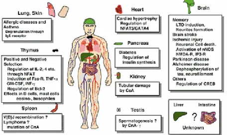
In conclusion, within the past few years PP2B has been implicated in a wide variety of extremely important biological responses including lymphocyte activation, neuronal and muscle development, neurite outgrowth, morphogenesis of vertebrate heart valves, learning and memory (Fig. I.8).
Figure I.8: Calcineurin as a multifunctional regulator (Shibasaki et al., 2002).
I.3.2
The PPM Family
The PPM protein phosphatases belong to a large and diversified family of protein phosphatases expressed in both eukaryotes and prokaryotes that dephosphorylate serine and threonine residues, as does the PPP family. PPC relates to the main enzyme subtype of the PPM, including Arabidopsis ABI1 (Leung et al., 1994), Arabidopsis KAPP-1 (Stone et al., 1994), pyruvate dehydrogenase phosphatase (Lawson et al., 1993) and Bacillus subtilis SpoIIE phosphatase (Barak et al., 1996). Within the PPM family, the PP2C domain occurs in numerous structural contexts that reflect functional diversity. For example, PP2C domain of the Arabidopsis ABI1 gene is fused with EF- hand motifs (Leung et al., 1994; Meyer et al., 1994), whereas in KAPP-1, a kinase-interaction domain that associates with a phosphorylated receptor precedes the phosphatase domain (Stone et al., 1994). Other examples include the Ca2+-stimulated mitochondrial pyruvate dehydrogenase phosphatase, which contains a catalytic subunit sharing 22% sequence identity with mammalian PP2C (Lawson et al., 1993) and SpoIIE phosphatase, which has ten membrane-spanning regions preceding the PP2C-like catalytic domain (Fig. I.9).
ABI1
Leung et al., 1994KAPP-1
Stone et al., 1994 Mitocondrial Pyruvate dehydrogenase Phosphatase Lawson et al., 1995SpoIIE
Duncan et al., 1995Yeast adenylate cyclase
Kataoka et al., 1985
PPM Family
PP2C domain EF-hand motifs
PP2C domain
Kinase interactio domain
22% identical to mammalianPP2C
PP2C domain
10 membrane sppaning regions
Cyclase catalytic domain
Similar to PP2C domain
Figure I.9: The PPM family.
PP2C was originally identified as a Mg2+-dependent protein phosphatase. The enzyme is monomeric with a molecular mass of 43-48 kDa (Cohen, 1989) and is resistant to okadaic acid. To date the existence of six distinct PP2C genes (PP2Cα, PP2Cβ, PP2Cγ, PP2Cδ, Wip1 and FIN13) has been reported in mammalian cells (Tamura et al., 1989; Wenk et al., 1992; Fiscella et al., 1997; Guthridge et al., 1997; Travis and Welsh, 1997; Tong et al., 1998). In addition, mouse PP2Cβ has been found to have five distinct isoforms (β1, β2, β3, β4, β5), which are splice variants from a single pre-mRNA (Terasawa et al., 1993; Hou et al., 1994; Kato et al., 1995).
Considerable information on the function of mammalian PP2C has accumulated over the last decade. PP2C can be activated by polyunsaturated fatty acids such as arachidonic acid. Upon addition of fatty acids PP2C no longer requires unphysiologically high Mg2+ concentrations for activity, and Ca2+ ions become inhibitory in the micromolar range. The stimulation of PP2C in vitro by fatty acids suggests that this enzyme may be regulated by an endogenous lipid messenger in vivo (Klumpp et al., 1998). It was shown (Murray et al., 1999) that PP2Cγ is physically associated with the spliceosome in vitro and is required during the early stages of spliceosome assembly for efficient formation of the A
MKK4, MKK6b and MKK7 activities in mammalian cells (Hanada et al., 1998). Recent research further revealed that PP2C might be involved in the control of cell apoptosis (Wolf et al., 1997), gene expression and other cellular functions (Bohmann, 1990; Kurosawa, 1994; Schonthal, 1995). More recently, PP2Cα has been implicated in Wnt signaling, being a positive regulator and exerting its effects through the dephosphorylation of axin (Strovel et al., 2000), a role that was first attributed to PP2A (Willert et al., 1999). PP2C influences the dynamic interactions between the actin cytoskeleton and membrane constituents linked to moesin (Hishiya et al., 1999). Members of the moesin protein family are localized membrane structures rich in actin filaments that act as linkers between the plasma membrane and the actin cytoskeleton. Furthermore, PP2Cα and PP2Cβ2 are responsible for the inactivating dephosphorylation of Cdk2/Cdk6 (Cheng et al., 2000b). The dephosphorylation of Cdk2 and Cdk6 by PP2C isoforms is inhibited by the binding of cyclins. PP2Cβ was recently implicated in the regulation of the TAK1 signaling pathway: TAK1 is a stress-activated protein kinase that is activated by interleukin-1, transforming growth factor-β or stress. PP2Cβ negatively regulates the TAK1 signaling pathway by direct dephosphorylation of TAK1 (Hanada et al., 2001).
I.4
PHOSPHATASE INHIBITORS
One of the most significant advances in the study of Ser/Thr-PPs, and the elucidation of the cellular events they control, was the identification of several naturally occurring toxins as powerful and specific phosphatase inhibitors. Among these are okadaic acid (Fujiki and Suganuma, 1993), cantharidin (Laidley et al., 1997), calyculin A (Ishihara et al., 1989), microcystins (Carmichael, 1992; Fujiki and Suganuma, 1993; Carmichael, 1994) and tautomycin (MacKintosh and Klumpp, 1990; Hori et al., 1991) to name a few examples. Another class of phosphatase inhibitors are the protein inhibitors readily available inside the cell. The specificity of these inhibitors for a given phosphatase has placed the former as key tools in the study of phosphorylation dependent processes.
I.4.1
Toxins as natural inhibitors of PPs
I.4.1.1
Okadaic acid and derivatives
Okadaic acid (OA) is a polyether compound with a C-38 structure and was isolated from the black sponge Halichondria okadai (Fujiki and Suganuma, 1993) (Fig. I.10).
Figure I.10: Structure of okadaic acid (R1=H, R2=H, R3=CH3).
OA is a potent phosphatase inhibitor in smooth muscle preparations (Bialojan et al., 1988), and was reported to inhibite PP1, PP2A and PP2B with IC50 values of 272, 1.6, and
3,600nM, respectively (Bialojan and Takai, 1988). PP2C, phosphotyrosyl phosphatase, acid phosphatase and alkaline phosphatase were not inhibited by up to 10µM OA. It is currently thought that tumor promotion by OA and related compounds like calyculin A and microcystin LR is due to inhibition of PP1 and PP2A [a review can be found in (Fujiki and Suganuma, 1993)]. OA in concentrations <2nM inhibits also other PP2A related family members such as PP4, PP5, and PP6 (Cohen, 1997).
I.4.1.2
Cantharidin and analogs
Cantharidin (CT), cantharidic acid, palasonin and endothall are structural analogs (Laidley et al., 1997). CT is the vesicant in blister beetles, palasonin is an anti-helmintic in seeds of a medical tree and endothal is a synthetic herbicide. The toxic action of CT is
cantharidic acid exhibited similar inhibitory activity (Li et al., 1993). Endothal inhibits both PP1 and PP2A with IC50 values of 5,000 and 1,000nM (Li et al., 1993). Neither
compound inhibits PP2B (>30,000nM) or PP2C (>1mM). CT is a terpenoid and is cell membrane permeable. It is less potent and selective than OA, but much cheaper. Mutational analysis indicates that OA and CA might act on different amino acids on PP1 (Zhang et al., 1994b) (Fig. I.11).
Fig. I.11: Structure of Cantharidin.
I.4.1.3
Calyculin A:
Calyculin A (CA) was isolated from another marine sponge, Discodermia calyx. It is an octamethylpolyhydroxylated C-28 fatty acid linked to two γ-amino acids and esterified with phosphate. It is cell membrane permeable. CA is equally potent against PP1 and PP2A (Ishihara et al., 1989; Fujiki and Suganuma, 1993) with IC50 values from 1 to
14nM. OA and CA seem to compete for the same inhibitory site on PP2A (Takai et al., 1995). Mutational analysis suggests that CA inhibits the enzyme via interaction with amino acid tyrosine-272 on PP1 because its IC50 is changed from 0.5 to 3,000nM in the Y272K
mutant (J. Zhang et al., 1996) (Fig. I.12).
I.4.1.4
Microcystins
Whereas OA and CA are fatty acid derivatives, microcystins (Mic) and nodularin (Nod) are peptide toxins. Mic are of toxicological and public health relevance. They cause death in cattle and humans exposed to water contaminated by certain algae. These include colonical and filamentous algae and cyanobacteriae like Microcystis aeruginosa (Carmichael, 1992; Fujiki and Suganuma, 1993; Carmichael, 1994). Mic are cyclic heptapeptides containing five constant (some of which are unique) and two variable amino acids. The variable amino acids in Mic are indicated in the one-letter code. Hence, their short-hand notation is microcystin-LR, -YR and –RR. Mic-LR inhibits PP1 and PP2A with IC50 values of 0.1nM each (MacKintosh et al., 1990). It inhibits PP2B with an IC50 of
0.2µM and does not inhibit PP2C up to 4µM (MacKintosh et al., 1990). PP4 and PP5 are inhibited by <2nM Mic. Mic is the most potent (and toxic) PP inhibitor, but being a peptide it is not cell permeable. The 3D structure of Mic-LR bound to PP1 has been determined (Goldberg et al., 1995; Bagu et al., 1997), and mutation of the critical cysteine-273 in PP1 to alanine impeded covalent binding of Mic to PP1 (Fig I.13).
Figure I.13: Structure of Microcystin.
I.4.1.5
Nodularin
Nodularin (Nod), a cyclic pentapeptide, was isolated from the toxic cyanobacterium Nodularia spumigenia (Carmichael, 1992). Like Mic it is toxic to the liver. Nod R and its
an IC50 of 8.7µM but does not affect PP2C (Honkanen et al., 1991). The IC50 values are
comparable to those of Mic-LR. In contrast to Mic, Nod R or V do not covalently bind to PP1 or PP2A (Craig et al., 1996). Mutational analysis suggests that Nod also inhibits PP activity via interaction with amino acid Tyr-272 on PP1 because its IC50 changed from 0.5
to 150nM in the Y272S mutant (J. Zhang et al., 1996). The 3D solution structure of Nod closely resembles that of Mic-LR (Annila et al., 1996) (Fig. I.14).
Figure I.14: Structure of Nodularin.
I.4.1.6
Tautomycin
Tautomycin (Tau) was isolated from Streptomyces spiroverticillatus as an antibiotic because it is toxic to yeast and fungi. Its structure as a polyketide resembles somewhat that of OA (MacKintosh and Klumpp, 1990; Hori et al., 1991). It inhibits PP1 and PP2A with IC50 values of 0.2-0.7 and 0.4-0.65nM, respectively (MacKintosh and Klumpp, 1990;
Fujiki and Suganuma, 1993). Others reported IC50 values of 0.4 and 34nM for PP1 and
PP2A, respectively (Takai et al., 1995). It does not inhibit PP2C and its IC50 for PP2B is
100µM. OA prevents the interaction of Tau with the catalytic subunit of PP2A (Fig. I.15).
I.4.1.7
Cyclosporin
Ciclosporin (CsA) or cyclosporin A (a cyclic undecapeptide), and FK506 (a macrocyclic lactone) inhibit PP2B (Parsons et al., 1994). This inhibition is not direct as was already mentioned. First CsA and FK506 bind to cyclophilin and a FK506-binding protein (FKBP), respectively. Then, they interact with the latch region of PP2B B (Clipstone et al., 1994; Milan et al., 1994), leading to inhibition of PP2B A. The 3D structure of the PP2B-FK506 and FKBP complex supports this notion (Griffith et al., 1995); (Kissinger et al., 1995). Rapamycin, an immunosupressant fungal metabolite, also binds to FKBP but does not inhibit PP2B because it cannot interact with PP2B B for steric reasons (Griffith et al., 1995). In contrast, it targets a rapamycin-associated protein (FRAP) that leads to inactivation of some specialized protein kinases like p70s6k (Price et al., 1992; Alberts et al., 1993).Work with CsA is complicated by its poor aqueous solubility. However, it is cell membrane permeant (Fig. I.16).
Figure I.16: Structure of Cyclosporin.
I.4.1.8
Cypermethrin
PP2B, PP1 or PP2A (MacKintosh and MacKintosh, 1994), are thought to be due to artifactual reasons.
I.4.1.9
Apomorphine
Apomorphine and SKF-38393 inhibited PP2A from rat brain with IC50 values of 1
and 50µM, respectively. In contrast, apocodeine was inactive. It has apparently not been reported to inhibit other PPs (Kawai, 1991). Apomorphin is cell membrane permeant.
I.4.1.10 Fostriecin
The antitumor antibiotic fostriecin (FT) is a type II DNA topoisomerase-directed anticancer drug, like doxorubicin or etoposide (Boritzki et al., 1988). FT is a naturally occurring compound from Streptomyces pulveraceus subspecies fostreus, an actinomycete found in a Brazilian soil sample. It is a water soluble polyene lacton with a phosphate ester. FT inhibited both PP1 and PP2A with IC50 values of 4µM and 40nM, respectively, but it
did not inhibit tyrosine phosphatases (Roberge et al., 1994). It has been suggested that the PP inhibition at least contributes to its efficacy against solid tumors in humans.
I.4.1.11 Heparin
Heparin binds and inhibits to PP1 but not PP2A (Gergely et al., 1984; Erdodi et al., 1985a, b). Heparin can actually stimulate PP2A. This property led to a procedure to separate these phosphatases using a heparin-based affinity column chromatography. Spermine inhibits both PP1 and PP2A with similar potency (Shenolikar and Nairn, 1991).
I.4.1.12 Thyrsiferyl 23-acetate
This compound is, like OA, a polyether fatty acid and contains a squalene carbon skeleton. It was isolated from the red alga L. obtuse. It is unique because it is a selective inhibitor of PP2A. It does not inhibit PP1, PP2B, PP2C or tyrosine phosphatase activity at concentrations up to 1mM. Its IC50 for PP2A is 4µM. It is several orders of magnitude less
potent than OA or CA. It is expected to be cell membrane permeant (Matsuzawa et al., 1994).
I.4.1.12 Oscillamide B and Oscillamide C
Two new phosphatase inhibitors, oscillamide B and C, were isolated from the cyanobacteria Planktothrix (Oscillatoria) agardhii and P. rubescens. These inhibitors are ureido-containing cyclic peptides and inhibit PP1 and PP2A (Sano et al., 2001).
I.4.2
Protein inhibitors
I.4.2.1
Inhibitor 1 (I-1)
I-1 and I-2 were identified by Huang and Glinsmann (F. L. Huang and W. H. Glinsmann, 1976; Huang et al., 1976). They share unusual physical properties: both are heat stable and are not precipitated by 1% trichloroacetic acid, in contrast to most other proteins. I-1 comprises 171 amino acids (Aitken et al., 1982; Elbrecht et al., 1990; Endo et al., 1996). Only in the C-terminal were differences between rabbit, rat and human noted (Endo et al., 1996). I-1 from rabbit skeletal muscle and human brain has a calculated molecular mass of 18.7 and 19.2kDa, respectively (Aitken et al., 1982; Endo et al., 1996), but the apparent molecular mass on SDS-PAGE is 26kDa. This discrepancy has been explained by a low degree of order in the protein. I-1 binds to and inhibits PP1 only after being phosphorylated on Thr-35 by cAMP-dependent protein kinase or cGMP-dependent protein kinase (Hemmings et al., 1984b). It is highly selective for PP1, inhibiting PP1 and PP2A with IC50 values of 1.1 and 21,000nM, respectively (Endo et al., 1996). I-1 is a
cytosolic protein; it has been used as a tool to study whether a process involves PP1. Amino acids 9-12 KIQF are conserved in rat, rabbit and human and seem to be crucial for binding and inhibition of PP1 (Egloff et al., 1997).
I.4.2.2
Inhibitor 2 (I-2)
I-2 from rabbit skeletal muscle comprises 204 amino acids and has a calculated mass of 22.9kDa (Holmes et al., 1986a). Similarly to I-1, its apparent molecular mass on
(L. Zhang et al., 1996). I-2 inhibition of PP1 can be reversed by GSK3 phosphorylation of I-2.
I.4.2.3
DARPP-32
DARPP-32 (dopamine and cyclic AMP-regulated phosphoproteins, Mr 32,000Da) is similar to I-1 in function but derived from a different gene, and is mainly expressed in the brain (Hemmings et al., 1984a). It is cytosolic and a predicted molecular mass of 22.6kDa, but again a higher apparent molecular mass of 32kDa on SDS-PAGE (Williams et al., 1986). The same Thr residue on DARPP-32 is phosphorylated by cAMP-dependent protein kinase and also by cGMP-dependent protein kinase. Phosphorylation of DARPP-32 changes its IC50 for PP1 from 1µM to 2nM (Desdouits et al., 1995a; Desdouits et al.,
1995b), underscoring its high selectivity. Under physiological conditions (mM Mn2+ in the
assay) I-1 and DARPP-32 are dephosphorylated and inactivated by PP2A and even better by PP2B (Hemmings et al., 1984a; Hemmings et al., 1990; Desdouits et al., 1995c). The dephosphorylation by PP2B is dependent on the presence of calcium. It was suggested that this might be a way for Ca2+ levels to control protein phosphorylation (Hubbard and Cohen, 1989). Thiophosphorylated I-1 or DARPP-32 is not dephosphorylated and has been successfully used to study the physiological role of PP1-mediated phosphorylation in muscle contraction.
I.4.2.4
Inhibitor 1 PP2A
Inhibitor 1 PP2A has been isolated from bovine kidney. It is thermostable and not inactivated by 1% trichloroacetic acid. Its apparent molecular mass is 30kDa (Li et al., 1995). Identified as putative class II human histocompatibility leukocyte-associated protein (PHAP) I (Li et al., 1996a), it seems to inhibit the catalytic subunit directly. It is not known whether its activity is regulated by a posttranslational modification like I-1 of PP1 (Li et al., 1995).
I.4.2.5
Inhibitor 2 PP2A
This inhibitor has been isolated from bovine kidney. It is also thermostable and not inactivated by 1% trichloroacetic acid. Its apparent molecular mass was initially reported as 20kDa (Li et al., 1995). Protein sequencing revealed that the protein had been described before as SET (Li et al., 1996b), PHAP II (Vaesen et al., 1994) and template activating factor-1β (Nagata et al., 1995). These last two inhibitors might be useful tools to study the physiological function of PP2A. It has been speculated that I-1 and I-2 of PP2A might be involved in signal transduction, particularly to mediate the effects of insulin on PP2A (Li et al., 1995).
I.4.2.6
Simian virus 40 small tumor antigen (SV40)
SV40 is a member of the papova family of small DNA tumor viruses. Its lytic cycle takes place in permissive monkey cells. SV40 infection leads to the production of proteins that are immunogenic and that were called tumor antigens. One such antigen is the SV40 small tumor antigen. It can inhibit PP2A activity with an IC50 of 10-15nM. SV40 inhibits
PP2A trimeric complex (Yang et al., 1991). In cells the interaction of small tumor antigen with PP2A leads to deinhibition and thus activation of MAP kinase and MEK which induces cell proliferation (Sontag et al., 1993).
I.4.2.7
Inhibitor 4
Kikuchi and coworkers isolated a human cDNA for a novel PP1 inhibitory protein, named I-4, from a cDNA library of germ cell tumors (Shirato et al., 2000). I-4, composed of 202 amino acids, is 44% identical to I-2. I-4 conserves functionally important structure of I-2 and exhibited similar biochemical properties. I-4 inhibited activity of the catalytic subunit of PP1 with IC50 of 0.2nM, more potently than I-2 with an IC50 of 2nM. Gel
overlay experiments showed that I-4 binds PP1c directly through a multiple-point interaction.