i
Faculdade de Engenharia da Universidade do Porto
Effect of immobilized α6β1 synthetic ligands on
the behavior of oligodendrocyte progenitor cells
(OPCs) in 3D fibrin hydrogels
Joana Catarina Santos Leite
M
ASTERT
HESISIntegrated Masters in Bioengineering
Supervisor: Professor Isabel de Freitas Amaral
Co-Supervisor: Helena Sofia de Azevedo Domingues
Resumo
As doenças desmielinizantes, como a esclerose múltipla e a lesão da espinal medula, são condições patológicas devastadoras, levando frequentemente a lesão axonal, deficiência neuronal e perda de função. Embora as células progenitoras de oligodendrócitos endógenas (OPCs) sejam prevalentes nas lesões de desmielinização, estas não são capazes de se diferenciarem em oligodendrócitos mielinizantes devido ao microambiente inibitório local. Apesar dos esforços para desenvolver novas terapias capazes de restaurar até alguma extensão a função neuronal, a regeneração do sistema nervoso central (SNC) continua a ser um desafio devido à complexidade dos seus componentes e dos mecanismos subjacentes ao processo de desmielinização.
O transplante de OPCs surgiu como uma terapia viável para promover a remielinização de axónios não lesados e em regeneração. Ainda assim, o transplante de OPCs sob a forma de suspensões celulares apresenta desvantagens, tais como baixa sobrevivência e baixas proliferação celular e diferenciação celulares. Neste sentido, a combinação da entrega de OPCs com um biomaterial como suporte pode ser vantajosa, nomeadamente para proporcionar um nicho para que as OPCs transplantadas possam sobreviver e diferenciar-se em oligodendrócitos mielinizantes. Devido às suas propriedades, os hidrogéis podem ser concebidos para imitar fisicamente o tecido nativo, proporcionando um microambiente altamente permissivo para a sobrevivência celular e crescimento tecidular. Apesar de vários hidrogéis terem sido desenvolvidos para entrega de drogas e células para o SNC, apenas alguns foram explorados para entrega de OPCs, entre eles a fibrina.
Ambos as OPCs e os oligodendrócitos expressam um repertório limitado de integrinas, entre elas a integrina α6β1. A interacção entre a laminina-211 expressa no SNC e a integrina α6β1 tem mostrado desempenhar um papel importante na sinalização dos mecanismos que estimulam a formação da membrana de mielina pelos oligodendrócitos.
Deste modo, o objectivo da presente dissertação consistiu em desenvolver um hidrogel capaz de incorporar ligandos sintéticos que se ligam especificamente à integrina α6β1, para entrega de OPCs no SNC. Para este efeito, os hidrogéis de fibrina foram funcionalizados com péptidos sintéticos com afinidade reportada para a integrina α6β1: (1) o péptido T1 do indutor angiogénico CCN1 (GQKCIVQTTSWSQCSKS) e (2) o péptido sintético HYD1 (KIKMVISWKG). É esperado que a funcionalização com ligandos para esta integrina melhore a bioespecificidade da fibrina para as OPCs, através da promoção da extensão de processos e da formação de membrana de mielina num microambiente 3-D.
O efeito da concentração de fibrinogénio na viabilidade e diferenciação de OPCs em hidrogéis 3-D de fibrina foi primeiramente avaliado. Embora a concentração de fibrinogénio não tenha afectado significativamente a viabilidade celular, esta revelou um impacto na extensão de processos pelas OPCs, um indicador de diferenciação, com géis de fibrina preparados com concentrações de fibrinogénio intermédias de 4 e 6 mg/mL a mostrarem o maior número de processos por célula, após 9 dias de cultura. Uma vez que a concentração de
fibrinogénio de 4 mg/mL mostrou o melhor desempenho biológico global, esta foi seleccionada para utilização nos estudos de funcionalização.
A funcionalização de fibrina com HYD1, T1, e com uma combinação de HYD1 com T1, que se ligam especificamente à integrina α6β1, promoveu a extensão de processos pelas OPCs em hidrogéis 3-D de fibrina, sem afectar a formação de membrana de mielina. Uma concentração de 5 µM de péptido foi suficiente para aumentar o número de processos por célula após 5 dias de cultura, em todos os géis funcionalizados. Além disso, a funcionalização com 5 µM de péptidos também levou a valores maiores de comprimento máximo, embora não tenham sido encontradas diferenças estatisticamente significativas. Ainda, o aumento da concentração de péptido para 10 µM ou 20 µM não levou a um aumento da extensão de processos, independentemente do péptido utilizado. Em adição, nenhum efeito aditivo/sinérgico foi observado em termos de impacto na extensão de processos com a combinação dos dois péptidos (5 µM de cada péptido).
Os resultados obtidos sugerem assim que os géis de fibrina funcionalizados com ligandos para a integrina α6β1 são interessantes candidatos para cultura e diferenciação de OPCs num microambiente 3-D, e, finalmente, para utilização como um veículo para a entrega de OPCs no SNC.
Palavras-Chave: Desmielinização, Células progenitoras de oligodendrócitos, oligodendrócitos, hidrogéis de fibrina, integrina α6β1, laminina 322, péptidos HYD1 e T1.
Abstract
Demyelinating disorders such as Multiple Sclerosis (MS) and Spinal Cord Injury (SCI) are devastating pathological conditions, often leading to axonal damage, neuronal disability and loss of function. Although endogenous oligodendrocyte progenitor cells (OPCs) are prevalent in demyelinated lesions, they are not able to differentiate into myelinating oligodendrocytes (OLs), due to the local inhibitory microenvironment. Despite the efforts made to develop new therapies capable of restoring at some extent neuronal function, regeneration of the central nervous system (CNS) remains a challenging issue due to the complexity of its components and of the mechanisms underlying the process of demyelination.
Transplantation of OPCs has emerged as a feasible therapy to promote remyelination of spared and regenerating axons. Still, transplantation of OPCs in the form of cell suspensions presents drawbacks such as low cell survival and poor proliferation and differentiation. In this sense, the combination of OPC delivery with a suitable biomaterial-based vehicle may be advantageous, namely to provide a supportive niche for transplanted OPCs to survive and differentiate into myelinating OLs. Due to their properties, hydrogels may be designed to physically mimic the native tissue while providing a highly permissive microenvironment for cell survival and tissue ingrowth. Despite several hydrogels have been developed for cell and drug delivery into the CNS, only a few were explored for OPC delivery, and among them fibrin. Both OPCs and OLs express a limited repertoire of integrins, among them the α6β1 integrin. The interaction between laminin-322 expressed in the CNS and α6β1 integrin has shown to play an important role in signaling mechanisms that stimulate myelin membrane formation by OLs. Therefore, the aim of this thesis was to develop a hydrogel incorporating synthetic ligands engaging specifically the α6β1 integrin, for delivery of OPCs into the CNS. For this purpose, fibrin hydrogels were functionalized with synthetic peptides with reported affinity for α6β1 integrin: (1) the T1 peptide of the angiogenic inducer CCN1 (GQKCIVQTTSWSQCSKS) and (2) the HYD1 synthetic peptide (KIKMVISWKG). The functionalization with α6β1 ligands was expected to improve fibrin biospecificity towards OPCs, by promoting process outgrowth and myelin membrane formation, in a 3-D microenvironment.
The effect of fibrinogen concentration on OPC viability and differentiation in 3-D fibrin hydrogels was first assessed, with fibrinogen concentrations ranging from 2 mg/mL to 8 mg/mL Although fibrinogen concentration did not significantly affect cell viability, this was found to impact OPC process outgrowth, an indicator of differentiation, with fibrin gels prepared with intermediate fibrinogen concentrations of 4 and 6 mg/mL showing the highest number of processes per cell after 9 days of cell culture. Since the fibrinogen concentration of 4 mg/mL showed a better overall biological performance, this was selected for use in functionalization studies.
The functionalization of fibrin with HYD1, T1, and with a combination of HYD1 with T1, specifically binding to α6β1 integrin has shown to promote OPC process extension in 3-D fibrin hydrogels, without hindering myelin membrane formation. A peptide input concentration of 5 µM was sufficient to increase the number of cellular processes per cell after 5 days of cell culture, in all functionalized gels. Additionally, functionalization with 5 µM of peptides also led to higher values of maximum length, although statistically significant differences in maximum length were not found. Still, increasing the peptide input concentration from 5 µM to 10 µM or 20 µM did not result in an increase in process extension, regardless of the peptide. In addition, no additive/synergetic effect resulted from the functionalization of fibrin with the combination of the two peptides (5 µM of each peptide), in terms of their impact on process outgrowth.
The results obtained suggest that fibrin gels functionalized with α6β1 ligands are interesting candidates for OPC culture and differentiation in a 3-D microenvironment, and ultimately, for use as a vehicle for OPC delivery into the CNS.
Key-words: Demyelination, Oligodendrocyte progenitor cells, Oligodendrocytes, Fibrin hydrogel, α6β1 integrin, Laminin-322, HYD1 and T1 peptides
Acknowledgements
Esta lista de agradecimentos não é de todo suficiente para agradecer a todas as pessoas que se cruzaram comigo e que de algum modo me ensinaram diferentes formas de viver e que me viram crescer e mudar a todos os níveis. Ao terminar agora mais uma fase importante, não poderia deixar de agradecer a algumas dessas pessoas com especial carinho.
Antes de qualquer outro agradecimento, não poderia deixar de agradecer aos meus pais , por me terem ensinado desde sempre que com coragem e perseverança, é possível ultrapassar todos os obstáculos.
Ao Ivo, por me fazer acreditar que quem não vive desassossegado, não vive sequer. Obrigada por todo o apoio, pelo carinho, pelos telefonemas longos (e tardios) mas, acima de tudo, por me (desa)ssossegares todos os dias.
À Brígida de Sousa, por ser a melhor amiga que alguma vez poderia ter, por mesmo à distância me ter feito sempre acreditar que dias melhores iriam finalmente chegar.
A todos os amigos que conheci ao longo do curso e que de algum modo me marcaram, cada um à sua maneira. Um obrigado especial à Bruna e ao Nuno, por me fazerem sempre acreditar que não estava sozinha com os meus pensamentos e por me mandarem para A Roda acalmar quando era preciso.
Um dos meus agradecimentos mais especiais é dedicado à Rita Bento, por me ter ajudado sempre em tudo o que precisei durante a realização do trabalho que deu origem a esta dissertação. Obrigada Rita, por estares sempre disponível para me ajudar com tudo (mesmo quando te era quase impossível). Obrigada pelas horas infindáveis de confocal, sempre acompanhadas de boas conversas e momentos de riso.
Obrigada à Joana Silva, que me deu o impulso e apoio iniciais para que começasse a ambientar-me com o tema do trabalho.
Obrigada também à Ana Rita Ferreira por se ter disponibilizado sempre para me ajudar quando precisei.
Obrigada aos Salsichinhas, que fizeram todos os dias com que as catacumbas do INEB não parececem tão sombrias.
Gostaria de agradecer à Professora Isabel Amaral, que orientou todo este trabalho, por me ter dado a oportunidade de trabalhar neste projecto e por ter estado sempre presente e disponível, mas acima de tudo, por tudo o que me ensinou ao longo deste período. Obrigada também à Sofia Domingues, que co-orientou o trabalho, pela sua disponibilidade em esclarecer todas as dúvidas e por me ter dado a oportunidade de realizar procedimentos que enriqueceram o meu trabalho.
Não poderia terminar sem agradecer a toda a equipa do INEB, especialmente à NeuroTeam, na qual fui integrada, por me ter recebido desde o início com a maior simpatia.
“I read somewhere how important it is in life not necessarily to be strong…but to feel strong .” Chris McCandless, in ‘Into the Wild’
Table of Contents
Resumo ... iii
Abstract ... vi
Acknowledgements ... ix
Table of Contents ...xii
List of figures ...xiv
List of tables ...xvi
Abbreviations ...xvii
Chapter 1 - Introduction
... 11.1. Demyelinating disorders affecting the Central Nervous System (CNS) .. ...2
1.1.1. Myelination in the CNS ... 2
1.1.2. Demyelination mechanisms ... 3
1.1.2.1. Multiple Sclerosis...4
1.1.2.2. Spinal Cord Injury...4
1.1.3. The inhibitory microenvironment and remyelination failure...5
1.2. Transplanted OPCs as a source of myelin-producing cells...6
1.2.1. OPC differentiation into myelin-producing OLs ... 6
1.2.2. Therapeutic effect of OPCs in remyelination ... 8
1.2.3. Role of the ECM and OPC integrin receptors in myelin- membrane formation... 9
1.2.4. The cytoskeleton in myelin-membrane formation ... 12
1.3. Vehicles for OPC delivery to the CNS...13
1.3.1. Overview...13
1.3.2. Hydrogels as vehicles for OPC delivery into demyelinated lesions ...13
1.4.Fibrin hydrogels as versatile vehicles for cell delivery...16
1.4.1. Fibrin structure ... 16
1.4.2. Fibrin as a 3-D matrix for the differentiation of OPCs ... 18
1.4.3. Fibrin functionalization with α6β1 integrin ligands ... 19
Objective...22
Chapter 2 – Materials and Methods ... 23
2.1. Cell culture ... 24
2.2. OPC culture in fibrin hydrogels... 26
2.3. Immunocytochemistry...28
2.4. Process outgrowth quantification...30
2.5. Apoptotic and dead cell quantification...31
Chapter 3 - Results
...33
3.1. Optimization of OPC 3-D culture in fibrin hydrogels ...34
3.2. Effect of fibrinogen concentration on OPC behavior...35
3.2.1. Effect of fibrinogen concentration on OPC process outgrowth...35
3.2.2. Effect of fibrinogen concentration on OPC viability...39
3.3. Effect of immobilized α6β1 ligands on OPC behavior in 3-D fibrin hydrogels...41
3.3.1. Effect of immobilized α6β1 ligands on OPC process outgrowth...41
3.3.2. Effect of immobilized α6β1 ligands on myelin membrane formation...45
3.3.3. Effect of immobilized α6β1 ligands on OPC apoptosis...47
Chapter 4 – Discussion ... 50
Chapter 5 – Conclusion and Future Perspectives ... 56
5.1. Conclusion ...57 5.2. Future Perspectives...58
References
... 59Supplementary Data
... 66 Appendix A ... 68 Appendix B ... 69 Appendix C ... 70 Appendix D ... 71 Appendix E ... 72 Appendix F ... 73 Appendix G ... 75List of figures
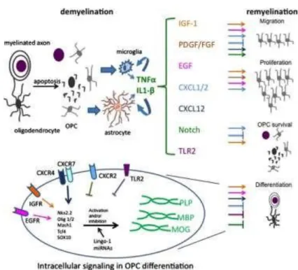
Figure 1. Cellular and molecular events occurring during demyelination... 3 Figure 2. Remyelination mechanisms: role cytokines, chemokines, growth factors, transcription
factors and other proteins in OPCs
fate...7
Figure 3. Integrins, adaptor proteins, and signaling pathways. ...10 Figure 4. Integrin regulation of Rho-GTPases during stages of cell spreading and process
outgrowth...11
Figure 5. Immunocytochemical localization of microtubules and MBP. Immature OLs (a -c)
and mature OLs (d-f) derived from rat brain and cultured for 6 days in vitro can be observed. Immunofluorescence with antibodies against tubulin (a,d) and MBP (b,e). Nuclei stained withDAPI(c,f)...13
Figure 6. Scheme representing fibrinogen structure, its conversion to fibrin and
thrombin-mediated conversion of the factor XIII to XIIIa...17
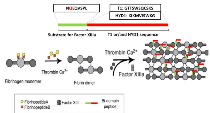
Figure 7. Functionalization of fibrin hydrogels with bi-domain peptides (HYD1, T1) with α6β1
ligands at the carboxyl terminus nd th factor XIIIa substrate domain (NQEQVSPL) at the amino terminus, through the action of factor XIIIa. ...21
Figure 8. Steps of the dissection of the neonatal rat cortex, from the skull cut (a) until the
cortex is meninges-free and totally isolated (d)...24
Figure 9. Summary of dissection, plating and culture of neonatal rat cortices until OPC isolation
and culture...25
Figure 10. One-drop system for preparing 30µL Fibrin drops...27 Figure 11. Manual OPC process counting and measurement using Freehand Line (in yellow) and
Measure function from ImageJ software...31
Figure 12. Distribution of OPCs within 3-D fibrin gels after 7 days of cell culture, as a function
of the cell seeding density...34
Figure 13. Immunocytochemistry analysis of OPC process extension in fibrin hydrogels as a
Figure 14. Quantitative analysis of process outgrowth of OPCs cultured in fibrin hydrogels, as
a function of fibrinogen concentration...38
Figure 15. Cell viability of OPCs cultured in fibrin hydrogels, as a function of fibrinogen
concentration. ...40
Figure 16. Immunocytochemistry analysis of OPC process extension in 4 mg/mL fibrin hydrogels
functionalized with bi-domain peptides (HYD1, T1 and HYD1+T1), as a function of peptide input concentration in polymerizing gels. ...43
Figure 17. Process outgrowth quantitative analysis of OPCs cultured in fibrin hydrogels (4
mg/mL of fibrinogen) functionalized with bi-domain peptides, as a function of peptide input concentration in polymerizing gels. ...44
Figure 18. Process outgrowth quantification of OPCs cultured in 4 mg/mL fibrin hydrogels
functionalized with 5 µM of bi-domain peptides (HYD1, T1 and HYD1+T1)...45
Figure 19. Immunocytochemistry analysis of OL myelin production in 4 mg/mL fibrin hydrogels
functionalized with bi-domain peptides (HYD1, T1 and HYD1+T1), as a function of peptide input concentration in polymerizing gels. ...46
Figure 20. (A) Apoptotic analysis of OPCs cultured in unmodified 3-D fibrin hydrogels (4 mg/mL
of fibrinogen) or fibrin hydrogel functionalized with 5 µM of HYD1, T1 and HYD1+T1; (B and C) Percentage of apoptotic and dead cells, as assessed by image analysis of the 2-D projections of CLSM stacks. ...48
List of tables
Table I. Major intrinsic and extrinsic factors controlling the differentiation of OPCs into
OLs. ...7
Table II. Hydrogels used for neural tissue engineering: mechanisms of gelation and
advantageous (+) and disadvantageous (-) characteristics... 14
Table III. . Effect of fibrinogen and thrombin concentration on fibrin hydrogels mechanical properties. ...18
Abbreviations
BBB Blood-Brain Barrier BMP Bone Morphogenic Protein CNS Central Nervous System
CNP 2’-3’-cyclic nucleotide 3’-phosphohydrolase DS Donkey serum
ECM Extracellular Matrix
ERK Extracellular Signal-Regulated Kinases FAK Focal Adhesion Kinase
FDA Food and Drug Administration FGF Fibroblast growth factor GalC Galactocerebroside HA Hyaluronan
IGF Insulin-like Growth Factor MAP Microtubule-associated Protein MAPK Mitogen-activated Protein Kinases MBP Mielin-basic Protein
MMP Matrix Metalloproteinases
MOG Myelin/Oligodendrocyte Glycoprotein MS Multiple Sclerosis
NGS Normal Goat Serum NRG Neuroreguline NT-3 Neurotrophine-3 OL Oligodendrocyte
OPC Oligodendrocyte progenitor cell PAI Plasminogen Activator Inhibitors PDGF Platelet-derived growth factor PEG Poly-(ethyleneglycol)
PGA Poly-(glycolic acid)
PI3K Phosphatidylinositol 3 kinase PKB (Akt) Protein Kinase B PLA Poly-(lactic acid)
PLP Proteolipid Protein
PNS Peripheral Nervous System ROCK Rho kinase
SCI Spinal Cord Injury SFKs Src Family Kinases
TGF-β Transforming growth factor
Chapter 1
Introduction
1.1. Demyelinating disorders affecting
the Central Nervous System (CNS)
1.1.1. Myelination in the CNS
The myelin-membrane formation is a distinctive characteristic of glial cells in vertebrates. During the myelination process, maturing axons are wrapped by multiple concentric membranous layers of myelin produced either by oligodendrocytes (OLs) in the CNS or by Schwann cells in the peripheral nervous system (PNS). The myelin sheath enables not only the rapid and accurate conduction of electrical impulses along the nerve fiber, but also axonal protection and integrity. Besides, there is a mutual regulation mechanism between OLs and axons: during development, glial cells ensure neuronal cells survival and determine both axolemma domains organization and axons diameter, while in turn, axons provide crucial signals for glial proliferation, survival and differentiation, consequently regulating myelin-membrane formation as well. In adult life, myelinating OLs are responsible for maintaining axons diameter, axolemma organization and also neuronal integrity and function, whereas axonal signals regulate oligodendrocyte progenitor cells (OPCs) differentiation into OLs and maintain myelin integrity [1].
The myelin membrane is composed of a specific set of lipids and proteins, whose assembly must occur at a precise and controlled time and space: the glial cells must associate with the axons at the appropriate developmental time, so signaling mechanisms can be established to deliver newly synthesised myelin-membrane components to the axon [2]. More precisely, proliferative OPCs migrate into the white matter regions, exit the cell cycle and undergo differentiation, turning into mature OLs. At this point, they begin to express a subset of myelin-associated proteins. The mechanism starts with the OLs process extension, followed by targeting and adhesion to axonal receptors. Once this connection is established, myelin components are synthetized and taken to the appropriate sites within the sheath, axons become wrapped and myelin membrane is finally compacted, where the sheaths are almost fused and absent of cytoplasm. Nonetheless, this membrane must be maintained throughout adulthood. This is achieved by a continuous turnover of myelin associated to a high expression of myelin genes, which continues even after the myelination developmental process.
Myelin is composed by approximately 70% lipid and 30% protein. The lipid composition includes cholesterol, phospholipids and glycosphingolipids, while the major proteins present in myelin are myelin basic protein (MBP) and proteolipid protein (PLP), but other proteins such as myelin-associated glycoprotein (MAG) and myelin/oligodendrocyte glycoprotein (MOG) are also present [3,4]. This composition gives it insulating properties, which are crucial for rapid and efficient propagation of neuronal impulses [5].
1.1.Demyelinating disorders affecting the Central Nervous System (CNS)
As myelin formation requires the synthesis of both lipids and myelin specific proteins , a complex cellular mechanism is required to properly synthesize and localize these components
[6]
.
1.1.2. Demyelination mechanisms
A demyelinating disease is any disease of the nervous system in which the myelin sheath of neurons is damaged. When this occurs, axons lose their myelin sheaths as a consequence of OLs death in the site of primary lesion. Furthermore, OLs also undergo apoptosis at considerable distances from the lesion, which leads to the hindrance of action potential propagation by surviving axons and, consequently, to the loss of neuronal function.
The breakdown of the blood-brain barrier (BBB) facilitates the infiltration of macrophages and myelin-specific T-cell lymphocytes into the CNS, which attack the myelin-producing OLs. As a consequence of myelin debris, both microglia and astrocytes are activated. After clearing the debris, macrophages, activated microglial cells and reactive astrocytes work together to secrete growth factors, cytokines and matrix metalloproteinases (MMPs), that degrade ECM components and increase inflammation. These growth factors and cytokines also activate quiescent OPCs, thus increasing their proliferation rate. In addition, OPCs respond to pro-migratory signals provided by demyelinated axons. As a result of these demyelination events, OPCs proliferate and migrate to demyelinated areas, in an attempt to differentiate into myelinating OLs to repair the lesion (Figure 1) [7].
Figure 1. Cellular and molecular events occurring during demyelination. Adapted from: [7]
The main diseases associated to demyelination in the CNS are multiple sclerosis (MS), stroke and traumatic lesions, including spinal cord injury (SCI) and traumatic brain injury (TBI). MS and SCI will be discussed in detail in the next sub-sections.
1.1.2.1. Multiple Sclerosis
MS is the most common nontraumatic disorder of the CNS. It affects around 2.5 million globally, 0.5 million in European Union and 50 000 in Portugal, being more incident in developed countries. MS is a chronic inflammatory demyelinating and degenerative disease, affecting mainly young adults and causing progressive and significant disability. In addition to motor and sensory deficits, there is cognitive deterioration in as many as 65% of patients.
The disease course greatly varies from patient to patient, but it generally presents a relapsing/remitting form, in which episodes of acute demyelination and neurological dysfunction are followed by remyelination and functional recovery. The breakage of the blood-brain barrier leads to the infiltration of macrophages and T-cell lymphocytes, which react against cells expressing myelin-specific antigens, i.e. oligodendrocytes. This leads to destruction of myelin sheaths, axonal demyelination and, consequently, neuronal death [8]. Remyelination occurs spontaneously in the first stages of the disease,
when the lesions present an acute nature. However, remyelination is commonly incomplete or not efficient and chronically demyelinated lesions eventually fail to remyelinate. After several episodes, the failure of remyelination leads to augmented axonal degeneration and progressive disability [9].
The only current treatment for MS consists of FDA-approved medications based on anti-inflammatory drugs, which are expected to prevent immune and inflammatory episodes during the relapsing/remitting phase of the disease. However, with the disease progression and accumulated axon damage, these medications become not effective in advanced stages [10].
Remyelination of axons is therefore necessary for functional recovery. Proliferation and migration of OPCs near MS plaques have been the target of several studies, however an inability of OPCs to differentiate has also been reported. This inability may be a result of inhibitory mechanisms or loss of axonal function, since axons normally provide differentiation signals for OPCs.
1.1.2.2. Spinal Cord Injury
Globally, it is estimated that around 250 000 and 500 000 people suffer a SCI. There is no reliable estimate of global prevalence, but the estimated annual global incidence is 40 to 80 cases per million. In the United States, there are approximately 12 000 new cases per year with an additional 232000–316 000 people currently living with SCI [11].
SCI is a result of a lesion caused by a compressive or stretch injury in the spinal cord. It presents complex hurdles to regeneration due to the multifaceted nature of inhibition conditions that occur to cord parenchyma and stroma after trauma. In addition,
1.1.Demyelinating disorders affecting the Central Nervous System (CNS)
from a clinical sight, SCI is aggravated by its heterogeneity in size, shape and extent of injury. The primary injury caused by initial mechanical trauma is often followed by secondary injury events, in which the lesion extends from grey to white matter and may cause blocking of the propagation of action potential along axons [12,13]. This secondary lesion includes several related events such as BBB dysfunction, local inflammation and axonal demyelination, which eventually end up in neuronal death and breakdown of neurological pathways [14]. Furthermore, OLs are very sensitive to SCI injury, thereby undergoing both necrotic and apoptotic cells death [15].
SCI is well characterized by the development of a glial scar by reactive astrocytes, which presents the major hurdle to neuronal regeneration after injury. The glial scar associated with the production of neurite outgrowth inhibitors form an inhibitory microenvironment, thus preventing neuronal function recovery [16].
Current treatments include surgery (when the spinal cord is totally compromised) and methylprednisolone administration, however none of them is effective. Ongoing therapeutic approaches, including the delivery of neurotrophic factors, antagonists of neurite outgrowth inhibitors and modulation of inflammatory response have shown to restore some of the lost functionality [11,16].
1.1.3. The inhibitory microenvironment and remyelination
failure
Remyelination requires the generation of new mature OLs, which are believed to derive from a population of adult precursor cells, often referred to as adult OPCs.
In response to injury, local OPCs switch from a quiescent to a proliferative and responsive phenotype. This involves both changes in morphology and upregulation of genes associated with the generation of OLs during development. This is followed by the repopulation of the demyelinated area with OPCs. Once recruited, OPCs must differentiate into functional OLs.
However, the adult CNS has a limited regenerative capacity, due to the fact that, although endogenous OPCs are prevalent in demyelinated lesions, they are not able to differentiate into OLs and remyelinate due to the local inhibitory microenvironment. The formation of a glial scar, composed of astrocytes and connective tissue elements, is one of the barriers to regeneration of CNS axons. The molecular composition of the scar (connective tissue components such as collagen and elastin), the inhibitory molecules (tenascin, semaphorin 3, ephrin-B2, slit proteins and chondroitin sulfate proteoglycans) released by astrocytes and active microglia, as well as genetic factors, sex and age, are all contributing factors for regenerative failure [17,18].
1.2.
Transplanted OPCs as a source of
myelin-producing cells
1.2.1. OPC differentiation into myelin-producing OLs
OPCs were first identified in the early 80s by Martin Raff and his team as proliferating cells that could differentiate both into OLs or type 2 astrocytes, the reason of their original name O-2A cells Nonetheless, further studies confirmed that in most of the cases their differentiation was associated to an OL fate [19].
During development, the first OPCs are originated from the neuroepithelial precursor cells (NEPs) from the ventricular zone of the spinal cord. The first wave of OPC production starts in the ganglionic eminence and eventually a second and third waves originate from the lateral and caudal ganglionic eminences, respectively, giving rise to adult OLs. OPCs then migrate to other areas, populating the future brain and ultimately myelinating the CNS [20].
The development of myelin-producing OLs involves two interdependent stages. The first one is the differentiation of OPCs into OLs expressing markers of differentiation, including galactocerebroside (GalC) and myelin basic protein (MBP), while in the second stage, these OLs subsequently undergo morphological changes associated with the production of myelin sheaths
[21].
Several intrinsic and extrinsic factors are involved in differentiation of OLs and the major ones are summarized in Table I.
Downstream signaling of transcription factors that lead to increased expression levels of Olig1 and Olig2 promotes OPC differentiation into OLs and consequently, myelination of the axonal tracts. A factor that controls the formation of oligodendrocytes is platelet-derived growth factor (PDGF) (Figure 2). PDGF stimulates the proliferation of OPCs and in its absence, OPCs exit the cell cycle and may differentiate into OLs prematurely. Also, this factor avoids neural stem cells (NSCs) differentiation in other cell types, such as neurons and astrocytes, thereby ensuring an OPC fate [22]. Thyroid hormones, particularly T3, induce proliferation of
OPCs and promote their differentiation into OLs by triggering cell-cycle exit, thereby enhancing the morphological and functional changes involved in their maturation [23].
Nonetheless, a balance must be kept between promoters and inhibitors of OL differentiation, so it occurs in a controlled time and space fashion [24].
In the CNS (Figure 2), myelination is controlled by three main types of signals: axon- derived signals (e.g. TNFα), ECM (e.g. Fyn signaling) and soluble factor signals (e.g. IGF-1, PDGF, CXCL12, Notch family) and finally, intracellular signaling cascades (e.g. downstream signaling from receptors that increase the expression of Olig1 and Olig2) inside myelinating OLs. Both positive and negative effects should be considered. The control of myelination by ECM will be discussed in detail in the section 1.2.3.
1.2. Transplanted OPCs as a source of myelin-producing cells
Figure 2. Remyelination mechanisms: role cytokines, chemokines, growth factors, transcription
factors and other proteins in OPCs fate. [25]
Table I. M ajor intrinsic and extrinsic factors controlling the differentiation of OPCs into OLs. Based
on: [7]
Extrinsic factors Intrinsic factors
T3 Triggers cell-cycle exit. Olig1 In response to demyelination,
promotes the transcription of myelin-specific proteins.
PDGF Withdrawal of this growth factor acts on intracellular factors to trigger cell-cycle exit.
Olig2 In response to demyelination,
regulates Nkx2.2 expression.
IGF-1 Promotes cell-cycle exit and OPC differentiation.
Nkx2.2 - Lineage specification;
- Promotes the transcription of myelin-specific genes.
TGF-β1 Promotes cell-cycle exit and OPC differentiation.
M yt1 Controls the expression of
myelin-specific proteins.
Sox2 Promotes cell-cycle exit and OPC differentiation.
Fyn* Activates the transcription of
myelin-specific genes.
PI3K-AKT1 Activates mTOR (final differentiation of OPCs into OLs).
*discussed in detail in sub-section 1.2.3.
In the adult CNS, new OLs are derived from adult OPCs dispersed throughout both grey and white matter and in the subventricular zone (SVZ). Although adult OLs differentiation show slower rates of migration and longer cell cycle times, it presents some similarities with developmental OLs [8]. It was previously mentioned that when myelin is damaged, local OPCs undergo an activation step, thereby switching from a quiescent state to a regenerative phenotype. This activation step involves the upregulation of several genes associated with
OLs differentiation, including Olig2, NKX2.2, MYT1 and Sox2. OPCs are firstly recruited in response to factors released by microglia and astrocytes and start to proliferate. The differentiation phase of OPCs into OLs comprises three major steps: developing contact with the axons, expressing myelin genes and finally producing a myelin membrane and wrapping it around axons [20].
1.2.2. Therapeutic effect of OPCs in remyelination
As previously mentioned, OPCs are glial cells that differentiate into myelinating OLs during embryogenesis and early stages of post-natal life. However, a number of OPCs remain in an undifferentiated state, hence their abundance in the adult brain.
Since local OPCs cannot differentiate due to the inhibitory microenvironment, transplantation of OPCs as emerged as a feasible therapy to promote remyelination of spared and regenerating axons.
Adult OPCs can be induced to proliferate and migrate in vitro by the platelet-derived growth factor (PDGF) and fibroblast growth factor (FGF), which are both upregulated during remyelination [8].
A few studies provided proof-of-principle for clinical glial cell transplantation. Archer et al. (1997) performed OPC transplantation into shaking dog pups, with PLP mutations that trigger Pelizaeus-Merzbacher disease) and demonstrated the ability of the transplanted OPCs to myelinate axons in the demyelinated lesions of these animals. [26] In 2008, Windrem et al.
studies showed that transplantation of highly enriched preparations of human glial progenitor cells into multiple sites in mice carrying mutations in MBP enables widespread myelination throughout the CNS and recovery from myelin dysfunction in some cases. The transplanted mice also exhibit prolonged survival [27].
In a study performed by Keirstead et al. (2005), transplantation of human embryonic stem cell (hESC)-derived OPCs into a rat model of SCI resulted in enhanced remyelination and improved motor function. However, enhanced remyelination and locomotor recovery were only observed when OPCs were transplanted at early time points after SCI (7 days after injury). In animals receiving OPCs 10 months after injury, there was no enhanced remyelination or locomotor recovery, possibly due to the presence of inhibitory molecules associated to the astroglial scar [17].
1.2.3. Role of the ECM and OPC integrin receptors in myelin-
membrane formation
It is known that the interactions between cells and their surrounding microenvironment are crucial for cell survival, growth and differentiation. The organization of the ECM in the developing CNS is still poorly understood. However, Colognato et al. (2005) studies have shown that ECM interactions are relevant for glial cell development [28].
1.2. Transplanted OPCs as a source of myelin-producing cells
The ECM is a complex association of extracellular molecules, including structural proteins (i.e. collagen), glycosaminoglycans (GAGs) and specially glycoproteins such as fibronectins and laminins, which activate intracellular signal pathways through interactions with integrins
[29].
Integrins are transmembrane protein receptors that bind to ECM proteins, thus mediating cellular adhesion and signalling cascades between the extracellular and intracellular environments. When a ligand binds to the extracellular domain of a specific integrin, integrin clustering occurs and the actin filaments as well as signalling proteins are recruited to the intracellular domain of the integrin.
Integrins structure is heterodimeric, thus consisting of two different chains, α and β subunits. The α chains are responsible for determining ligand specificity, while β subunits have a cytoplasmatic tail that can bind to several intracellular anchor proteins, such as talin, α-actinin and filamin. These anchor proteins can then bind to actin, thus allowing the actin association to integrins, which leads to integrin clustering and consequently, to the formation of focal adhesions between cells and the ECM [30,31]
.
Integrins have been demonstrated to be important for myelination. OPCs and OLs express a limited repertoire of integrins, including αvβ1, αvβ3, αvβ5, αvβ8 and particularly the laminin receptor α6β1 integrin. Each one of these integrins is regulated and expressed depending on the differentiation stage since they play different roles in development: αvβ1 promotes migration, αvβ3 proliferation, αvβ5 differentiation and α6β1 differentiation and survival of new formed OLs [32]. Moreover, α6β1 integrin has shown to enhance cell survival of myelinating oligodendrocytes and promote myelination of axonal tracts in vivo [17,33,34].
It has been previously referred that upon differentiation into OLs, OPCs undergo a phenotype switch, characterized by a dramatic change of morphology and the formation of a large network of branching processes. When OPCs exit the cell cycle in order to differentiate into myelinating OLs, the survival of these developing cells depends on the contact with axonal receptors and on the effect of soluble factors, such as PDGF and neuroregulin-1 (NRG-1). While these factors act as mitogens and inhibit differentiation at earlier stages of development, upon OL maturation they are fundamental for cell survival and differentiation. Hence, the same factors that stimulate proliferation and hinder differentiation may become crucial for survival, by switching their phenotype at the precise time and space where OLs undergo final stages of differentiation. Although the mechanisms underlying this switch are still not well understood, they are thought to be regulated by interactions between integrin receptors and ECM ligands [35]. But how do receptors transmit signals from the ECM to OPCs in order to change
their phenotype?
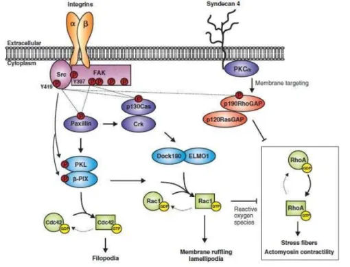
Several downstream effectors and pathways may be involved in ECM signaling for myelin- membrane formation, including Src Family Kinases (SFKs), small Rho GTPases, Focal Adhesion Kinase (FAK), ILK and the phosphatidylinositol 3-kinase (PI3K) and mitogen-activated protein kinase (MAPK)/Extracellular Signal-Regulated Kinases (ERK) pathways (Fig. 3).
Figure 3. Integrins, adaptor proteins, and signaling pathways. [36]
The signaling cascade begins with integrins and other ECM receptors connecting to the downstream pathways through SRKs. SRKs are non-receptor tyrosine kinases tethered to the inner side of the cell membrane that play an important role in the transduction of external signals into changes in intracellular signaling.
Both Fyn and Lyn belong to the SRKs family and regulate integrin-guided pathways. Lyn associates with the PDGFR-αvβ3 integrin complex and its activation contributes to the proliferation of OPCs. On the other hand, OPC differentiation into OLs depends on Fyn. Fyn is expressed throughout the brain and particularly highly expressed in OLs and its activation and expression levels are upregulated during the final stages of differentiation. It is activated by signals triggered by laminin through integrins (particularly α6β1) [37,38].
SRK pathways regulate the activation of Rho GTPases (e.g. Rho, Rac and Cdc42) through Fyn (Fig. 4). After differentiation, Fyn phosphorylates and activates p190RhoGAP, which then inactivates Rho activity, thus causing modifications in OL morphology. Furthermore, Fyn activates both Cdc42 and Rac, which promote differentiation of OLs. Rho GTPases, either through activation or inactivation, regulate the polymerization of actin, and consequently control cytoskeleton structure and cellular motility [39].
FAK is activated in differentiating OPCs and, such as Fyn, it also integrates signals from the ECM, inducing cytoskeleton changes that ultimately contribute to myelination. Both Fyn and FAK pathways mediate laminin interactions with the cytoskeleton.
Laminins are heterotrimeric proteins of the ECM that consist of a α-chain, a β-chain and a -chain. The trimeric proteins form a cross-like structure that can bind to other cell membrane and ECM molecules, thereby playing an important role in cell adhesion, migration, proliferation and differentiation [40]. Laminins are found in myelinating axon tracts stimulating soluble factors such as PDGF and NRG [38].
The interaction between laminin-211 expressed in the CNS and oligodendrocyte α6β1 integrin has shown to play an important role in signaling mechanisms that enhance the
1.2. Transplanted OPCs as a source of myelin-producing cells
morphological changes, stimulating the myelin membrane formation by OLs [34]. Studies from Buttery and ffrench-Constant (1999), Relvas et al. (2001) and Colognato et al. (2004) have shown that laminin-integrin interactions affect oligodendroglial process ex tension in both length and branching [20,33,38]. Moreover, in a study performed by Zhao et al. (2009), laminin was reported to re-appear in demyelinated lesions that are starting to repair. Laminins potentiate the ability of newly differentiated OLs to survive at limited quantities of trophic factors. On the other hand, fibronectin was shown to reduce the ability of OLs to form branched processes by stimulating OPCs to continue to proliferate [28].
Figure 4. Integrin regulation of Rho-GTPases during stages of cell spreading and process outgrowth. [41]
The previously referred Fyn pathway is also required to enhance PDGF signaling to promote myelin-membrane formation and to change NRG signaling course from a PI3K to a MAPK pathway, this way changing the development stage from proliferation to differentiation. PI3K is thus activated by NRG-1 type III produced by naked axons, subsequently activating the PI3K-AKT1 pathway by growth factors and ECM molecules. This pathway will eventually induce differentiation of OPCs into myelin-producing OLs, as well as cytoskeleton reorganization [42]. In a study from Colognato et al. (2002), the activation of ERK1 and ERK2 was enhanced as a response to NRG when OLs were cultured on laminin-2 [38]. Therefore, besides enhancing survival, MAPK/ERK signaling has shown to promote process extension and differentiation of OLs. Furthermore, it has revealed that a switch from a PI3K-dependent to a MAPK-dependent signaling is necessary in order to promote differentiation.
1.2.4. The cytoskeleton in myelin-membrane formation
During OL development, signals relevant to process formation mus t be transduced into adequate changes in cytoskeletal organization. OPCs have a bipolar morphology, however, as
the OLs mature, they become multipolar through the extension of several processes that eventually extend many orders of branches. Therefore, OL cytoskeleton plays an essential role on their interaction with axons and also in the regulation of branching and process outgrowth [44].
OLs present a complex cytoskeleton architecture, which is characterized by an organized and dynamic network of microtubules and microfilaments, however absent from intermediate filaments. Myelin-constituting proteins were shown to be involved not only in OL differentiation, but also in process extension. In 2006, Galiano et al. reported MBP as a microtubule-stabilizing protein in OLs undergoing differentiation [45]. Furthermore, 2’-3’- cyclic
nucleotide 3’-phosphohydrolase (CNP) was associated to process outgrowth by copolimerization with tubulin [46].
Microtubules are polarized dynamic structures which are formed by the polymerization of heterodimeric complexes of α-tubulin and β-tubulin. Microtubule growth and integrity are regulated by microtubule-associated proteins (MAPs) that bind to the microtubules through tubulin domains. Several MAPs can be found in OLs, including MAP1B, MAP4, MAP2c and tau proteins. The latter are specifically present at branching areas and ends of the cellular process extensions. Upon differentiation, there is a decrease in tau phosphorylation, enhancing their binding to microtubules.
Also Fyn kinase interacts with OL cytoskeleton by coupling with tau and tubulin, thus contributing for the initiation of process outgrowth and myelination. Moreover, The OL morphology during differentiation may occur via ECM pathways from integrins to Fyn and finally, to Rho GTPases, which leads to the formation of the motility structures referred [45].
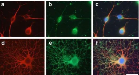
In Figure 5, it is possible to observe the differences in branching and process outgrowth associated with different stages of differentiation (OPCs and OLs).
Figure 5. Immunocytochemical localization of microtubules and M BP during OL differentiation.
OPCs (a-c) and OLs cultured for 6 days in vitro (d-f) derived from rat brain can be observed. Immunofluorescence with antibodies against tubulin (a,d) and M BP (b,e). Nuclei stained with DAPI and merged with tubulin (red) and actin (green) staining(c,f). Scale bar=25 µm. [44]
1.4.Fibrin hydrogels as versatile vehicles for cell delivery
1.3. Vehicles for OPC delivery to the CNS
1.3.1. Overview
Despite being a source of potential myelin-producing cells, transplantation of OPCs by direct injection of cell suspensions presents drawbacks such as low cell survival and poor proliferation and differentiation [43]. This is mainly due to the lack of mechanical and biochemical support associated to the inhibitory microenvironment generated either by the glial scar or the inflammation mechanisms occurring upon demyelination [8]. Tissue engineering provides an alternative approach to overcome these limitations and thus increase the efficacy of OPC transplantation therapies in the CNS, namely by combining cellular transplantation with 3-D biomaterial based-vehicles. These 3-3-D matrices may protect transplanted cells from the local hostile tissue environment, thereby creating a supportive niche for OPC survival, proliferation and differentiation.
1.3.2. Hydrogels as vehicles for OPC delivery into
demyelinated lesions
Cells can sense and respond to the biophysical properties of the microenvironment through mechanotransductional mechanisms. In particular, adhesion ligands binding to integrins and other cell surface receptors, serve as mechanical transducers between the surrounding environment and the cytoskeleton [44], allowing cells to sense and respond to matrix stiffness. The stiffness of the matrix affects cell spreading, morphology and function, and generally, cells show better in vitro behavior when cultured in matrices with mechanical properties similar to those of their native microenvironment.
Therefore, apart from being biocompatible and biodegradable (by cell-secreted proteolytic enzymes, ideally) the engrafted biomaterials should have mechanical properties closely matching those of the target tissue.
Hydrogels have proven to fulfill these requirements, as these can be prepared to present different stiffnesses (by varying the polymer concentration and the cross-linking degree). Furthermore, hydrogels are hydrophilic polymer networks with high water content (similar to the ECM) and high permeability, allowing exchange of oxygen, nutrients and metabolites, thus providing a highly permissive microenvironment for cell survival and growth. Finally, hydrogels can be decorated with biochemical or biophysical cues (cell adhesive or growth factor binding ligands) to better mimic the native ECM [34].
In situ forming hydrogel systems are of particular interest in tissue engineering applications. Injectable hydrogels are likely the most investigated materials for local drug and cell delivery to the CNS. It was previously referred that hydrogels already present a set of advantages that make
them suitable for these applications. In addition, injectable hydrogels have the ability to provide a minimally invasive, localized and filling platform for therapeutic purposes. Upon injection into the spinal cord tissue, these polymers undergo a rapid transition from liquid to gel phase, thereby adapting to the specific lesion site and ultimately integrating with the native tissue.
Also, like any hydrogel-based scaffold design for the CNS, injectable hydrogels can be formulated to present mechanical properties closely matching those of the spinal cord ECM. [47].
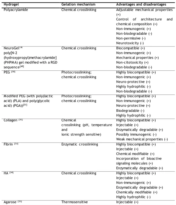
A list of hydrogels used for neural tissue engineering is presented on Table II. The mechanism of gelation is indicated and the main advantages/disadvantages discussed.
Table II. Hydrogels used for neural tissue engineering: mechanisms of gelation and advantageous (+) and
disadvantageous (-) characteristics. Adapted from: Li X. et al. (2012) [48]
Hydrogel Gelation mechanism Advantages and disadvantages
Polyacrylamide Chemical crosslinking Adjustable mechanical properties
(+)
Control of architecture and chemical composition (+) Non-immunogenic (+) Non-biodegradable (-) Non-permissive (-) Neurotoxicity (-) NeuroGelTM poly[N-2 (hydroxypropyl)methacrylamide] (PHPM A) gel modified with a RGD sequence[49]
Chemical crosslinking Biocompatible (+)
Non-immunogenic (+) M echanical properties (+) Non-citotoxicity (+) Non-biodegradable (-) PEG [50] Photocrosslinking; chemical crosslinking Highly biocompatible (+) Non-immunogenic (+) Neuro-protective (+) Highly hydrophilic (-) Non-biodegradable (-) M odified PEG (with poly(lactic
acid) (PLA) and poly(glycolic acid) (PGA))[51] Photocrosslinking; chemical crosslinking Highly biocompatible (+) Non-immunogenic (+) Neuro-protective (+) Biodegradable (-) Highly hydrophilic (-) Collagen [52] Chemical crosslinking (pH, temperature and
ionic strength sensitive)
Highly biocompatible (+) Injectable (+)
Enzymatically degradable (+) Possibly immunogenic (-) Weak mechanical properties (-)
Fibrin [53] Enzymatic crosslinking Highly biocompatible (+)
Injectable (+)
Chemical modifiable (+) Incorporation of bioactive signaling molecules (+) Enzymatically degradable (+)
HA [54] Chemical crosslinking Highly biocompatible (+)
Injectable (+) Non-immunogenic (+) Enzymatically degradable (+) Chemically modifiable (+) Highly hydrophilic (-)
1.4.Fibrin hydrogels as versatile vehicles for cell delivery
Non-neuronal permissive (-) Lacking of cell binding domain (-) Non-naturally enzymatically degradable (-)
Alginate [56] Ionic crosslinking Easy crosslinking (+)
Injectable (+)
Inhibition of protein adsorption (-) Non-enzymatically degradable (-)
Chitosan [57] Ionic (polyanions) and/or
chemical crosslinking, thermosensitive Highly biocompatible (+) Easy modification (+) Injectable (+) Enzymatically degradable (+) Only soluble in dilute acid (-) Inflammatory response (-)
M ethylcellulose [58] Thermosensitive Injectable (+)
Limited protein adsorption (-) Non-enzymatically degradable (-)
M atrigel ® [59] Thermosensitive Injectable (+)
Highly biocompatible (+) Enzymatically degradable (+) From mouse tumor cells (-) Immunogenic (-)
Doubt about exact composition (-) Polysialic acid (PolySia)-based
Hydrogels [60]
Diepoxyoctane crosslinking Bioresorbable (+)
Non-immunological (+)
Highly specific degradation (+) Xyloglucan [61] Thermosensitive Injectable (+) Difficult production (-) Enzimatically degradable by glycoside hydrolases (GH) Peptide hydrogel: - PuraM atrixTM
(Peptide RADA 16) - β-sheet forming ionic selfcomplementary peptide [62]
- Self-assembling peptide amphiphile (PA) [63,64]
pH, temperature and ionic strength sensitive
Injectable (+)
Highly biocompatible (+) Low mechanical properties (-)
Although hydrogels have already been used for cell delivery to the CNS [30], at the best of our knowledge, only two studies using hydrogels for OPC delivery into the CNS were reported to date. Li et al. (2013) developed an injectable biocompatible hydrogel based on thiol- functionalized hyaluronic acid (HA-S) and thiol-functionalized gelatin (Gtn-S), which were crosslinked by poly-(ethylene glycol) diacrylate (PEGDA). Transplant of OPCs within these hydrogels showed enhanced survival and differentiation into OLs, as well as the ability to remyelinate demyelinated axons [65]. In the same year, Asmani et al. performed the first study reporting fibrin hydrogels as a suitable 3-D support for culture and differentiation of OPCs derived from endometrial stromal cells, which not only allows cell-cell interaction, but also mechanical stimulation similar to that of ECM. This study will be discussed in detail in the section 1.4.2. [66]
1.4. Fibrin hydrogels as versatile vehicles
for cell delivery
1.4.1. Fibrin structure
Fibrin is a FDA-approved hydrogel with widespread clinical use since it was first purified in large quantities in the 1940s. Due to its role in hemostasis and wound repair, fibrin has been used extensively in hemostatic materials, such as fibrin sealants (known as fibrin glue) and wound dressings [67].
As a naturally occurring clotting agent in human body, fibrin is biocompatible and susceptible to proteolytic degradation mediated mainly through plasmin and MMPs. In addition to its important role in hemostasis, fibrin serves as a provisional matrix for tissue repair following injury, constituting a key regulator of wound healing.
Fibrin sealants include fibrinogen (fibrin zymogen form) obtained from human pooled plasma by cryoprecipitation of fresh-frozen plasma or stored plasma [(Tisseel and Artiss (Baxter) and Evicel (Johnson & Johnson)] as well as bovine thrombin containing CaCl2 and antifibrinolytic
agents (aprotinin) [68].
Additionally, fibrin can be prepared from the patient’s own blood and used as an autologous scaffold without the potential risk of foreign body reaction and infection or, alternatively, from recombinant fibrinogen and thrombin [68].
Fibrin may thus be obtained from fibrinogen polymerization by the serine protease thrombin and then cross-linked by factor XIIIa in the presence of calcium chloride (CaCl2) to
form a protein mesh.
Fibrinogen molecules, the circulating precursors of fibrin monomers, are dimeric glycoproteins consisting of two sets of polypeptide chains, Aα-, Bβ- and , which are bound together by disulfide bridges (Fig. 6). Each molecule contains two outer D domains and a central E domain, connected to each other by a coiled-coil segment.
1.4.Fibrin hydrogels as versatile vehicles for cell delivery
Figure 6. Scheme representing fibrinogen structure, its conversion to fibrin and thrombin-mediated
conversion of the factor XIII to XIIIa. [69]
During the coagulation process, polymerization of fibrinogen molecules, which will eventually originate fibrin, initiates when thrombin cleaves the fibrinopeptide A (FPA) from fibrinogens Aα-chains. Associations between D and E domains allow the formation of double-stranded fibrils, that together with branching processes, form a clot-like network. This assembly promotes antiparallel alignment of -chain regions, which are covalently crosslinked into - dimers by the plasma transglutaminase IIIa (active form of factor XIII), in the presence of CaCl2 (Figure 6).
The inactive form, factor XIII, circulates in the blood with fibrinogen and becomes activated by thrombin cleavage. The factor XIIIa then stabilizes the fibrin gel by crosslinking of glutamine residues within the fibrin network to lysine residues , thereby forming the 3-D protein network referred above. Cells can then be directly embedded in the 3-D network upon gel formation.
The properties of fibrin hydrogels can be controlled by two main variables. The first one is the concentration of fibrinogen and thrombin, which affect the fibrin network structure and mechanical properties of the gels obtained. Normally, when the fibrinogen concentration is increased, the resulting gels are dense and turbid (composed of thick fibers). If fibrinogen concentrations are maintained constant, varying only the concentrations of thrombin, lower concentrations lead to the formation of fibrin hydrogels with thick fibers, few branch points and large pores, while in opposition, higher concentrations result in a less turbid formation of tightly packed thin fibers. Moreover, gel stiffness is also affected by fibrinogen and thrombin concentration, mainly by fibrinogen concentration than thrombin’s and normally with greater matrix stiffness corresponding to higher concentrations (Table III). The other variable consists in using protease inhibitors to prevent the degradation of fibrin by cell- secreted proteases [70].
Protease inhibitors that have been added to fibrin hydrogels in order to slow proteolysis include the serine protease inhibitor aprotinin and pharmacological MMP inhibitors [68].
Table III. Effect of fibrinogen and thrombin concentration on fibrin hydrogels mechanical properties.
[68]
Fibrinogen concentration Thrombin concentration
Fibrinogen concentration
Fibrin gels dense and turbid, with thick fibers;
Greater stiffness;
Fibrinogen concentration
Fibrin gels soft and permissive, with thin fibers;
Lower stiffness.
Thrombin concentration
Fibrin gels with tightly packed thin fibers and less turbid
Thrombin concentration
Fibrin gels with thick fibers, few branch points and large pores;
1.4.2. Fibrin as a 3-D matrix for the differentiation of OPCs
Fibrin mechanical and biological properties make it an attractive biopolymer to be used as a cell carrier. This fibrous, non-globular protein polymerizes in situ without adverse effects on co-transplanted cells and with gelation times that allow the use of minimally- invasive procedures for the implantation of the cell-loaded matrix. Moreover, fibrin structural and mechanical properties can be easily adjusted by varying the concentration of fibrin components in fibrin polymerizing solution to prepare suitable environments for OPC culture and neurite outgrowth [71,72]. Fibrin gels obtained with low fibrinogen concentrations are more permissive to cell infiltration, however these are too soft and thus do not present enough mechanical strength and robustness. In this sense, a fibrin scaffold should be both permissive allowing cell infiltration and regeneration and hold appropriate mechanical properties to a specific application [68].
The advantages of using fibrin as a scaffold include the extremely elastic resistance to deformation without breaking and the presence of natural ligands for cell adhesion (two pairs of RGD sites and a pair of AGDV sites), proteolytic enzymes (e.g. plasmin), growth factors (e.g. FGF-2, FGF-5 and FGF-7, vascular endothelial growth factor (VEGF-B), AB, BB, PDGF-DD, TGF-β1, TGF-β2, BMP-2 and BMP-2/7 and neurotrophin-3 (NT-3) and brain- derived neurotrophic growth factor (BDNF)) [73], ECM proteins (e.g. fibronectin), and protease inhibitors (including aprotinin, -aminocaproic acid, plasminogen activator inhibitors PAI-1 and PAI-2, α2-macroglobulin and thrombin activatable fibrinolysis inhibitor) [68]. Recently, Asmani et al. (2013) performed the first study reporting fibrin hydrogels as a suitable 3-D support for culture and
1.4.Fibrin hydrogels as versatile vehicles for cell delivery
differentiation of OPCs derived from endometrial stromal cells, which not only allows cell-cell interaction, but also mechanical stimulation similar to that of ECM. In this study, hydrogels with three different fibrinogen concentrations (2 mg/mL, 3 mg/mL and 4 mg/mL) were tested. Comparing the three gels, it was concluded that the hydrogel with a fibrinogen concentration of 3 mg/mL provided the best 3-D scaffold to mimic spinal cord environment. The analysis of OPCs cell body and the process extensions confirmed the interaction between cells and the fibrin scaffold, thereby indicating that OPCs had successfully attached and incorporated into the matrix
[66]
.
1.4.3. Fibrin functionalization with α6β1 integrin ligands
In opposition to synthetic hydrogels, fibrin is not a passive and non-interactive cell delivery matrix, thus allowing specific binding and functionalization with cell delivery domains and growth factors in order to enhance their bioactivity and specificity [67].
Peptide adhesion domains covalently incorporated within 3-D fibrin matrices can greatly increase the bioactivity of the matrix. In addition, the immobilization of these peptides by substrates for factor XIIIa seems to provide an effective approach for fibrin gels functionalization. Schense et al. (2000) incorporated peptides from laminin and N-cadherin either alone or in combination with fibrin hydrogels. In this study, each gel was modified with a different bi-domain peptide, containing a factor XIIIa substrate sequence in one domain and a different bioactive peptide in the other domain, which included RGD, IKVAV, YIGSR and RNIAEIIKDI (derived from the ECM laminin) and HAV (derived from N- cadherin). Neurite extension in vitro was enhanced when exogenous peptides were included, with an improvement reaching 75%. When tested in vivo, the fibrin derivative containing laminin-derived peptides induced an 85% enhancement in the regeneration of the rat dorsal root compared to non-modified fibrin gels. Therefore, these results demonstrated the possibility to enhance the bioactivity of fibrin gels by the enzymatic incorporation of exogenous oligopeptide domais of morphoregulatory proteins (e.g. laminin, N-cadherin) [74].
As previously referred, growth factors might also enhance the bioactivity of fibrin hydrogels. Taylor et al. (2004) developed a heparin-based delivery system (HBDS) to immobilize NT-3 within fibrin gels, thus allowing the release of NT-3 in a controlled manner, as a therapy for SCI. The HBDS system consisted of an immobilized linker peptide which sequesters heparin within fibrin gels and then, the sequestered heparin binds NT-3, thereby avoiding its diffusion. Fibrin gels containing the HBDS system with NT-3 stimulated neural outgrowth from chick dorsal root ganglia up to 54% compared to unmodified fibrin. In a preliminary in vivo study, fibrin gels containing the HBDS and NT-3 demonstrated increased neural fiber density in spinal cord lesions when compared to unmodified fibrin at 9 days [75]. Sakiyama-Elbert and Hubbell (2000) have previously reported the development of an affinity- based drug delivery system designed to slow the diffusion of heparin-binding growth factors from fibrin gels. These systems were used to
![Figure 1. Cellular and molecular events occurring during demyelination. Adapted from: [7]](https://thumb-eu.123doks.com/thumbv2/123dok_br/15911317.1092704/21.894.188.745.648.1029/figure-cellular-molecular-events-occurring-demyelination-adapted.webp)
![Figure 3. Integrins, adaptor proteins, and signaling pathways. [36]](https://thumb-eu.123doks.com/thumbv2/123dok_br/15911317.1092704/28.894.205.674.103.396/figure-integrins-adaptor-proteins-signaling-pathways.webp)