Abcessos Renais - Revisão
António Murinello , Paula Mendonça , Ricardo Correia , Rocha Mendes
Margarida Albergaria , Jorge Neta , João Coelho , Sofia Lourenço
1 1 2 2,
3 4 1 1
1. Unit of Internal Medicine 1 2. Unit of Urology
3. Unit of Radiology 4. Unit of Histopathology Curry Cabral Hospital. Lisbon. Portugal
Resumo
Abstract
Baseados num caso de associação de abcessos renal, perirrenal e de quisto renal infectado, ocorrendo após biopsia prostática, os autores fazem revisão bibliográfica dos abcessos renais, classificados segundo características anatómicas – perinéfrico, cortical e cortico--medular (nefrite bacteriana aguda focal/multifocal, pielonefrite enfisematosa e pielonefrite xantogranulomatosa). Os abcessos renais são mais frequentes em diabéticos, menos vezes em alcoólicos, desnutridos e imunodeprimidos, estando associados geralmente a pielone-frites recorrentes, refluxo vesicoureteral, litíase urinária, ou resultam de êmbolos sépticos hematogéneos originados em focos extra-renais de infecção. A é responsável
pela maioria dos casos, e menos vezes a , e
. Descrevem-se a expressão clínica, complicações evolutivas e diagnóstico radiológico, enfatizando-se as infecções acompanhadas de formação de gás. Referem-se opções terapêuticas médico-cirúrgicas, cujas indicações dependem do diagnóstico precoce, complicações e factores predisponentes.
Abcesso renal / Abcesso perinéfrico / Abcesso de quisto renal
Based on a case of associated renal, perinephric and renal cyst abscesses, occurring after prostatic biopsy, the authors do a concise review of renal abscesses, classified by anatomic categories – perinephric, cortical and corticomedullary (acute focal/multifocal bacterial nephritis, emphysematous and xantogranulomatous pyelonephritis). Renal abscesses are frequent in diabetics, less so in alcoholics, undernourished and immunosuppressed patients, and commonly associated with recurrent pyelonephritis, vesicoureteral reflux, urinary lithia-sis, or resulting of hematogenous septic emboli from extra-renal sources of infection.
is responsible for most cases, other agents are
. Clinical presentation, complications and imagiology are con-sidered, emphasizing gas-forming infections. Indications for medical and surgical therapeutic options depend on early diagnosis, complications and predisposing factors.
Renal abscess / Perinephric abscess / Abscess of renal cyst Escherichia coli
Klebsiella Proteus, Enterobacter, Pseudomonas Estafilococo aureus
Esche-ricia coli Klebsiella, Proteus, Enterobacter, Pseu-domonas, Staphylococcus aureus
Palavras chave:
Key words:
Artigos de Revisão
Correspondência:
António Murinello Av. Eng. António Azevedo Coutinho, Lt. 8 r/c – dto. 2750-644 CASCAIS Portugal
E-mail: amurinello@iol.pt Tel. (351) 918 626 874
12
Introduction
Clinical Report
Renal abscesses are considered a rarity amongst intra-abdominal abscesses . Symptomatology can be varied and misleading, their course frequently insidious for a time, with frequent elusive diagnosis, causing sig-nificant degree of morbidity and mortality. Renal absces-ses are classified by anatomic categories – perinephric, cortical and corticomedullary abscesses .
usually are the result of rupture of renal abscess or infected hydronephrosis into the perirenal space, or more rarely from hematogenous seeding of septic emboli from extra-renal sources of infection.
occurs in confluent areas of the renal cortex and medulla, commonly as sequela of severe acute pyelonephritis, complication of vesicoureteral reflux, ureteral and pelvic calculi, being more frequent in diabetics .
results of hematogenous seeding of septic emboli from anywhere, being common in intravenous drug users, immunosuppression and patients with an extra-renal source of infection (endocarditis, perinasal sinusitis, skin or dental infections).
are rare as a cause of renal abscess, but infection of renal cysts is a possible etiology for a renal abscess .The authors present a patient with long history of bladder calculi and recurrent urinary infections, who after trans-rectal prostatic biopsy developed a clinical picture of serious urinary sepsis due to concomitant renal cortico-medullary, perinephric and renal cyst abscesses. Surgery also showed pyonephrosis associated with almost obs-tructive pyeloureteral calculi. Cultures of urine and pus of all abscess areas revealed the same infecting micro-organism ( -lactamase producing Partial neph-rectomy and post-operatory drainage and antibiothe-rapy were successful.
A 76 year old white man was admitted to our Unit ( ) on due to undiagnosed total hematuria. He had a long history of hypertension, atrial fibrillation, and chronic liver disease due to heavy alco-hol consumption until 10 years before. Urologic history began with a renal colic in 1980. From two years ago he had several episodes of painless hematuria, attributed to urinary stones. On ultrasonography (US) showed: (1) cortical cyst in the left kidney lower pole (7cm on long axis) and bilateral renal microlithiasis; (2)
1,2,3 2 4 5,6 Perinephric abscesses Re-nal corticomedullary abscesses
Renal cortical abscess (carbuncle) Infection of renal cysts I. Medicine 1 22 JUL 06 FEB 05 β E. coli).
several bladder stones; (3) prostatic hypertrophy (59 gr). From he had progressive dysuria, stranguria and frequent total hematuria. Blood tests revealed: no anemia, no leukocytosis, normal ionogram, glycemia and creatinine, urea 50 mg/dL (15.0-45.9); prostatic specific antigen (PSA) 10.98 ng/mL (N <4.00). Urinary sediment: 49 leukocytes/µL (1-10) and >1000 erythro-cytes/µL (1-29). Proteinuria 35.00mg/24h. Uroculture was negative. On an urologist in another Hos-pital tried an unsuccessful cistoscopy (pain and heavy hematuria). After that traumatic urologic intervention he had left renal colic several days later, being seen on
by an urologist in the Ambulatory Unit of our Hospital, who under prophylaxis with co-trimoxazole proceeded to transrectal prostatic biopsy, which re-vealed chronic prostatitis and no malignant tissue. Three days after prostatic biopsy the patient began to feel unwell, with nausea, anorexia, asthenia, and strong pain on the left lumbar region, with temporary amelioration with analgesics. Suddenly on the 10 day after prostatic biopsy ( ), he had intense shivering and worse-ning of lumbar pain with irradiation to the right lumbar region and to the anterior abdominal wall, being assisted in the Emergency Unit of our Hospital. He had no fever and abdominal examination disclosed diffuse abdominal tenderness. There was no urinary retention. Urinary sediment had many leukocytes and erythrocytes and nitrituria. Blood tests showed: no anemia, no leuko-cytosis 7.700x10 /L (with 87.8% neutrophils); platelets 64.000x10 /L (N 130.000-400.000); I.N.R. 1.2 (0.90--1.20); APTT 46” (26.0-36.0); total/conjugated bilir-rubin 3.6 mg/2.5 mg/dL (0.20-1.3/0.00-0.30); potassium 3.1 mEq/L (3.60-5.00); creatinine 2.7 mg/dL (0.70--1.20), urea 119 mg/dL; PCR 21 mg/dL (N <1.00). Abdominal plain film was not informative. He was dis-charged with a diagnosis of renal insufficiency and hy-pokalemia and treated with ciprofloxacin. Two days later however, the patient came again to the Emergency Unit complaining of intense left lombalgia and total he-maturia. Blood tests revealed: no anemia; leukoctes 6.800x10 /L (with 90.3% of neutrophils); platelets 34.000x10 /L; glycemia 160 mg/dL; creatinine 2.5 mg/dL; urea 126.2 mg/dL; potassium 3.2 mEq/L; -GT 129 U/L (15-73); alkaline phosphatase 286 U/L (38--126); normal AST/ALT; albumin 2.10 g/L (3.50-5.00); PSA 39.30 ng/mL. Urinary sediment revealed 500 leuko-cytes/µL, many erythrocytes and nitrituria. Abdominal US showed diffusely heterogenous hepatomegaly, gal-lbladder lithiasis, moderate splenomegaly, bilateral renal microlithiasis, kidneys measuring on long axis 13.3 cm (right) and 14.0 cm (left), this one with slight
pyelocali-FEB 06 JUN 06 10 JUL 06 20 JUL 06 th 9 9 9 9 γ Acta Urológica 2007, 24; 1: 11-18 Murinello, Mendonça, Correia, Mendes, Albergaria, Neta, Coelho, Lourenço
ceal ectasia and voluminous cortical cyst (8.4 cm of dia-meter) of the lower pole, with echogenic content at the bottom, attributed to intracystic hemorrhage. The patient was admitted to our Unit on with a provisory diagnosis of hematuria due to thrombocyto-penia. The patient appeared ill, was feverish (38.0 C), with atrial fibrillation (heart rate approximately 170/min) and respiratory rate 24/min. There were no signs of liver failure. On the abdomen a painful mass without definite limits and with firm consistency and lumber contact was palpable at the left flank. There was moderate bilateral edema of the legs, and rectal touch detected slightly painful prostate, diffusely and mode-rately enlarged. While waiting for uroculture result, the patient was treated with intravenous levofloxacin for three days, without amelioration. As uroculture show-ed significant colony growth (> 10 colony-forming units/mL) of ß-lactamase producing , only sensible to gentamicin and amoxiclav, the last one was prescribed
23 JUL 06 0
5
E. coli
due to renal failure. At the same day hemogram revealed hyperleukocytosis (30.700x10 /L with 91% neutrophi-lia) and abdominal non-contrast enhanced CT scan demonstrated a cyst (10x6 cm) on the lower pole of the left kidney with non pure intra-luminal content, with presence of gas pointing to infection with gas-forming bacteria. There was densification of the perirenal fat and also a fluid collection on the posterior perirenal space, contacting the psoas muscle and with a vertical exten-sion measuring 20 cm. Urinary lithiasis was evident in the lumbar portion of the left ureter causing pyelocali-ceal ectasia. The presence of gas in the calyxes was in favor of a concomitant process of pyelitis. A voluminous calcified stone was shown in the bladder (Figs. 1 A, B, C, D). On the same day surgical catheter was inserted posteriorly in perirenal space, with drainage of abundant pus with cultural growth of with identical antibio-tic susceptibility. Anaerobic culture was negative. After clinical stabilization and due to maintenance of
sup-9 E. coli Fig. 1 Fig. 1A Fig. 1B . Fig. 1 C Fig. 1 D
– Non-contrast enhanced abdominal CT scan: : gas in calyceal topography; suppurative collection in the posterior perirenal space causing anterior dislocation of the kidney and contacting the psoas muscle. : urinary stone with topography in the lumbar left ureter; perirenal fluid collection; gas-forming renal abscess; perirenal fluid collection; cystic formation with non pure content and with internal gas : similar plane, but better definition of perirenal collection.
14
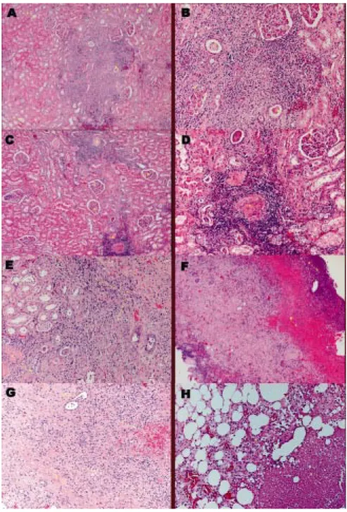
purative collections as seen in CT scan done on , operatory drainage was realized on . We proceeded to drainage of posterior perinephric space as well as of the purulent collection in the renal cyst, the wall of which was ressected. The parenchyma of the inferior pole of the left kidney was oedematous and enlarged, due to several areas of suppuration that were in contact with the cyst. Ureteropyelolithotomy to re-move voluminous obstructive left ureteral lumbar stone and blood clots and partial nephrectomy of the inferior pole of the left kidney was necessary. Urine was turbid, showing characteristics of pyonephrosis. Post-opera-tory drainage was necessary for seven days. Indwelling urinary Folley catheter was maintained and the patient was discharged for a period of convalescence, before proceeding to prostatectomy and surgical removal of the bladder, because he was considered not to have medical conditions to support another type of surgery at the same general anesthesia. Oral amoxiclav was main-tained until completing six weeks of therapy. One week after surgery blood tests revealed normal values of gly-cemia (101 mg/dL) and urea (39.2 mg/dL), and to-tal/conjugated bilirrubin 1.40/0,50 mg/dL. Meanwhile microscopical examination of the kidney revealed se-rious lesions of chronic interstitial nephritis with areas of acute pyelonephritis and suppuration in contact with a necrotic area of abscess. There were several cystic areas with structureless lining suggestive of gas spaces, one of which with voluminous hemorrhage and mixed inflam-matory infiltrate. Lesions of glomerular and vascular sclerosis were conspicuous. The perirenal fat tissue showed areas of hemorrhage, steatonecrosis and orga-nized panniculitis (Figs. 2 A, B, C, D, E, F, G, H).
is usually the result of a rup-ture of a renal abscess or of pyonephrosis to the peri-renal space . Usually there are predisposing urologic conditions (renal or ureteral calculi with partial or total obstruction, noncalculous hydronephrosis, polycystic renal disease or infected renal carcinoma). and
are the most common etiologic agents, but and can also be found . Rarely perinephric abscesses are due to septic embolization from an infection elsewhere or the origin is a rupture of an infected lesion of the colon (inflammatory bowel disease, diverticulitis, infected colon carcinoma) .
is considered a spectrum of disease encompassing various intra-renal
2 AUG
06 4 AUG 06
Perinephric abscess
Renal corticomedullary abscess
Discussion
7 8 9 7 E. coli Proteus K. pneumoniae Ps. aeruginosainflammatory processes, which include: and and Renal corti-comedullary abscess differs from renal cortical abscess because it develops from ascending rather than hema-togenous spread of bacteria, and it is usually associated with underlying urinary tract abnormality (eg, vesi-coureteral reflux, urinary tract obstruction), recurrent urinary tract infections and infected urinary lithiasis. Bacterial pathogens causing cystitis ascend to the upper urinary tract to infect renal medulla. Subsequent lique-faction of renal parenchyma and involvement of renal cortex is the postulated pathogenesis for development of corticomedullary abscess. Scarring of ureters and of ureteral-pelvic junction may occur, causing obstructive symptoms. Previous genitourinary instrumentation is a frequent initiating event leading to occurrence of corti-comedullary abscess .
are responsible for 75% of infections, 15--20% of cases are caused by
and species, the remaining are due to gram-positive bacteria ( , ) Obligatory anaerobes are ulti-mately being referred with greater frequency, isolated or in association with other gram-negative microbial agents, as responsible for genito-urinary
suppura-tions ( pigmented and
sp., anaerobic Gram-positive cocci and sp.) However, because of inconsistent use of adequate methods for their isolation and identifi-cation and also because of their fastidious nature with slow growth and also polymicrobial nature, anaerobic infection remains many times undiagnosed.
Gram-negative facultative anaerobes or obligatory anaerobes may produce gas by fermenting glucose in necrotic tissues . Facultative Gram-negative anaerobes
include and
. Patients with renal infections by these microorga-nisms are generally critically ill and their urologic problems compounded by serious medical problems, such as elec-trolyte imbalance, dehydration, acidosis, hyperglycemia. Diabetics have a high incidence of bacteriuria and pyelo-nephritis, as the hyperglycemia and ketoacidosis impairs host cellular defenses and protects bacteria from the bactericidal activity of lactic acid. Diabetes also predisposes to intrinsic vascular disease leading to pap-illary necrosis and renal infections, thereby reducing tissue absorption of gases produced by anaerobic bacteria and resultingingas-forming(pneumaturia)pyelonephritis .
is a well-localized inflammation of the kidney without frank abscess
forma-acute focal multifocal bacterial nephritis, emphysematous xanthogranulomatous pyelonephritis.
Acute focal bacterial nephritis 4 4 10,11 12 13.14 E. coli
Klebsiella, Proteus, Entero-bacter, Serratia Pseudomonas
Streptococcus faecalis Staphylococcus aureus .
Bacteroides fragilis, Prevotella Porphyromonas
Actinomyces
E. coli, Klebsiella, Proteus mirabilis, Citrobacter yeasts
Acta Urológica 2007, 24; 1: 11-18 Murinello, Mendonça, Correia, Mendes, Albergaria, Neta, Coelho, Lourenço
Fig. 2 Fig. 2 A Fig. 2 B Fig. 2 C Fig. 2 D Fig. 2 E Fig. 2 F Fig. 2 G Fig. 2 H – Histopathology: : HE (4x10): lower pole of left kidney – dilated and atrophic remnant tubules (A), some of it containing eosinophilic proteinaceous material (B). : HE (40x10): lower pole of left kidney – dilated and atrophic remnant tubules, some of it structureless, without epithelial lining, suggesting gas spaces (A), others with eosinophilic proteinaceous material in the lumen (thyroidization) (B), others with suppurative neutrophilic masses (C). : HE (10x10): lower pole of left kidney – hialinized glomerules (A) and related areas of necrotic tubules, contrasting with other areas of preserved glomerulus (B) : HE (40x10): lower pole of left kidney – a higher amplification of the same aspects described on Fig 2 C. : HE (40x10): lower pole of left kidney – areas of tubular progressive invasion and substitution by chronic inflammatory reactive cells (A), with posterior deposition of collagen (B) in the cicatrisation process. : HE (4x10): capsule of renal abscess - with areas of necrosis (A) and hemorrhage (B): : HE (40x10): capsule of the abscess – extensive inflammation of renal interstitium, partially occupied by fibrosis (A), with dilation (B) and obliteration (C) of renal tubules. Dilated tubules had epithelial lining cells and inflammatory cells in the lumen. Several areas of interstitial hemorrhage (D). : HE (40x10): perirenal abscess – perirenal fat with zones of panniculitis (A) and steatonecrosis (B)
16
tion and frequently progresses to the more severe , in which case there is heavy polymorphonuclear infiltrate through the kid-ney with areas of liquefaction and abscess formation.
(EPN) is an un-common severe necrotizing form of acute multifocal bacterial nephritis, which on abdominal radiography exhibits characteristic intraparenchymal (so-called “gas-gangrene of the kidney”) or perirenal gas and not, by definition, isolated within collecting system. The pre-sence of gas suggests the prepre-sence of gas-forming ana-erobic or facultative anaana-erobic pathogens. Although
is the most common microorganism associated with EPN, any lactose-fermenting organism may be involved. Patients with severe EPN often present in and require intensive medical treatment. The function of the affected kidney is often very poor. The disease occurs almost exclusively in patients with diabetes mellitus, with overall mortality of 45%. Occasionally it is seen in patients without diabetes mellitus but with obstruction of the renoureteral unit. Among diabetics with EPN, obstruction of affected kidney is seen in about 30% of the patients. It has also been reported in debilitated alcoholics and the severely immunocompromised . Pathogenesis of EPN involves four factors: gas-forming microorganisms, high tissue glucose levels in the majo-rity of cases, impaired tissue perfusion and defective immune response. Urinary obstruction aggravates these factors due to increase in pyelocalyceal pressure and compromised renal circulation. Pathological findings often include evidence of diabetic glomerulopathy, im-paired tissue perfusion in the form of vascular throm-bosis and infarction, glomerulosclerosis and papillary necrosis . Renal interstitium commonly show diffuse infiltrative processes by acute suppurative and chronic inflammatory infiltrate forming multiple abscesses and large areas of infarction and multiple cysts in the necrotic areas, the lining of which are structureless thus sugges-ting gas spaces .
is a chronic long-term infection of a renal unit often asso-ciated with a renal calculus. As a result of long-standing infection, the kidney enlarges and it fixes itself to the retroperitoneum by peritoneal fibrosis and extension of the granulomatous inflammation. Native tissue planes, such as the plane between , adjacent retro-peritoneal structures and peritoneum, are disrupted. Histologically, granulomatous tissue containing lipid-laden macrophages (ie, foam cells) destroys and repla-ces renal parenchyma. Renal calculi are usually present in 75% of patients with XGP (50% are staghorn calcul)i.
acu-te multifocal bacacu-terial nephritis
Emphysematous pyelonephritis Xantogranulomatous pyelonephritis (XGP) E. coli extremis Gerota fascia 15 15 15 Proteus mirabilis E. coli Staphylococ-cus epidermidis Staphylococcus aureus
E. coli Proteus, and Brucella Aspergillus . is the most common bacteria associated with XGP, but is also common. The histopatho-logical process may evolve from an area of renal involve-ment to widespread extension into the retroperito-neum .
is a multilocular abscess involving the renal parenchyma as a result of coales-cence of cortical microabscesses. Although
can be a causative agent,
is by far the commonest pathogen, spreading to the kidney through hematogenous dissemination from a focus elsewhere. The primary focus can be a carbuncle, a furuncle, a suppurative respiratory infection, or a cel-lulitis. Intravenous drug users are commonly affected by these infections as well as diabetics. Renal lesions usually coalesce to form a fluid-filled mass with a thick wall. The interval from the original staphylococcal infection and the onset of symptoms may be as long as several months, and the patient may have forgotten the primary focus of infection unless questioned closely. Other less commonly etiologic agents are and
much more rarely and Urinary symptoms are commonly absent in these types of abscess, as well as urinalysis abnormalities.
Occasionally renal abscesses can be complicated by extension to nearby structures. Besides the rupture to the perirenal space of renal corticomedullary and cortical abscesses, extension of the infectious process can also cause peritonitis, subphrenic abscess, pleural empyema , fistulous communication to skin, spleen, colon, pericardium or bronchus , sequelar calyceal di-verticulum due to rupture into the collecting system , papillary necrosis, loss of renal function, massive retro-peritoneal hemorrhage due to abscess erosion into the renal vasculature , psoas abscess , extension to the pelvis and groin, and epidural abscess . Bacteraemia due to these abscesses can result in spread of infection to other sites in the body.
Clinically renal abscesses can present as acute pye-lonephritis, subsequently failing to respond to appro-priate antibiotherapy. Alternatively, patients may have insidious onset of symptoms with absence of localizing signs such as flank pain, pyuria or bacteriuria, which most commonly occurs in patients with diabetes melli-tus, alcohol or drug abusers, immunocompromised pa-tients, and steroid therapy in patients with staghorn calculi. Generally clinical features of renal abscesses usually include fever, chills, and flank or abdominal pain. Dysuria and other urinary tract symptoms are variably present. Nonspecific constitutional symptoms (malaise, fatigue, weight loss) are common, as well as
gastroin-4 16 17 18 19 20 21 22 23 24 Renal cortical abscess
Acta Urológica 2007, 24; 1: 11-18 Murinello, Mendonça, Correia, Mendes, Albergaria, Neta, Coelho, Lourenço
testinal symptoms (nausea, vomiting). Signs of renal abscesses vary greatly and have no specific characteris-tics to aid in diagnosis. Most patients with renal infection appear ill and in distress, febrile, and may have signs of haemodynamic instability with tachycardia and hypoten-sion and a clinical picture of sepsis. A palpable flank mass is not consistently found, but costovertebral angle ten-derness is usually detected .
Most patients demonstrate leukocytosis with left shift. Urinalysis generally show signs of infection, but bacteriuria and pyuria may be absent if the ureter is completely obstructed and in cortical and perinephric abscess caused by bacteriemic emboli. Uroculture is frequently positive in corticomedullary and perinephric absceses, but not in cortical abscess. Blood cultures are positive in more than 50% of the patients with renal abscesses, and are particularly useful in patients with urosepsis.
Plain X-ray of the abdomen has a sensitivity of only 33% to diagnosis the presence of gas in renal paren-chyma. Renal abscesses can be evaluated with US, CT scan or MRI; however, CT is the imaging of choice. On US the abscess can appears similar to a cyst, but with some internal echoes and/or wall irregularity; as a solid mass mimicking a renal neoplasm; or the echo pattern of an abscess may be indistinguishable from adjacent renal parenchyma. US has no sensitivity to diagnose gas in the kidney. On CT, the abscess appears as a heterogenous low-attenuation mass. There is often an irregular, en-hancing wall secondary to hyperaemia or granulation tissue formation. A bulge in the renal cortex is typically present in peripheral abscesses. Inflammatory changes are seen in the adjacent fat. CT is the best imaging method to diagnose gas in the kidney. CT scans provide excellent anatomic details of renal or perirenal absces-ses collections to be surgically explored. Anaerobic me-tabolism of a microorganism produces insoluble gases, such as hydrogen, nitrogen and oxygen. Gases are pro-duced as a result of fermentation, denitrification and deamination by a microorganism. Gases in renal paren-chyma and perirenal space are well visualized in CT scan . On contrast-enhanced MRI, a liquefied portion of the abscess and enhancing wall are suggestive of the diagnosis .
The first line of treatment of renal abscesses inclu-des correction of hyperglycemia, dehydration or elec-trolyte imbalance. Appropriate antibiotics against the most likely pathogens are given intravenously after obtaining blood and urine cultures, and changed accor-dingly to antibiogram susceptibilities. Appropriate anti-biotics for use in treatment of infected renal cysts or
4
12
25
abscesses are agents that diffuse well into these closed sites, such as trimethoprim-sulfamethoxazole and fluo-roquinolones. When the patient’s general condition improves, the decision is then made either to wait the effect of antibiotherapy, to incise and drain the abscess percutaneously or to proceed to partial or total ne-phrectomy. When renal abscesses are yet small due to an early diagnosis, the preferred treatment would be image-guided percutaneous drainage, often performed under CT guidance. Additional microorganisms may be isolated at the time of drainage, in which case directed antibiotherapy may be necessary. Early diagnosis and aggressive medical antibiotherapy and surgical manage-ment of gas-forming infections of the genitourinary tract are vital .
Incision and drainage of a renal abscess is performed when the patient’s general situation is too serious to attempt a nephrectomy, to preserve as much as renal tissue as possible, particularly in those patients with marginal renal function or when the renal parenchyma is only partially involved in the abscess process. Should the decision be made to incise and drain a renal abscess, all intra-renal loculi and abscess cavities must be adequa-tely drained and also any complicating subphrenic abs-cess. A nephrectomy is performed when the renal parenchyma was replaced by an abscess cavity or when there is insufficient parenchyma worth conserving. Early diagnosis of EPN is necessary to try to avoid the ne-cessity of a nephrectomy, which frequently offers the best outcome. Diabetic patients are easier to control medically following nephrectomy or incision drainage of a renal abscess. It is also important to consider the addi-tional therapy of underlying disease (e.g., obstructive uropathy) .
Although most reported patients had an associated urological abnormality, such as stones, obstruction or anatomic abnormalities, an interesting series by Tung Shu refers to the possibility to find this type of absces-ses in a group of patients with otherwise anatomically normal urinary tracts, ant that with accurate diagnosis and minimally invasive therapy had excellent functional and anatomical outcomes.
Patients undergoing transrectal ultrasound-guided biopsies of the prostate are somewhat prone to bac-teriuria and bacteraemia, despite partial protection by antibiotic prophylaxis . There are several predisposing factors favoring the occurrence of post-biopsy infec-tions, namely former urinary tract infection, prostatitis, urinary catheterization, and diabetes mellitus . Our patient had a long history of previous urinary tract infections and urinary stones, and was submitted to
12
15
26
27
18
prostatic biopsy just preceding the development of the renal abscess, with rupture to perinephric space and a renal abscess in an old known renal cyst. It is however speculative to attribute the development of the process to prostatic biopsy, because the patient had strong rea-sons to develop a renal abscess (recurrent urinary tract infections, bladder stone, chronic prostatitis, ureteral stone with almost complete obstruction, old known vo-luminous renal cyst, chronic liver disease and impaired glucose regulation). Fortunately it was possible to pre-serve two thirds of the affected kidney at surgery and to adequately drain all suppurative collections, with reco-very to normal values of blood urea and creatinine.
Bibliography
1. Patterson JE, Andriole VT. Renal and perirenal abscess. Infect Dis Clin North Am 1987; 1: 907-26
2. Anderson KA, McAninch JW. Renal abscess: classification and review of 40 cases. Urology 1980; XVI: 333-8 3. Jaik NP, Sajvitha K, Mathew M, Sekar U, Kuruvilla S,
Abraham G, et al. Renal abscess. J Am Physicians India 2006; 54: 241-3
Willard TB, Teagule JL, Steinbecker K. Renal cortico-me-dullary abscess. Internet http://www.emedicine.com/ med/topic2848.htm, pg 1-20
5. Case Records of the Massachusets General Hospital (Case 38-1989) Scully RE, Marck EJ, McKneeley WF, McKneeley BU eds. New Engl J Med 1989; 321: 813-23
6. Schab SJ, Bander SJ, Klahr S. Renal infection in autosomal dominant polycystic kidney disease. Am J Med 1987; 82: 714-8
7. Salvatiera O Jr, Bucklew WB, Morrow JW. Perinephric abscess. A report of 71 cases. J Urol 1967; 98: 296-302 8. Roberts JA. Pyelonephritis, cortical abscess, and
perine-phric abscess. Urol Clin North Am 1999; 26: 753-63 9. Tsai SH, Peng YJ, Wang nc. Pyomyositis with hepatic and
perinephric abscesses caused by in a dia-betic nephropathy patient. Am J Med 2006; 331: 292-4 10. Apostopoulos C, Konstantoulaki S, Androulakis P,
Vito-poulu T, Varkarakis M. Isolation of anaerobic organisms from the kidney in serious renal infections. Urology 1982; 20: 479-81
11. Brook I. Urinary tract and genito-urinary suppurative infec-tionsduetoanaerobicbacteria.IntJUrol2004;11:133-41 12. Patel NP, Lavengood RW, Fernandes M, Ward JN, Walzak
M. Gas-forming infections in genitourinary tract. Urology 1992; XXXIX: 341-5
4.
Candida albicans
13. Kunin M. Bridging septa of the perinephric space: anato-mic, pathologic and diagnostic considerations. Genitouri-nary Radiology 1986; 158: 361-5
14. Korubkin M, Silverman PM, Quint LE, Francis IR. CT of the extraperitoneal space: normal anatomy and fluid collec-tions. AJR 1992; 159: 933-41
15. Abdul-Halim H, Kehinde EO, Abdeen S, Lashin I, Al-Hunayaa, Al-Awadi KA. Severe emphysematous pyelo-nephritis in diabetic patients. Urol Intern 2005; 75: 123-8 16. Onaran M. Sen I, Polat F, Irkilato L, Tunc L, Biri H. Renal
brucelloma: a rare infection of the kidney. Int J Urol 2005; 12: 1058-60
17. Heussel CP, Kanazor HV, Heussel G, Thelon M, Jahn B. Multiple renal abscess in an AIDS patient: con-trast-enhanced helical CT and MRI findings. Eur Radiol 1999; 9: 616-9
18. Granados Loarca EA, Quezada Ochon RE, Salazar Monterroso CP. Renal abscess perforated into the thorax. Actas Urol Esp 2004; 28: 129-32
19. Perrey M, Adder OB, Kaftori JK. Retroperitoneal peri-cardial fistula caused by perinephric abscess. Urol Radiol 1990; 12: 22-4
20. Doughney KB, Dineen MK, Venable DD. Nephrobronchial colonic fistula complicating perinephric abscess. J Urol 1986; 135: 765-7
21. Soulen MC, Soane R, Cullins MH. Sequelae of acute renal infections: CT evaluation. Radiology 1989; 173: 423-6 22. Murray HW, Soane R, Cullins MH. Fatal retroperitoneal
haemorrhage - an unusual complication of renal cortical abscess. JAMA 1979; 241: 1823-4
23. Curtin JJ, Ridley NT, Colbeck R. Case report: staghorn calculus complicated by psoas abscess presenting as flank mass in a teenager. Brit J Radiol 1993; 66: 844-6
24. Jubelt B. Spinal epidural abscess. In Rowland LP ed., Merrit’s Neurology 11 ed. Lippincott Williams Wilkins. Philadelphia 2005, pg 150-4
25. Hoddick W, Jeffrey RB, Goldberg HI, Federle MP, Laining FC. CT and sonography of severe renal and perirenal infections. AJR 1983; 140: 517-20
26. Shu Tung, Green J, Orihuela E. Renal and perirenal abs-cesses in patients with otherwise anatomically normal urinary tracts. J Urology 2004; 172: 148-50
27. Ruebush TV, McConville JH, Calia FM. A double-blind study of trimethoprim-sulfamethoxazole prophylaxis in patients having transrectal needle biopsy of the prostate. J Urol 1979; 69: 106-10
28. Shivde SR, Cooke CP, O’Neil WA, Cowie AG, Lawrence WT, Watson GM. Trimethoprim versus gentamicin for the prevention of bacteriuria following transrectal biopsy of the prostate – do patients need additional anaerobic cover? Urol Int 2002; 69: 106-10
Aspergillus
th
Acta Urológica 2007, 24; 1: 11-18 Murinello, Mendonça, Correia, Mendes, Albergaria, Neta, Coelho, Lourenço