155
Hypervascularization in bone metastases from renal cell carcinoma
Radiol Bras. 2009 Mai/Jun;42(3):155–157
Original Article • Artigo Original
Signs of hypervascularization at magnetic resonance
imaging in bone metastases from renal cell carcinoma*
Sinais de hipervascularização em imagens de ressonância magnética em metástases ósseas de carcinoma de células renais
Leonir Terezinha Feltrin1, José Renato Ferreira2, Augusto Elias Mamere1, Rafael Darahem de Souza Coelho3, Alexandre Oliveira Cecin1, Fabiano Rubião Lucchesi3, Marco Antônio Lopes Pinheiro4, Clóvis Simão Trad5
OBJECTIVE: To evaluate the frequency of hypervascularization by visualizing vascular structures inside or around bone metastases from renal cell carcinoma. MATERIALS AND METHODS: Magnetic resonance imaging studies of 13 untreated patients with diagnosis of renal cell carcinoma and 15 metastatic bone lesions were retrospectively evaluated. RESULTS: Signs of hypervascularization were found in 12 of the 15 bone lesions (80%), 6 of them localized in the lumbar spine, 3 in the hip, 3 in the thoracic spine, 1 in the ulna and 1 in the tibia. CONCLUSION: The high frequency of hypervascularization of bone metastases from renal cell carcinoma found in the present study may suggest that the renal etiology is a useful parameter in the evaluation of a usual clinical presentation of a single bone lesion with unknown primary neoplasm.
Keywords: Bone metastases; Renal cell carcinoma; Magnetic resonance imaging; Hypervascularization.
OBJETIVO: Avaliar a frequência de hipervascularização pela visualização de vasos no interior ou ao redor de metástases ósseas de carcinoma de células renais. MATERIAIS E MÉTODOS: Foram avaliados, retrospecti-vamente, exames de ressonância magnética de 13 pacientes com diagnóstico de carcinoma de células re-nais, com 15 lesões ósseas metastáticas, que não haviam sido submetidos a nenhum tratamento. RESUL-TADOS: Foram encontrados sinais de hipervascularização em 12 das 15 lesões (80%), sendo 6 na coluna lombar, 3 na bacia, 1 na coluna torácica, 1 na ulna e 1 na tíbia. CONCLUSÃO: A alta frequência de hiper-vascularização em metástases ósseas de carcinoma de células renais encontrada neste trabalho pode suge-rir a etiologia renal, tornando-se muito útil na apresentação clínica usual de lesão óssea única com neoplasia primária desconhecida.
Abstract
Resumo
* Study developed at Hospital de Câncer de Barretos – Fun-dação Pio XII, Barretos, SP, Brazil.
1. Masters, MDs, Radiologists, Hospital de Câncer de Barre-tos – Fundação Pio XII, BarreBarre-tos, SP, Brazil.
2. MD, Resident, Department of Radiology – Hospital de Cân-cer de Barretos – Fundação Pio XII, Barretos, SP, Brazil.
3. PhDs, MDs, Radiologists, Hospital de Câncer de Barretos – Fundação Pio XII, Barretos, SP, Brazil.
4. MD, Radiologist, Hospital de Câncer de Barretos – Funda-ção Pio XII, Barretos, SP, Brazil.
5. PhD, Professor at Faculdade de Medicina de Ribeirão Preto da Universidade de São Paulo (FMRP-USP), Ribeirão Preto, SP, Brazil.
Mailing address: Dra. Leonir Terezinha Feltrin. Avenida Ante-nor Duarte Villela, 1331. Barretos, SP, Brazil, 14784-400. E-mail: [email protected]
Received March 11, 2008. Accepted after revision April 20, 2009.
hypervascularization may suggest the renal etiology of a bone metastasis whose pri-mary tumor is unknown.
The present study is aimed at evaluat-ing the frequency of this sign observed at magnetic resonance imaging (MRI) stud-ies of patients with bone metastases from RCC.
MATERIALS AND METHODS
The present study retrospectively evalu-ated MRI examinations showing 15 lesions in 13 patients — 4 women and 9 men, age range between 37 and 69 years (mean, 52 years) — with renal cell neoplasm and bone metastases. The diagnoses of bone me-tastases were based on previous radiologi-cal and histopathologiradiologi-cal findings of RCC. MRI studies were performed in a 1.5 T Magnetom Symphony (Siemens; Erlangen,
Feltrin LT, Ferreira JR, Mamere AE, Coelho RDS, Cecin AO, Lucchesi FR, Pinheiro MAL, Trad CS. Signs of hypervascularization at magnetic resonance imaging in bone metastases from renal cell carcinoma. Radiol Bras. 2009;42(3):155–157.
Bone metastases are found in 20–60% of patients with RCC(3) and may be the first sign of this neoplasm in 48% of patients(4). In cases where the spine is involved, the bone lysis may be associated with mechani-cal instability, severe pain, radiculopathies and symptoms of nervous compression. Surgical decompression and stabilization constitute the treatment of choice for radio-therapy-resistant lesions(5). Long bones with metastatic lesions are also frequently submitted to surgical management, espe-cially in cases where fractures are present. RCC metastases are highly vascular-ized(6) and sometimes result in intense in-traoperative bleeding(7). Thus the observa-tion of this intense vascularizaobserva-tion on im-aging studies may be useful to the surgeon performing a biopsy or surgical resection as a warning sign regarding the possibility of bleeding. Additionally, the presence of
0100-3984 © Colégio Brasileiro de Radiologia e Diagnóstico por Imagem
INTRODUCTION
156
Feltrin LT et al.
Radiol Bras. 2009 Mai/Jun;42(3):155–157 Germany) equipment, with T1- and
T2-weighted TSE sequences and 3 mm-thick slices for all of the patients. Seven patients received intravenous contrast agent (gado-linium) for pre- and post-contrast, multi-planar, T1-weighted acquisitions.
All the lesions were evaluated before surgery, chemotherapy, arterial emboliza-tion or radiotherapy.
The lesions were independently and retrospectively evaluated by two radiolo-gists with experience in bone lesions, con-sidering the signal intensity and presence/ absence of hypervascularization. The pres-ence of hypervascularization at MRI was defined as the visualization of multiple spots and tubular structures with hyposig-nal intensity on all the sequences, corre-sponding to vessels within or surrounding the lesion.
RESULTS
Among the 13 patients with bone me-tastasis from renal cell carcinoma, 15 le-sions were found, as follows: 6 in the lum-bar spine, 3 in the thoracic spine, 3 in the hip, 1 in the humerus, 1 in the ulna, and 1 in the tibia. In two cases lesions were found in both the lumbar spine and hip.
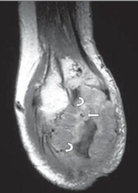
Vessels (sign of hypervascularization) were found within the lesions (Figures 1, 2 and 3) and in the periphery (Figures 2 and 3) in 12 of the 15 lesions (80%), 6 of them in the lumbar spine, 3 in the hip, 1 in the thoracic spine, 1 in the ulna, and 1 in the tibia.
Two patients with metastasis to the lum-bar spine were submitted to surgery (be-cause of medullary compression), with in-tense intraoperative bleeding. One of these
patients presented hypovolemic shock. The other patients were treated with radio-therapy.
DISCUSSION
Signs of hypervascularization (flow void) as an imaging finding frequently observed in RCC metastases have been described by Choi et al.(8), being reported in 76.7% of 20 cases studied. Uchino et al.(9) have evaluated MRI studies of patients with single intracranial metastasis of RCC, reporting intra- and/or peritumor vessels in five of among seven cases. Nguyen et al.(10) have reported a case of RCC with visible vessels within a thrombus invading the in-ferior vena cava and the right atrium.
In spite of being very frequently found, the presence of hypervascularization is not
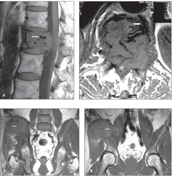
Figure 1.A: Sagittal MRI T1-weighted images of the lum-bar spine of a male, 69-year-old patient demonstrating hypointense lesion at L1, with tubular images suggesting the presence of vessels (arrow). B:
Axial MRI T1-weighted image demonstrating the presence of large-caliber vessels within and in the periphery of the lesion (arrow).
A B
Figure 2.A,B: Coronal MRI T1-weighted image of the hip of a male, 43-year-old patient dem-onstrating the presence of an expansile lesion in the wing of the right iliac bone, with hypervascularization within (straight arrows) and in soft tis-sues surrounding the lesion (curved arrow).
157
Hypervascularization in bone metastases from renal cell carcinoma
Radiol Bras. 2009 Mai/Jun;42(3):155–157 RCC-specific. Hypervascularization is also found in other less common lesions, such as arteriovenous malformations, high-flow hemangiomas and alveolar soft part sar-coma(11,12). Kawahara et al.(13) have re-ported a case of hepatocellular carcinoma metastasis to calvaria presenting signs of hypervascularization (flow void) at MRI.
It is known that tumors such as thyroid melanomas and carcinomas present hyper-vascularization. However, no report has
been found in the literature about observa-tion of vessels within metastases of these tumors.
Similarly to data in the literature, the authors of the present study have found signs of hypervascularization in 80% of cases. Although these signs have been de-scribed in studies with small samples, their high frequency allows the authors to affirm that the presence of hypervascularization within or surrounding bone lesions must suggest the evaluation of the kidney as the primary site of the neoplasm. In cases where the primary tumor is already known and a surgical treatment is indicated, the surgeon must be warned about the presence of these large, newly formed vessels(14), to prevent eventual intraoperative complica-tions (hemorrhage).
CONCLUSION
The presence of hypervascularization (flow void) within or in the periphery of multiple or single bone lesions should be considered as an indication for investiga-tion of the kidney as primary site of the tumor. The surgeon must be warned about the presence of these vessels, in order to prevent complications resulting from intra-operative hemorrhage.
REFERENCES
1. Boring CC, Squires TS, Tong T, et al. Cancer sta-tistics, 1994. CA Cancer J Clin. 1994;44:7–26.
2. Thrasher JB, Paulson DF. Prognostic factors in re-nal cancer. Urol Clin North Am. 1993;20:247–62.
3. Saitoh H. Distant metastasis of renal adenocarci-noma. Cancer. 1981;48:1487–91.
4. Nielsen OS, Munro AJ, Tannock IF. Bone me-tastases: pathophysiology and management policy. J Clin Oncol. 1991;9:509–24.
5. Sundaresan N, Scher H, DiGiacinto GV, et al. Sur-gical treatment of spinal cord compression in kid-ney cancer. J Clin Oncol. 1986;4:1851–6. 6. Prando A. Achados da tomografia
computadori-zada em metástases pancreáticas do carcinoma de células renais. Radiol Bras. 2008;41:225–8.
7. King GJ, Kostuik JP, McBroom RJ, et al. Surgi-cal management of metastatic renal carcinoma of the spine. Spine. 1991;16:265–71.
8. Choi JA, Lee KH, Jun WS, et al. Osseous metasta-sis from renal cell carcinoma: “flow void” sign at MR imaging. Radiology. 2003;228:629–34.
9. Uchino A, Hasuo K, Mizushima A, et al. Intra-cranial metastasis of renal cell carcinoma: MR im-aging. Radiat Med. 1996;14:71–6.
10. Nguyen BD, Westra WH, Zerhouni EA. Renal cell carcinoma and tumor thrombus neovascularity: MR demonstration with pathologic correlation. Abdom Imaging. 1996;21:269–71.
11. Rak KM, Yakes WF, Ray RL, et al. MR imaging of symptomatic peripheral vascular malforma-tions. AJR Am J Roentgenol. 1992;159:107–12.
12. Suh JS, Cho J, Lee SH, et al. Alveolar soft part sarcoma: MR and angiographic findings. Skeletal Radiol. 2000;29:680–9.
13. Kawahara I, Tsutsumi K, Hirose M, et al. Skull metastasis of hepatocellular carcinoma: case re-port. No Shinkei Geka. 2005;33:903–9.
14. Baroni RH. Novos paradigmas na avaliação por imagem dos tumores parenquimatosos renais. Radiol Bras. 2008;41:v–vi.