John Wiley & Sons. Printed in Singapore
J
ournal ofCutaneous Pathology
Intraepidermal epidermotropic
metastatic melanoma: a clinical
and histopathological mimicker
of melanoma
in situ
occurring
in multiplicity
The distinction between primary melanoma and melanoma metastatic to the skin has major prognostic implications. We report a case of a 67-year-old male with a diagnosis of a superficial spreading melanoma (stage IB) rendered 6 years earlier who presented clinically with an atypical nevus on his left thigh. Histopathological examination showed an intraepidermal melanocytic proliferation that was interpreted as melanoma in situ. Subsequently, 45 additional pigmented macules appeared in crops over a 9-month period. Clinically and
dermoscopically, these lesions were extremely polymorphic.
Histopathological findings were compatible with melanoma in situ, as each lesion consisted of a wholly intraepidermal proliferation of
markedly atypical melanocytes arranged singly and in nests. A complete gastrointestinal study showed multiple pigmented metastatic lesions throughout the stomach and small bowel, which supported a diagnosis of metastatic melanoma with gastrointestinal and epidermotropic skin involvement. Monosomy of chromosome 9 and a BRAF V600E mutation were detected in the primary tumor sample and in
macro-dissected secondary lesions. No CDKN2A or CDK4 germline mutations were found. Intraepidermal epidermotropic metastases of melanoma have been rarely described in literature. In this case, histopathology alone was insufficient to distinguish metastatic melanoma from multiple in situ melanomas. The recognition of
epidermotropic metastases should be based on the correlation between clinical, dermoscopic, histopathological and molecular findings.
Keywords: atypical features, cutaneous metastases, dermoscopy,
epidermotropism, malignant melanoma
Lestre S, Jo˜ao A, Ponte P, Peixoto A, Vieira J, Teixeira MR, Fidalgo A. Intraepidermal epidermotropic metastatic melanoma: a clinical and histopathological mimicker of melanoma in situ occurring in
multiplicity.
J Cutan Pathol 2011; 38: 514–520.© 2011 John Wiley & Sons A/S.
Sara Lestre1, Alexandre Jo˜ao1,
Pedro Ponte1, Ana Peixoto2,
Joana Vieira2, Manuel R.
Teixeira2,3and Ana Fidalgo1
1Dermatology Department, Hospital Santo
Ant´onio dos Capuchos, Centro Hospitalar de Lisboa-Central, Lisbon, Portugal
2Genetics Department, Portuguese Oncology
Institute, Porto, Portugal, and
3Biomedical Sciences Institute (ICBAS),
Porto University, Porto, Portugal
Sara Lestre,
Dermatology Department, Hospital Santo Ant´onio dos Capuchos,
Alameda Santo Ant´onio dos Capuchos, 1169-050 Lisbon, Portugal Tel: +35121 313 63 00 Fax: +35121 356 22 08 e-mail: saralestre@gmail.com
Cutaneous melanoma represents a lethal skin cancer that is responsible for 80% of skin cancer mortality.1 Metastatic melanoma progression typically evolves from local cutaneous growth to regional metastasis involving lymph node(s) or in-transit metastasis, and later disseminated metastatic disease may involve skin or any visceral organ.2 Cutaneous melanoma metastases usually involve the dermis and subcutaneous tissue and spare the overlying epidermis.3Rarely, metastatic melanomas can show prominent epidermotropism, mimicking primary melanoma. Epidermotropic metastatic melanoma represents a recognized pathological variant. Its recognition is particularly challenging as its clinical and histopathological appearance can simulate primary melanoma.4 This distinction is crucial in the prognosis and treatment approach to affected patients. In 1978, Kornberg and colleagues proposed histopathological criteria for the diagnosis of epidermotropic metastatic melanoma,5which are still used at the present time. However, in the past few decades, the morphological spectrum of epidermotropic metastatic melanoma has broadened and additional criteria have been added.4,6 – 8
Herein, we report a patient whose melanoma recurred with 46 epidermotropic metastases that were wholly intraepidermal. The patient also suf-fered from multifocal asymptomatic gastrointestinal metastases.
Case report
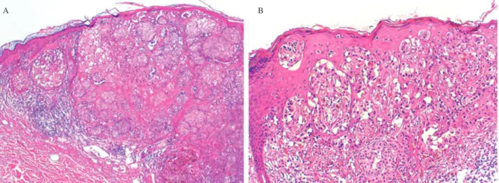
In 2002, a 67-year-old male was diagnosed with a non-ulcerated superficial spreading melanoma (stage IB, T2 N0 M0, Breslow’s thickness 1.3 mm, Clark’s level III) involving the lower back (Fig. 1). Sentinel lymph node evaluation was not performed. After wide excision (1 cm), adjuvant immunotherapy with
A B
Fig. 1. Histopathology of the primary melanoma of the back, which was excised in November 2002. The proliferation was broad and
markedly asymmetrical (A: hematoxylin and eosin,×40). There was a population of atypical melanocytes with pagetoid spread and epidermal consumption (B: hematoxylin and eosin,×100).
Fig. 2. Histopathology of a pigmented lesion of the thigh that
appeared 6 years after the excision of the primary melanoma. There was a wholly intraepidermal proliferation of atypical melanocytes with pagetoid spread, indistinguishable from melanoma in situ (hematoxylin and eosin,×40).
interferon-alpha (3 millions IU, thrice weekly) was started and maintained over 12 months. He was regularly followed and remained free from disease recurrence up until February 2009, when an irregularly shaped and highly pigmented macule appeared on his left thigh. His environmental exposure history was significant for several sunburns before 20 years of age and family history was remarkable for melanoma in two siblings.
Histopathological examination of the new pig-mented lesion was interpreted as melanoma in situ (Fig. 2). Subsequently, several small pigmented mac-ules appeared in crops on the back (Fig. 3). The most irregular ones were excised and all biopsy specimens showed markedly atypical melanocytes arranged in irregular nests and solitary units at all levels of epi-dermis, mimicking melanoma in situ. Neither dermal involvement nor invasion of vascular or lymphatic spaces was detected (Fig. 4). Those judgments were supported by Melan-A and CD31 stains.
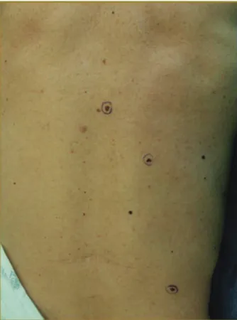
Fig. 3. The clinical appearance of several pigmented lesions on the
back is depicted.
New crops of pigmented lesions appeared on the back, arms and legs. Most of them were small (less than 5 mm of diameter), brownish, well-defined, sym-metric and macular. A total of 46 lesions were ex-cised during a 9-month period. Histopathological findings were similar in all specimens, consisting of an exclusively intraepidermal proliferation of large atypical melanocytes with increased nuclear to cytoplasmatic ratio and prominent nucleoli. Those findings were compatible with melanoma in situ, although many lesions were small, symmetric and circumscribed (Fig. 5).
Dermoscopically, the lesions were extremely poly-morphic. Most of the excised macules had a light-brown symmetric homogeneous pattern with only few darker dots at the periphery. Frequently, a reg-ular starburst pattern was also observed. In some lesions, melanoma-specific criteria were present, par-ticularly blue-white structures and irregular blotches (Fig. 6).
The patient underwent staging with inguinal nodal ultrasonography; computed tomography (CT) of the head, chest, abdomen and pelvis; brain magnetic resonance (MR) and positron emission tomography/CT (PET-CT). Distant metastases were not identified by this testing. Results of routine laboratorial evaluation, including lactate
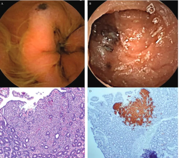
dehydrogenase (LDH) and S-100 protein, were unremarkable. Mucosal and uveal involvement by melanoma was also excluded. Even though the patient was asymptomatic, an upper endoscopy was performed that showed pigmented macules and ulcerated nodules involving the stomach and duodenum mucosa. The biopsy specimens revealed metastatic melanoma within superficial mucosa (Fig. 7). This diagnosis was supported by positive immunostaining with HMB-45. Surgical resection of the stomach and proximal duodenum was considered, but surgery was rejected after detection of multifocal metastatic melanoma throughout the entire length of the jejunum and ileum by capsule videoendoscopy.
Molecular analysis was also performed. Genomic DNA was extracted from peripheral blood nucleated cells using conventional procedures. No germline point mutations or exonic rearrangements were found in the CDKN2A gene or point mutations in
CDK4 ( codon 24). Fluorescence in situ
hybridiza-tion (FISH) analysis was completed using paraffin-embedded sections of the primary lesion and a com-mercially available probe to target the gene CDKN2A in chromosome band 9p21 and the centromere of chromosome 9 (LSI CDKN2A /CEP 9 Probe -Vysis/Abbott). Monosomy of chromosome 9 (includ-ing loss of a copy of the CDKN2A gene) was found in the primary tumor. We also performed mutation analysis of BRAF (codon 600) in paraffin-embedded sections of the primary tumor sample and in freshly collected biopsy specimens from seven presumed sec-ondary lesions of the skin. A BRAF V600E mutation was detected in the primary tumor sample but not in the seven skin biopsy specimens analyzed. Given this unexpected finding, we looked for monosomy 9 and BRAF codon 600 in paraffin-embedded tis-sue sections of five distinct and separate secondary skin lesions (molecular analysis was completed after macrodissection to avoid contamination with normal cells) and indeed found that the primary tumor and all the analyzed secondary lesions shared the chro-mosome 9 monosomy and BRAF V600E mutation.
During ongoing follow up, albeit with the con-tinuous appearance of new pigmented lesions until November 2009, the patient remained asymptomatic with good general status. A multidisciplinary team decided to delay initiation of systemic chemother-apy and/or chemoimmunotherchemother-apy and wait for the beginning of a new clinical trial recruiting patients with stage IV malignant melanoma.
In February of 2010, the patient died as a result of a large aortic thrombus with an extensive systemic embolization affecting the brain, heart, spleen and kidney. The patient’s family refused an autopsy.
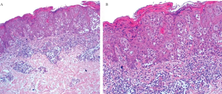
A B
Fig. 4. The histopathology of a pigmented macular lesion from the lower back (April 2009) is illustrated. There is an atypical intraepidermal
melanocytic proliferation with an associated lymphocytic infiltrate in the superficial dermis (A: hematoxylin and eosin,×40). Further detail shows atypical melanocytes arranged in singly and in nests (B: hematoxylin and eosin,×100).
Fig. 5. The histopathological features of a pigmented skin removed
from the right thigh (July 2009). There is a small, symmetric and well-circumscribed lesion composed of nests of atypical melanocytes confined to the epidermis.
Discussion
Cutaneous metastases of melanoma have a variable appearance. Papules, nodules and tumors commonly involve the dermis and subcutis and less frequently involve the overlying epidermis.3 Metastases with epidermal involvement have been designated epidermotropic metastatic melanoma and usually present as multiple, symmetric, small lesions occurring in crops located in the same region as the primary melanoma. Less frequently, widely disseminated lesions can be seen.3 – 6
Histopathological criteria for defining epider-motropic metastatic melanoma have evolved over the years. The distinction between metastatic and primary melanoma was initially based on the absence of involvement of the overlying epithelium.9 In
1978, Kornberg and colleagues developed spe-cific histopathological features that favored the diagnosis of epidermotropic metastatic melanoma, including: (a) dermal involvement equal or greater
than epidermal involvement; (b) junctional epider-mal involvement not extending beyond the edge of the dermal component; (c) atypical melanocytes fill-ing the papillary dermis with thinnfill-ing of epidermis; (d) widening of papillae with the formation of an epithelial collarette; and (e) the identification of atyp-ical melanocytes in vascular and lymphatic spaces.5 Subsequently, epidermotropic metastatic melanoma with pagetoid spread of atypical melanocytes beyond the lateral limits of the dermal component of der-mal melanoma was described.6,10,11 Several reports
also suggested that angiotropism is suggestive of metastatic disease.7,12
Immunohistochemistry may be useful as a tool for distinguishing epidermotropic metastatic melanoma and multiple primary melanomas in situ, but the approach lacks specificity: metallothionein expression is strongly expressed by epidermotropic metastatic melanoma but is also expressed by Spitz nevi. In epidermotropic metastases, the intensity of HMB-45 staining is greater than that of S-100, but this phenotype does not exclude primary melanoma.4,13
Abernethy et al. reported two patients with widespread epidermotropic metastatic melanoma, including classic lesions with intraepidermal exten-sion beyond the intradermal component and 10 lesions with only epidermal involvement simulating melanoma in situ.13 In contrast to previous reports, our patient’s lesions all showed identical histopathol-ogy with exclusively epidermal involvement and no detectable dermal disease. In this setting, the distinc-tion between epidermotropic metastatic melanoma and multiple primary melanomas is virtually impos-sible by light microscopy. However, the diagnosis of
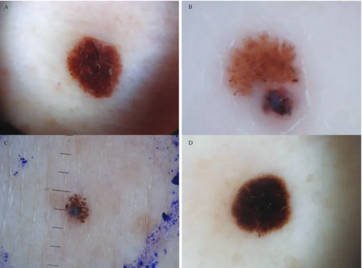
A B
C D
Fig. 6. Heterogeneous clinical and dermoscopic findings are depicted. There is a uniform diffuse brown lesion with a few darker dots
throughout and at its periphery (A). A multicomponent pattern lesion has significant asymmetry of color and structure; blue-white structures, irregular dots and irregular blotches are seen (B). Depicted is a globular pattern lesion with variously sized dark brown globules; a blue-white veil is also noted at the periphery (C). A regular and symmetric starburst pattern is also recognizable (D).
epidermotropic metastatic melanoma was suggested by clues expressed by most of our patient’s lesions, including: (a) multiple disseminated lesions appear-ing in crops; (b) lesions of small size; and (c) lesional symmetry and circumscription.
Regression of the dermal component is unlikely to be the explanation of this epidermal-only pattern. Rather, the phenomenon of epidermotropic metas-tasis probably represents hematogenous spread with only transient or minimal dermal involvement. The survival of patients with melanoma is heavily depen-dent on the stage of the malignancy, and patients with epidermotropic metastases have a poor prog-nosis and distant metastases involving other organs are a frequent finding.14 PET combined with CT imaging (PET-CT) has become a major diagnos-tic tool in the detection of metastadiagnos-tic disease, as the technique improves the identification of deep soft tissue, lymph node and visceral metastases.15,16 PET-CT has been also proposed as the method of choice in detecting gastrointestinal melanoma
metastases, which frequently are underdiagnosed by standard endoscopic techniques.17,18However, small metastatic deposits need to achieve a certain size to be detected by PET-CT.15In this case, the diagnosis of metastatic melanoma was particularly challeng-ing. While the presence of widely disseminated small pigmented lesions appearing in crops in a patient with a previous diagnosis of melanoma suggested epidermotropic metastatic melanoma as a possibil-ity, a staging evaluation (including a PET-CT) was negative for distant metastatic disease. In addition, histopathology was unable to discriminate epider-motropic metastases from multiple melanomas in situ and dermoscopy also failed to show unequivocal dermoscopic melanoma criteria. Indeed, few stud-ies are available regarding the dermoscopic features of cutaneous metastatic melanoma.19 – 22 In a study with 130 cutaneous melanoma metastases, the sac-cular and vassac-cular patterns as well as the presence of pigmentary halo and peripheral gray spots have been proposed as the most significant dermoscopic
A B
C D
Fig. 7. Gastrointestinal melanoma metastases are depicted. The endoscopic appearance of gastrointestinal lesions shows dark-brown macules
and ulcerated nodules. The histopathological features of a gastric melanoma metastasis include a proliferation of atypical melanocytes arranged in confluent nests in the superficial mucosa (hematoxylin and eosin,×40); positive immunostaining with HMB-45 was also observed. elements supporting interpretation as metastatic
melanoma.19 To the best of our knowledge, the dermoscopic results of epidermotropic metastatic melanoma have not been previously described. In our case, the dermoscopic findings were polymor-phic and included reticular, homogeneous, globular and spitzoid patterns simulating melanocytic nevi or primary cutaneous melanoma. These heterogeneous findings suggest that the diagnosis of epidermotropic metastatic melanoma cannot reliably be made on the basis of dermoscopic features alone.
Genetic analysis of melanoma can provide addi-tional information of use in the distinction of metastatic lesions from independent primaries.23 – 26 Although genetic intra- and intertumoral hetero-geneity is a frequent feature in melanoma, it would be expected that a primary melanoma and its metastases would share one or more genetic alterations. Loss of
heterozygosity (LOH) and X-chromosome inactiva-tion analysis studies suggest that metastatic lesions in most melanoma patients are clonally related to their primary lesions.23,25,27 In our patient’s tissue, the somatic mutation V600E in the BRAF gene and monosomy 9 were identified in the primary melanoma. Mutations in BRAF have been described in 40–80% of melanomas and V600E is the most common observed mutation. Mutations in BRAF appear to represent an early event in melanoma progression and are usually retained in secondary lesions.23,28 In our case, the V600E mutation was not initially detected in any of the seven freshly collected metastases, which were analyzed without macrodissection (and thus contaminating normal tissue may have obscured the molecular results). However, the subsequent use of FISH in paraffin-embedded tissue sections and molecular analysis
after macrodissection to avoid contamination allowed the demonstration of a molecular rela-tionship between five presumed epidermotropic metastases and the original primary melanoma. No evidence of hereditary predisposition associated with germline mutations of CDKN2A or CDK4, as can be seen in familial and multiple primary melanoma, was found.29,30.
We therefore conclude that the 46 pigmented lesions affecting our patient represent intraepider-mal epidermotropic metastatic melanoma and not
multiple examples of primary melanoma in situ. The clinical appearance of the lesions in con-junction with atypical dermoscopic findings, the presence of multiple gastrointestinal metastases, and the genetic findings strengthen this assertion. Our case shows that histopathology alone can be insuf-ficient to distinguish intraepidermal epidermotropic metastatic melanoma from multiple primary lesions. Its recognition should be based on correlation with clinical, dermoscopic, histopathological and genetic findings.
References
1. Miller AJ, Mihm MC. Melanoma. N Engl J Med 2006; 355: 51.
2. Zbytec B, Carlson JA, Granese J, Ross J, Mihm MC, Slominski A. Current concepts of metastasis in melanoma. Expert Rev Derma-tol 2008; 3: 569.
3. Plaza JA, Torres-Cabala C, Evans H, Diwan HA, Suster S, Preito VG. Cutaneous metas-tases of malignant melanoma: a clinicopatho-logic study of 192 cases with emphasis in the morphologic spectrum. Am J Dermopathol 2010; 32: 129.
4. White WL, Hitchcock MG. Dying dogma: the pathological diagnosis of epidermotropic metastatic melanoma. Semin Diagn Pathol 1998; 15: 176.
5. Kornberg R, Harris M, Ackerman AB. Epidermotropically metastatic malig-nant melanoma: Differentiating maligmalig-nant melanoma metastatic to the epidermis from malignant melanoma primary in the epider-mis. Arch Dermatol 1978; 114: 67. 6. Bengoechea-Beeby MP, Velasco-Os´es A,
Fern´andez FM, Reguil´on-Rivero C, Rem´on-Garijo L, Casado-P´erez C. Epidermotropic metastatic melanoma: Are the current histo-logic criteria adequate to differentiate primary from metastatic melanoma? Cancer 2006; 72: 1909.
7. Gerami P, Shea C, Stone M. Angiotropism in epidermotropic metastatic melanoma: another clue to the diagnosis. Am J Dermatopathol 2006; 28: 429.
8. Piana S, Valli R, Ricci C. Epidermotropic metastasis of melanoma: report of a case of metastatic melanoma with previously unreported morphological features. Am J Dermatopathol 2009; 31: 301.
9. Allen AC, Spitz S. Malignant melanoma: a clinicopathological analysis of the criteria for diagnosis and prognosis. Cancer 1953; 6: 45. 10. Heenan PJ, Clay CD. Epidermotropic metastatic melanoma simulating multiple primary melanomas. Am J Dermotopathol 1991; 13: 396.
11. Moulin G, Franc M-P, Barrut D, Coiffet J, Viornery P. M´etastases ´epidermotropes et jonctionnelles de m´elanome malin. Ann Der-matol Venereol (Paris) 1980; 107: 1061. 12. Moreno A, Espanol I, Romagosa V.
Angiotropic malignant melanoma. J Cutan Pathol 1992; 19: 325.
13. Abernethy JL, Soyer P, Kerl H, Jorizzo J, White WL. Epidermotropic metastatic malig-nant melanoma simulating melanoma in situ: a report of 10 examples from two patients. Am J Surg Pathol 1994; 18: 1140.
14. Savoia P, Fava P, Nard`o T, Osella-Abate S, Quaglino P, Bernengo MG. Skin metas-tases of malignant cutaneous melanoma: a clinical and prognostic survey. Melanoma Res 2009; 19: 321.
15. Kottschade LA, Suman VJ, Val Lowe, Morkovic SN. Positron emission tomogra-phy in early detection of relapse in high-risk melanoma. PET-TC relapse melanoma. Community Oncol 2009; 6: 344.
16. Strobel K, Dummer R, Husarik DB, Lago MO, Hany TF, Steinert HC. High-risk melanoma: accuracy of FDG PET/CT with added CT morphologic information for detection of metastases. Radiology 2007; 244: 566.
17. Tatlidil R, Mandelkern M. FDG-PET in the detection of gastrointestinal metastases in melanoma. Melanoma Res 2001; 11: 297. 18. Reinhardt MJ, Joe AY, Jaeger U, et al.
Diag-nostic performance of whole body dual modal-ity FDG PET/CT imaging for N- and M-staging of malignant melanoma: experience with 250 consecutive patients. J Clin Oncol 2006; 24: 1178.
19. Bono R, Giampetruzzi AR, Concolino F, et al. Dermoscopic patterns of cutaneous melanoma metastases. Melanoma Res 2004; 14: 367.
20. Ferrari A, Peris K, Piccolo D, Chimenti S. Dermoscopic features of cutaneous local recurrent melanoma. J Am Acad Dermatol 2000; 43: 722.
21. Schultz H. Epiluminescence microscopy fea-tures of cutaneous malignant melanoma metastases. Melanoma Res 2000; 10: 273. 22. Pizzicheta MA, Canzonieri V, Gatti A, et al.
Skin lesions in melanoma and Kaposi’s sar-coma. Case 2: dermoscopic features of metas-tases from cutaneous melanoma mimicking benign nevi and primary melanoma. J Clin Oncol 2002; 20: 1412.
23. Rodolfo M, Daniotti M, Vallacchi V. Genetic progression of metastatic melanoma. Cancer Lett 2004; 214: 133.
24. Edinger JT, Radfar A, Judik DM. Two cuta-neous malignant melanomas at the same anatomic site: a case report with molecular evaluation. J Cutan Pathol 2009; 36(Suppl. 1): 74.
25. Orlow I, Tommasi D, Bloom B, et al. Evalu-ation of the clonal origin of multiple primary melanomas using molecular profiling. J Invest Dermatol 2009; 129: 1972.
26. Morita R, Fugimoto A, Hatta N, Take-hara K, Takata M. Comparison of genetic profiles between primary melanomas and their metastases reveals genetic alterations and clonal evolution during progression. J Invest Dermatol 1998; 111: 919.
27. Bahrami S, Cheng L, Wang M, Jones TD, Malone JC, Billings SD. Clonal relation-ships between epidermotropic metastatic melanomas and their primary lesions: a loss of heterozygoty and X-chromosome inactivation-based analysis. Mod Pathol 2007; 20: 821.
28. Rao UNM, Jones MW, Finkelstein SD. Genotypic analysis of primary and metastatic cutaneous melanoma. Cancer Genet Cyto-genet 2003; 140: 37.
29. Blackwood MA, Holmes R, Synnestvedt M, et al. Multiple primary melanoma revisited. Cancer 2002; 94: 2248.
30. Hayward NK. Genetics of melanoma predis-position. Oncogene 2003; 22: 3053.