Universidade de Aveiro Ano 2014/2015
Departamento de Biologia
Susana Margarida
da Silva Correia
Alteração da resposta regulatória do Rab7a e do
Rab9 no subtipo MM1 e VV2 da doença
Creutzfeldt-Jakob
Altered regulatory response of Rab7a and Rab9 in
MM1 and VV2 subtype of Creutzfeldt-Jakob disease
Universidade de Aveiro Ano 2014/2015
Departamento de Biologia
Susana Margarida
da Silva Correia
Alteração da resposta regulatória do Rab7 e do Rab9
no subtipo MM1 e VV2 da doença Creutzfeldt-Jakob
Altered regulatory response of Rab7a and Rab9 in
MM1 and VV2 subtype of Creutzfeldt-Jakob disease
Dissertação apresentada à Universidade de Aveiro para cumprimento dos requisitos necessários à obtenção do grau de Mestre em Biologia Molecular e Celular, realizada sob a orientação científica da Professora Doutora Etelvina Ferreira, Professor auxiliar do Departamento de Biologia da Universidade de Aveiro e da Professora. Doutora Inga Zerr, do departamento de Neurologia da Universidade Georg-August, Göttingen.
Dedico este trabalho aos meus avós João Silva e Teresa Gonçalves por todo o amor e educação que nos deram. Dedico também ao meu padrinho João Luis Silva que é mais de um pai para mim, à minha mãe Madalena Silva e à minha irmã Ângela Correia por todo o apoio que me foi dado ao longo dos anos dos quais sem eles não seria a pessoa que sou hoje.
o júri
presidente Prof. Doutor Helena Silva
Professor Auxiliar do departamento de Biologia da Universidade de Aveiro
Prof. Doutor Etelvina Maria de Almeida Paula Figueira
Professor Auxiliar do departamento de Biologia Etelvina Figueira da Universidade de Aveiro
Prof. Doutor Sandra Maria Tavares da Costa Rebelo
Professor Auxiliar Convidada da Secção Autónoma das Ciências da Saúde da Universidade de Aveiro
agradecimentos A realização desta dissertação de mestrado contou com importantes apoios e incentivos sem os quais não se teria tornado uma realidade e aos quais estarei eternamente grata.
À Professora Doutora Inga Zerr, do departamento de Neurologia da Universidade Georg-August, o meu agradecimento pela oportunidade de ingressar no seu grupo de investigação.
Ao Doutor Franc Llorens e Matthias Schmitz, do departamento de Neurologia
da Universidade Georg-August, o meu profundo agradecimento pela amizade e apoio que me deram ao longo deste ano.
À Professora Doutora Etelvina Figueira, orientadora da tese de mestrado, o meu agradecimento pelo apoio, preocupação e disponibilidade ao longo do ano.
À Professora Doutora Maria de Lourdes Gomes Pereira, Professora associada com agregação da Universidade de Aveiro, o meu agradecimento pela preocupação e disponibilidade ao longo deste ano.
Aos meus familiares o meus profundos agradecimentos por todo o apoio e motivação despendidos.
palavras-chave CJD-MM1, Rab7, Rab9, PrPc, PrPSc.
resumo O presente estudo foi levado a cabo com o intuito de identificar possíveis proteínas que interajam com PrPC que possam fornecer novos conhecimentos sobre as suas funções fisiológicas assim como no papel patológico. O presente estudo incidiu na investigação dos níveis das proteínas endossomais Rab7a e Rab9 em amostras do córtex frontal e cerebelo de pacientes diagnosticados com sCJD-MM1 e sCJD-VV2. Observamos um aumento significativo dos níveis intracelulares do Rab7a em pacientes diagnosticados com sCJD-MM1 e sCJD-VV2 quando comparadas com amostras de pacientes na mesma faixa etária sem a doença (controlo). As interações entre PrPC e as proteínas Rab7a e Rab9 foram posteriormente estudadas com o auxílio à microscopia confocal. Os resultados da imunofluorescência sugeriram potenciais interações entre as proteínas Rab7a com o PrPC. O siRNA-Rab7 foi usado para diminuir a expressão da proteína Rab7a (“Knockdown”) na cultura primária de células do córtex de ratos saudáveis. Na cultura de células do córtex tratadas com o siRNA-Rab7, foi observado um emparelhamento da via endocítica. Seguidamente investigamos possíveis interações da proteína Tau com a proteína Rab7a, recorrendo ao western blot e a microscopia confocal. Culturas de células do córtex de ratos saudáveis foram tratadas com siRNA-Tau, siRNA-Rab7a e siRNA. Os resultados da imunofluorescência das diferentes culturas celulares sugeriram uma potencial interação entre as proteínas Tau e Rab7a. Nas linhas celulares tratadas com siRNA-Tau os níveis intracelulares das proteínas Rab7a e Rab9 aumentaram significativamente, assim como, foi também observada a alteração da sua localização. Nas células tratadas com siRNA-Rab7a observamos uma diminuição da acumulação da proteína Tau na região citosólica. Em conclusão, este trabalho pode ajudar a perceber e a caracterizar a progressão da doença nos subtipos específicos, no caso de sCJD. Contudo, este trabalho poderá ser também um passo à frente para o desenvolvimento de novas estratégias terapêuticas para os subtipos sCJD-MM1 e sCJD-VV2.
keywords CJD-MM1, Rab7, Rab9, PrPc, PrPSc.
abstract The present study was undertaken to identify proteins interacting with PrPC that could provide new insights into its physiological functions and pathological role. We performed a target search for lysosomal network protein, Rab7a and Rab9, in frontal cortex and cerebellum of human brain from patients with sCJD-MM1 and sCJD-VV2. The intracellular level of Rab7a was increased significantly, when compared with healthy age-matched control. Interactions of PrPC and Rab7a/Rab9 were further investigated by using confocal laser scanning microscopy. Immunofluorescence results suggested potential interactions of Rab7a and PrPC. siRNA against the Rab7a gene was used to knockdown the expression of Rab7a protein in primary cell culture of cortical neurons from wild type mice. This depleted Rab7a resulted an impairment of PrPC trafficking leading to an accumulation of PrPC in the endocytosis pathway. Furthermore, interactions of Tau and Rab7a were investigated by using western blot analysis and confocal laser scanning microscopy. Cell cultures of cortex of wildtype mice were treated with siRNA-Tau, siRNA-Rab7 and control siRNA followed by immunofluorescence. The results of immunofluorescence suggested potential interaction of Tau and Rab7a. Cells lines treated with siRNA-Tau, the intracellular levels of Rab7a and Rab9 significantly increases and their localization is also modified. When we transfected this cells lines with siRNA-rab7a the accumulation of Tau decreases in cytosolic region and their localization was also modified when compared with control cells. In conclusion, this study may help to understand and characterize the subtype specific disease progression in CJD cases. Furthermore, it could be a step ahead to development of new treatment strategies for diseases subtype specific manner.
Index
I Introduction ...1
1 Cellular prion protein (PrPC) ...1
1.1 Structure of cellular prion and prion disease ...3
1.2 PrP-res form and prion diseases ...4
2 Creutzfeldt-Jakob disease (CJD) ...5 2.1 sCJD cognitive type ...7 2.1.1 sCJD MM1 and sCJD MV1 ...7 2.1.2 sCJD 129 MM2 ...7 2.1.3 sCDJ 129 VV1 ...8 2.2. sCDJ ataxia type ...9 2.2.1 sCDJ VV2 ...9 2.2.2 sCDJ MV2 ...9
3 Prion disease and PrPC Cellular trafficking ... 10
3.1 Vesicular trafficking ... 10
3.2 Rab GTPases proteins ... 11
3.2.1 Rab7a protein ... 15
3.2.2 Rab9 protein ... 15
4 Microtubules and Tau protein ... 16
II The aims of the present study ... 18
III Materials ... 19
1 Antibodies ... 19
2 Antibiotics ... 19
3 Culture media ... 20
4 Chemicals ... 20
5 Instruments and other materials ... 20
6 Kits ... 22
7 siRNA Duplex ... 22
8 Software ... 22
9 Stock solutions ... 23
IV Methods ... 24
1 Biological samples methods ... 24
1.1 Human brain ... 24
1.2 sCJD MM1 mice brain ... 24
2 Immunoblot ... 25
3 Immunofluorescence ... 25
4 Primary culture of mouse cortex ... 26
6 Cell lysis ... 27
7 Protein concentration estimated by Bradford assay ... 28
8 Immunofluorescence and quantification analysis ... 28
9 Statistical analysis ... 30
10 Ethics Statement ... 30
V Results ... 31
1 Intracellular levels of Rab7a and Rab9 proteins in frontal cortex and cerebellum of human brain ... 31
2 Intracellular levels of Rab7a and Rab9 in sCJD-Mice ... 32
3 Co-localization of PrPc and Rab7a by Immunofluorescence ... 33
4 Intracellular levels of PrPc and Rab7a/Rab9 in primary culture of mouse cortex ... 34
5 siRNA downregulation of Rab7a and Tau ... 37
6 Tau and Rab7a ... 37
VI Discussion ... 40
1 PrPc and Rab7a/Rab9 ... 40
2 Total-Tau and Rab7a ... 41
List of Figures
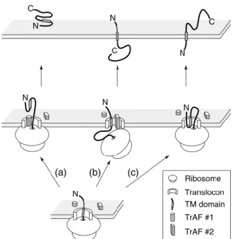
Figure 1 Potential mechanism for generating different topological form of PrPC ...2
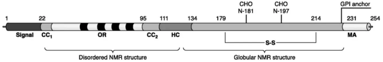
Figure 2 Primary structure of PrPC ncluding posttranslational modifications ...3
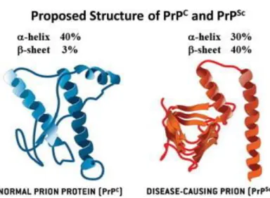
Figure 3 Structure of PrPC and PrPSc ...4
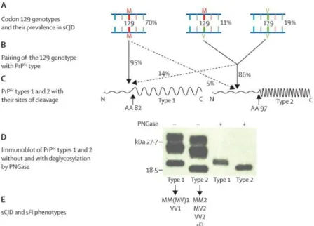
Figure 4 Correlations between the PrP genotype, as determined by the MV polymorphism at codon 129, and PrPSc types 1 and 2 ...6
Figure 5 General model of vesicular transport pathways ... 10
Figure 6 Role of ARF in the formation of COP-coated vesicles ... 11
Figure 7 The intracellular localization and associated vesicle transport pathway(s) of several Rab GTPases in eukaryotic cell ... 13
Figure 8 Activation cycle of small G proteins and their regulatory proteins ... 14
Figure 9 Hypothetical model for Rab-mediated vesicle docking ... 14
Figure 10 Microfilaments, microtubules and intermediate filaments in the nervous system 16 Figure 11 Intracellular levels of Rab7a and Rab9 proteins in frontal cortex and cerebellum human samples sCJD MM1, VV2 and AD patients... 31
Figure 12 Intracellular levels of Rab7a and Rab9 in sCJD-Mice at pre-symptomatic and Symptomatic stages ... 32
Figure 13 Co-localization of PrPc and Rab7a... 34
Figure 14 Co-localization of PrPc with Rab7a/Rab9 ... 35
Figure 15 Effect of Rab7a depletion on PrPc localization ... 36
Figure 16 Western blot of primary culture cortex of Wild type mice ... 37
Figure 17 Intracellular levels of Total-Tau and Rab7a in primary culture of wild type mice treated with siRNA-Rab7a and siRNA-Tau ... 38
List of Tables
Table 1 Transmissible spongiform encephalopathies (TSEs) in humans and animals ...4
Table 2 List of antibodies and their application in present study ... 19
Table 3 List of antibiotics ... 19
Table 4 List of culture media ... 20
Table 5 List of instruments used in this study ... 20
Table 6 List of the kits used in this study ... 22
Table 7 List of siRNA Duplex ... 22
Abbreviations oC Celsius μg Microgram μl Microliter μm Micro meter aa Amino acid AD Alzheimer’s disease
ATP Adenosine triphosphate
BFA Brefeldin A
bp Base pair
BSA Bovine serum albumin
BSE Bovine spongiform encephalopathy CSF Cerebrospinal fluid
dH20 Distilled water
ddH2O Double distilled water
DMEM Dulbecco's Modified Eagle Medium
DTT Ditiotreitol
ECL Enhanced chemiluminescence
ER Endoplasmic reticulum
ERAD Endoplasmic reticulum associated degradation ERGIC ER–Golgi intermediate compartment
FBS Fetal bovine serum
FCS Fetal calf serum
h Hour IAA Iodoacetamide kDa Kilodalton M Mol mg Milligram min. Minute ml Milliliter
PBS Phosphate buffered saline PVDF Polyvinylidene fluoride
PrPC Cellular prion protein
PrPSC Infectious isoform of prion protein
rpm Revolutions per minute Rab7a Ras-related protein Rab-7a Rab9 Ras-related protein Rab-9
RT Room temperature
s second
sCJD Sporadic Creutzfeldt-Jakob disease
SDS-PAGE Sodium Dodecyl Sulphate Polyacrylamide Gel Electrophoresis siRNA Small interfering RNA
TBS Tris buffered saline
TBST TBS with 0.1% Tween
TE Tris EDTA
TEMED N, N, N´, N´-tetramethylethylenediamine Tris Tris-(hydroxymethyl)-aminomethane
TSEs Transmissible spongiform encephalopathies
I Introduction
1 Cellular prion protein (PrPC)
The cellular form of the prion protein (PrPC) is a
glycosyl-phosphatidyl-inositol (GPI)-anchored glycoprotein, highly expressed in the neurons of the brain and spinal cord region (Stahl et al. 1987). However, the physiological function of PrPC is still not clear, in recent years, some putative functional aspects were
attributed to PrPC, synaptic regulatory response (as a receptor or receptor-related
in GABAA-ergic inhibitory synapses) (Puoti et al. 2012), sleep and circadian
rhythms regulator, inhibitory regulation of N-methyl-D-aspartate (NMDA) receptors, interaction with stress inducible protein 1 (STI1), regulation of cell differentiation and apoptosis, maintenance of peripheral nerve myelin, role in neuronal growth and survival (protection against bax-mediated cell death); signal transduction (caveolin1-dependent coupling to tyrosine kinase Fyn); signaling receptor (binding to neural cell adhesion molecule); oxidative stress, copper binding (cooper serving as a cofactor for an undetermined PrPc enzyme activity) (Lorca et al. 2011; Martins
et al. 2010), receptor for amyloid beta in Alzheimer’s disease and possibly for other amyloids (Laurén et al. 2009; Resenberger et al. 2011) and it is known that the conformal change of PrPc can cause serious neurodegenerative diseases
(Nicolas et al. 2009).
The PrPc is synthesized in the endoplasmic reticulum (ER), processed in
golgi apparatus and then transferred to the plasmatic membrane in full length form. During its synthesis the cellular prion can take various forms in the ER. The PrPc can be found at least in two transmembrane topologies (Hegde & Lingappa
1999):
1. ctmPrP = The ctmPrP form is when the c-terminal of PrPc is in the ER
lumen,
2. NtmPrP = The NtmPrP form is when the N-terminius of PrPc is in the ER
Figure 1. Putative mechanism for generating different topological forms: The PrPc is translocated to the endoplasmic reticulum (ER) by ribosomes. The nascent protein chain containing a transmembrane (TM) domain (shaded rectangle) can have different orientations into the ER membrane. This process depends on a particular translocation accessory factor (TrAF #1) that might shield the hydrophobic TM domain from being recognized by the translocon, allowing it to be translocated into the ER lumen leading to cyPrP form. If TM domain is recognized by translocation machinery ((b) or (c)), another translocation accessory factor (TrAF #2) interacts with nascent chain leading to NtmPrP form (b) or if there is no interaction of the translocation accessory factor (TrAF #2) with, the nascent chain, the least energetically demanding orientation, is favoured the ctmPrP form (c). The final topology resulting from each of these possibilities is shown at the top of the diagram (Take from Hegde e Lingappa, 1999).
The transportation of PrPc leads to unglycosylated and glycosylation (mono-
and di- glycosylated) forms formed in RE and it is transported to the golgi apparatus for further modifications (Figure 2). Therefore the cellular prion is transported to the cellular membrane under its mature form, PrPc (Harris 2003).
The PrPc can still be found in the cytosol, cyPrP. The cyPrP can be the result of
controlled mechanisms of ER. When the protein is misfolded or under ER stress conditions, proteins undergo retrograde transport to the cytosol, become polyubiquitinated, and are degraded by the proteasome through a process called ER-associated degradation, ERAD (Lin et al. 2013).
Figure 2. Primary structure of PrPc including posttranslational modifications: A secretory
signal peptide resides at the extreme NH2 terminus. CC1 and CC2 define the charged clusters. OR indicates the octapeptide repeat, and four are present. HC defines the hydrophobic core. MA denotes the membrane anchor region. S-S indicates the single disulfide bridge, and the glycosylation sites are designated as CHO. The numbers describe the position of the respective amino acids (Take from Aguzzi e Calella, 2009).
1.1 Structure of cellular prion and prion disease
The structure of PrPc is composed of three regions; alpha-helix (~ 40% of
protein) separated by loop/turn regions and by two small regions of beta-sheet (~ 3% of protein). This conformation of PrPc is sensitive to proteolysis, PrP-sen.
However, when PrPc undergoes structural changes, it leads to the increase of
beta-sheets ~ 40%, and PrPc becomes misfolded and resistant to proteases
attack, PrP-res (Prusiner 2004; Novakofski et al. 2005).
The conversion of PrP-sen to PrP-res can occur when PrP-res binds to PrPc
or when the PrP-sen conformation is altered into PrP-res by a poorly understood pathway (Figure 3). Its conversion on the cell surface leads to PrP-res accumulation extracellularly, and along endocytic pathway leads to accumulation of PrP-res in the lysosome (Moore et al. 2009; Lee et al. 2013). The accumulation of PrP-res in the lysosomes leads eventually to lysosome´s burst and the release of all lysosomal enzymes together with the PrP-res in the cytoplasm, leading to cells apoptosis (Araújo 2013).
Figure 3. Structure of PrPc and PrPSc: Normal and disease-causing prions structures (Take from
Lee et al., 2013).
1.2 PrP-res form and prion diseases
PrP-res accumulates intra and/or extracellularly leading to the neurodegenerative diseases called transmissible spongiform encephalopathies (TSEs),also called prion diseases. The aggregates of PrP-res yield neurological dysfunctions and cells death. The brain lost neurons and with advancing disease the brain looks like a sponge form leads to death in humans and other mammals (Belay 1999). In mammals, the TSE occurs as scrapie disease ( sheep), transmissible mink encephalopathy (mink), chronic wasting disease (deer), bovine spongiform encephalopathy (cattle) and feline spongiform encephalopathy (cats). The most common prion disease present in humans is Creutzfeldt-Jakob disease (Lee et al. 2013; Moore et al. 2009).
Table 1. Transmissible spongiform encephalopathies (TSEs) in humans and animals
(Liemann & Glockshuber 1998; Puoti et al. 2012).
Disease name Animal species Etiology
Scrapie Sheep Infection
Transmissible mink encephalopathy
Mink Infection
Chronic wasting disease Deer Infection
Bovine spongiform encephalopathy
Feline spongiform encephalopathy Cats Infection Bovine spongiform encephalopathies Cattle Infection Creutzfeldt-Jakab disease Iatrogenic Sporadic Inherited New variant Humans Infection Unknown Mutation in PrP gene Infection from bovine prion?
Gerstmann-Stränssler-Scheinker syndrome
Humans Mutation in PrP gene Fatal familial insomnia Humans Mutation in PrP gene
2 Creutzfeldt-Jakob disease (CJD)
The sporadic Creutzfeldt Jakob disease (sCJD) is the most common form of prion disease (85% to 95%) of all human prion disease (Ladogana et al. 2005; Masters et al. 1979). In sCJD, the source of contamination is unknown. The disease is not associated with any mutation in PRNP allele or by any exposure to Prion disease. However, studies have been documented that people with a medical history of psychosis, family history of CJD, history of multiple surgical procedures and residence for more than 10 years on a farm have a significant risk to develop sCJD (Villemeur 2013).
The sCJD is the prion disease with the highest degree of phenotypic heterogeneity. This phenotype heterogeneity has been associated with polymorphisms of codon 129 of the prion protein gene (PRNP) and to the size of protease resistant core of the prion protein (PrPSc), ~21 KDa (type 1) and ~19KDa
(type 2). The codon 129 of PRNP consists in an amino acid methionine (M) or valine (V) in each allele. The the resistant core of PrPSc patient genotype can be
MM, MV or VV with type 1 or type 2 of and therefore, they have a different phenotype of prion disease (Figure 4) (Parchi, Capellari, et al. 1999). The sCJD is subdivided in 6 subtype, sCJD MM1, sCJD MM2, sCJD MV1, sCJD MV2, sCJD VV1 and sCJD VV2. And these subtypes were grouped in two groups, sCDJ cognitive type and sCDJ ataxic type. Each group and subtypes are characterized
by different clinical and histopathological features (Cali et al. 2009; Puoti et al. 2012).
Figure 4. Correlations between the PrP genotype, as determined by the MV polymorphism at codon 129, and PrPSc types 1 and 2: (A) Diagrammatic representation of each of the three 129
genotypes (MM, MV, and VV) with their average relative prevalence in all subtypes of sCJD. (B) PrPSc type 1 is associated with the 129MM genotype in about 95% of cases, whereas MV and VV genotypes are associated with PrPSc type 2 in about 86% of cases. (C) Diagrammatic representation of PrPSc types 1 and 2; each consists of an amino-terminal region (N) of different sizes that is protease-sensitive and is digested down to amino acid (aa) 82 in type 1 and to amino acid 97 in type 2 (arrows). The different cleavage site is thought to be the result of the different conformation in PrPSc types 1 and 2. (D) Types 1 and 2 PrPSc have distinct electrophoretic mobilities because of the different size of their respective protease-resistant fragments (type 2 being smaller than type 1) and are easily distinguished by their different migration on electrophoresis, especially after cleavage of the sugars by the enzyme peptide N glycosidase F (PNGase). (E) Both 129 genotype and PrPSc types are thought to act as determinants of the phenotypes of sporadic prion diseases that are commonly identified with letters and numbers to indicate the associated genotype and PrPSc type. PrPSc=scrapie prion protein. M=methionine. V=valine. sCJD=sporadic Creutzfeld-Jakob disease. sFI=sporadic fatal insomnia (Take from Puoti et al., 2012).
2.1 sCJD cognitive type
2.1.1 sCJD MM1 and sCJD MV1
These sCJD subtypes account for approximately between 60 to 70% of sCJD cases and are observed in individual with genotype MM or MV at codon 129 of the PrP gene and carrie type 1 of PrPSc. However, approximately 95% of
individuals in this subgroup belong to MM1 genotype in codon 129, while the individuals with genotype MV are rare (Gambetti et al. 2003). The MM1 and MV1 genotype are clinical and histopathology indistinguishable, the difference between both is the 129 genotype. Therefore, the MM1 and MV1 belong to the same subgroup (Takada & Geschwind 2013; Puoti et al. 2012). The mean age onset of disease is around 66 years old and this subtype has a short duration of illness. When patients have severe signs of the disease in a few weeks enter vegetative state and die, the mean duration of the disease is 4 month after onset (Parchi, Giese, et al. 1999; Parchi et al. 2009).
The phenotype associated with subtype MM1/MV1 is characterized by rapidly progressive cognitive decline, as memory loss and confusion/disorientation occasionally accompanied by cortical visual disturbances. In an initial stage the cognitive decline appears in approximately 70% of patients, and in a short time rises to 93%. In this subtype the patients also present ataxia, about 53% percent in advanced stage. Approximately 97% of patients have a spontaneous or induced myoclonus, and mild psychiatric symptoms as depression, anxiety and psychosis (Araújo 2013; Gambetti et al. 2003; Puoti et al. 2012).
The patients with sCJD MM1 phenotype have presence of 14-3-3 protein and Tau in cerebrospinal fluid (CSF) and they are associated with periodic sharp wave complexes (PSWC) on electroencephalogram (EEG). The presence of these three characteristics make a positive diagnostic of sCJD MM1 (Wieser et al. 2006; Gambetti et al. 2003; Puoti et al. 2012).
2.1.2 sCJD 129 MM2
This subtype is very rare and account for 2 to 10% of sCJD cases, is observed in individuals with genotype MM2 at codon 129 of the PrP gene and carry type 2 of PrPSc. The mean age of disease onset is around 66 years old and
the duration of illness is about 14 months after onset (Bishop et al. 2010; Parchi, Giese, et al. 1999).
The phenotype associated with this subtype is characterized by a cognitive decline in all patients and amnestic aphasia in an initial stage in 32% of patients . Through the course of the illness, amnestic aphasia increases to 72% of patients and the patients occasionally feature pyramidal sings, apraxia, Parkinsonism and myoclonus (Moda et al. 2012; Araújo 2013; Gambetti et al. 2003).
Between 24 to 44% of e patients with sCJD MM2 phenotype have periodic sharp wave complexes (PSWC) on electroencephalogram (EEG) in the intermediate and advanced stages of disease. The 14-3-3 and Tau CFS test are positive in approximately 50% of all cases of sCJD MM2. However, the magnetic resonance imaging (MRI) showed abnormal imaging in most (93%) of patients (Puoti et al. 2012).
2.1.3 sCDJ 129 VV1
This subtype is very rare and account between 1 to 4% of sCJD cases, it is the most uncommon prion disease. This subtype is observed in individuals with genotype VV1 at codon 129 of the PrP gene and carries type 1 of PrPSc. The
mean age onset of disease is around 43 years old and has a long duration of illness, about 19 months after onset. In the initial stage it can be difficult to distinguish from sCJD MM2 subtype. Both subtypes have a cognitive decline and patients can remain mono-symptomatic for several months (Krasnianski et al. 2006; Meissner et al. 2005). However, in patients with sCJD VV1 subtype the cognitive decline is slower than in patients with sCJD MM2 and is occasionally associated with personality changes. Through the course of the illness, all patients feature cognitive decline and patients occasionally feature psychiatric symptoms, pyramidal signs, Parkinsonism and myoclonus (Parchi, Giese, et al. 1999; Meissner et al. 2005).
The patients with sCJD VV1 phenotype have presence of 14-3-3 protein and Tau in cerebrospinal fluid (CSF) and the electroencephalography (EEG) shows non-specific slowing but not periodic sharp wave (PSW) complexes. The
MRI is abnormal in all patients (Gambetti et al. 2003; Puoti et al. 2012; Wieser et al. 2006).
2.2 sCDJ ataxia type 2.2.1 sCDJ VV2
This subtype accounts for approximately 16% of sCDJ cases and is observed in individuals with genotype VV at codon 129 of the PrP gene and carry PrPSc type 2. The mean age onset of disease is around 64 years old and the
duration of illness is about 6 months after onset (Parchi, Giese, et al. 1999; Parchi et al. 2009).
The phenotype associated with this subtype is characterized by ataxia in almost all patients in the initial state of disease. Through the course of the illness, all patients have cognitive decline and ataxia. In this subtype it is also characteristic myoclonus (approximately 66% of patients) and pyramidal signs (50% of patients) (Araújo 2013; Gambetti et al. 2003; Puoti et al. 2012).
The patients with sCJD VV2 phenotype have presence of 14-3-3 protein and Tau in the CSF. The MRI is abnormal in approximately 60% of patients (Puoti et al. 2012).
2.2.2 sCDJ MV2
This subtype accounts for approximately 9% of sCDJ cases and is observed in individuals with genotype MV at codon 129 of the PrP gene and carry PrPSc Type 2. The mean age onset of disease is about 64 years old and the
duration of illness is about 17 months after onset (Parchi, Giese, et al. 1999; Krasnianski et al. 2006).
The phenotype associated with this subtype is characterized by ataxia in almost all patients in the initial state of disease. Cognitive decline is also common. Through the course of the illness, all patients have cognitive decline and ataxia. The patients occasionally feature Parkinsonism, myoclonus and psychiatric signs (Araújo 2013; Gambetti et al. 2003; Puoti et al. 2012).
The patients with sCJD MV2 phenotype have presence of 14-3-3 protein and Tau in the CSF. The MRI is abnormal in all patients (Puoti et al. 2012).
3 Prion disease and PrPC Cellular trafficking
3.1 Vesicular trafficking
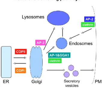
Eukaryotic cells require transport of lipids and proteins between extracellular and intracellular environments and transport of nutrients between organelles also occurs. This transport is accomplished by vesicle formation which bud from a donor compartment and merge with an acceptor compartment in the secretory and endocytic pathways (Chavrier et al. 1990). Coat proteins such as COPI, COPII and clathrin are necessary for membrane deformation on a specific donor compartment. Wherein, the coat protein COPII is necessary for formation of buds from the endoplasmic reticulum to the golgi apparatus. The coat protein COPI is necessary for formation of buds from cis-golgi for retrograde transport back to ER and the clathrin is necessary for formation of buds from plasma membrane and trans-Golgi network to the late endosomes (Figure 5) (Lodish et al. 2000b).
Figure 5. General model of vesicular transport pathways: Transport between organelles is
mediated by vesicles. Vesicle formation at various steps is driven by specific coat protein complexes (Take from Grant e Sato, 2006).
The binding of coat proteins to the membrane is regulated by small GTP-binding proteins called ADP-ribosylation factor (ARF) when is associated with COPI-coated vesicles and clathrin-coated vesicles. However, the small
GTP-binding proteins called Sar1 are associated with COPII-coated vesicles. The small GTP-binding proteins bind to membrane receptors. Only and when coat proteins are in the GTP-form they may lead to vesicle budding. Then the complexes of ARF-GTP/Sar1-GTP in the vesicle are hydrolyzed, leading to the conversion of ARF-GDP/Sar1-GDP form and the dissociation of coat proteins from the vesicle membrane (Figure 6). The non-coated vesicles are now a target for SNARES proteins. There are SNARES proteins that bind to non-coated vesicles called SNARES, and other that bind to target membrane called t-SNARES. The v-SNARES on the non-coated vesicle bind to the t-v-SNARES on the target membrane with the aid of Rab proteins, leading to membrane fusion (Gahan 2005).
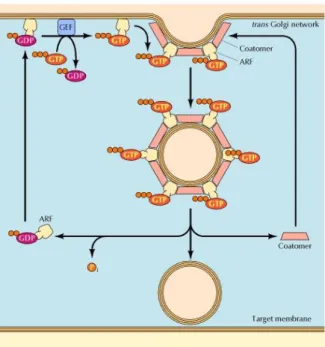
Figure 6. Role of ARF in the formation of COP-coated vesicles: ARF alternates between
GTP-bound and GDP-GTP-bound states. When GTP-bound to GDP, ARF associates with the membrane of trans Golgi network, where guanine nucleotide exchange factors (GEF) promote the exchange of the bound GDP for GTP. In its GTP-bound state, ARF promotes the binding of COPI coat protein (coatomer), leading to vesicle budding. Hydrolysis of the bound GTP then converts ARF to the GDP-bound state, leading to disassembly of the vesicle coat prior to fusion with the target membrane (Take from Gahan, 2005).
Rab GTPases belong to the largest branch of Ras superfamily of small GTPases that are necessary to regulate formation, transport, tethering and fusion of vesicles,. In Human cells were identified at least 63 different Rab family members and each Rab protein is localized in the membrane of a specific organelle (each organelle has at least one type of Rab protein) or in the cytosol (between 10 to 50% of Rab proteins can be detected in cytosol) (Figure 7) (Hutagalung & Novick 2011; Schimmoller et al. 1998; Zerial & McBride 2001). In cytosol Rab proteins are in an “inactive” form (GDP-bound) and a cytosolic protein called guanine nucleotide dissociation (GDI) bind to the Rab-GDP. Then, a membrane protein called GDI-displacement factor (GDF) displaces Rab from GDI and inserting Rab protein in the appropriate membrane. After insertion of Rab on the specific membrane, a protein called guanine nucleotide exchange factor (GEF) catalyzes the conversion of GDP-bound Rab to GTP-bound (Collins 2003). The Rab-GTP or “active” form is able now to interact with effector proteins located in that organelle and to regulate vesicle events, for instances, selecting the cargo, promoting vesicle movement and verifying the correct site of fusion (Pfeffer 2001). After, a GTPase accelerating protein (GAP) bind to the Rab-GTP form and catalyzes the hydrolysis of GTP-bound to GDP-bound. Thus, the GAP converts Rab back to its inactive form and is able now to interact with GDI that recruit Rab protein to the cytosol. This Rab protein can then undergo another cycle of GDP-GTP (Figure 8). The cycle between an active and an inactive form, GDP-GTP-bound and GDP-bound, regulates temporal and spacial membrane transport and the cycle is restricted to the membrane compartments where they are localized (Zerial & McBride 2001). For instance, Rab5 are localized in membrane of early endosomes and mediatefusion of clathrin-coated vesicles to form the early endosome. While Rab7 proteins are localized in the membrane of late endosomes and mediate transport between early to late endosome. The rate of vesicles fusion is controlled by absolute amount of Rab-GTP form. Thus, the cycle of some individual Rab proteins are essential for specific vesicles fusion and their interaction with V-SNARE proteins are critical to define which vesicle will be fuse with target membrane (Figure 9) (Lodish et al. 2008; Hutagalung & Novick 2011).
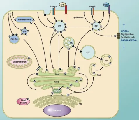
Figure 7. The intracellular localization and associated vesicle transport pathway(s) of several Rab GTPases in eukaryotic cell. Rab1 regulates ER-Golgi traffic while Rab2 is involved
in retrograde traffic from Golgi-ER. Rab6 regulates intra-Golgi traffic and several Rabs including Rab8, -10, and -14 regulate biosynthetic traffic from the trans-Golgi network (TGN) to the plasma membrane. Several Rabs are associated with endosomal traffic. Wherein, Rab5 regulated the formation of early endosome. Traffic can be directed towards the lysosome for degradation (regulated by Rab7) or to various recycling compartments to return factors to the plasma membrane (regulated by Rab15). Rab4 and Rab11 regulate fast and slow endocytic recycling, respectively. Specialized Rab functions include Rab18 controls the formation of lipid droplets. Rab33 together with Rab24 regulated the formation of autophagosomes. Rab21 and Rab25 regulate transport of integrins to control cell adhesion and cytokinesis. Rab13 regulates the assembly of tight junctions between epithelial cells. There are several Rabs that they function is poorly characterized or unclear. AP, autophagosome; ERGIC, ER-Golgi intermediate compartment; ER, endoplasmic reticulum; EE, early endosome; LD, lipid droplet; LE, late endosome; L/V, lysosome/vacuole; PAS, preautophagosomal structure; RE, recycling endosome; SV, secretory vesicle/granule (Take from Hutagalung e Novick, 2011).
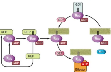
Figure 8. Activation cycle of small G proteins and their regulatory proteins. Rab proteins
include COOH-terminal prenylation signals. The Rab escort protein (REP) interacts with newly synthesized Rab and allows the Rab geranylgeranyl transferase (RabGGT) to add two geranylgeranyl lipid groups to the COOH terminus of Rab. Activation of Rab is mediated by a Guanine nucleotide exchange factor (GEF). GTP loading induces the translocation of Rab from the cytosol to the plasmatic membrane and permits the interaction with effectors. To “turn off” the cycle, a GTPase-activating protein (GAP) accelerates the intrinsic GTPase activity of Rab, allowing Rab to return to its inactive state in the cytosol. A guanine nucleotide-dissociation inhibitor (GDI) binds specifically to GDP-bound Rab, prolonging the inactive state and sequestering the GTPase in the cytosol. The insertion of Rab in the target membrane is mediated by a GDI dissociation factor (GDF) that releases the Rab from GDI (Take from Loirand, Sauzeau e Pacaud, 2013).
Figure 9. Hypothetical model for Rab-mediated vesicle docking. Transport vesicles carry Rabs
in their active, GTP-bound conformations. Active Rabs recruit specific docking factors from the cytosol. We propose that the docking factor·Rab complex can recognize protected t-SNAREs at the target membrane and in some way catalyze SNARE deprotection. In this manner, v- and t-SNARE pairing could take place (Take from Schimmoller, Simon e Pfeffer, 1998).
3.2.1 Rab7a protein
The rab7a protein belongs to the Rab family that regulates endosomal traffic. This protein is localized on the surface membrane of late endosomes and lysosomes, thus rab7a regulates the endosomal traffic between early endosome to the late endosome and late endosome to the lysosome (Bucci et al. 2000; Wu et al. 2005). This traffic is regulated by interaction of rab7a with various effectors such as Vps34/p150 PI3-Kinase complex, Rab interacting lysosomal protein (RILP), Rabring7 and proteasome (Dong et al. 2004; Mizuno et al. 2003; Stein et al. 2003). The interaction between Rab7a and a specific effector regulates a specific step. For instance, Rab7a GTP-bound form interacts with RILP protein to regulate the late endosome motility on microtubules to the lysosome (Cantalupo et al. 2001; Jordens et al. 2001). While, the interaction of Rab7a GTP-bound with hVPS34 via the p150 adapter protein activate PI3P formation, together, they regulate internal vesicle formation within multivesicular endosomes (late endosome) and the transport of epidermal growth factor receptor (EGFR) into intraluminal vesicles of MVB (Futter et al. 2001). However, the transport of EGFR into intraluminal vesicles is also regulated by recruitment of alpha subunit XAPC7 of 26S proteasome by Rab7a protein. Studies have shown that recruitment of XAPC7 is critical for the transport of cargo to the late endosome (such as the EGFR) and also its subsequent degradation (Mukherjee et al. 2005; Zafar et al. 2011; Dong et al. 2004).
3.2.2 Rab9 protein
The rab9 proteins are localized on the surface of late endosome and trans-Golgi network. Thus, Rab9 regulates endosomal traffic between late endosome and trans-Golgi network, retrograde transport. This traffic is regulated by interaction with several effectors such as TIP47, INPP5B, GCC185, PI3P PIKyve Kinase associated protein p40, NdeI, 14-3-3 protein theta and HPS4 (Hutagalung & Novick 2011). The interaction between Rab9 and a specific effector regulates a specific step. For instance, the Rab9 protein GTP-bound form interacts with TIP47 to recycling mannose-6-phosphate (MPRs) from late endosome to the trans Golgi network (Lombardi et al. 1993).
Studies have shown the impairment of Rab9 effect on late endosomal trafficking to trans Golgi network (Hanna et al. 2002).
4 Microtubules and Tau protein
The cytoskeleton is one of several elements that characterize the eukaryotic cell and consist in a network of fibrous elements. There are three types of fibrous elements, microtubules (MTs), microfilaments (MFs) and neurofilaments (NFs) that are found in the cytoplasm (Figure 10). The microtubules are responsible for the shape of the cell and by transport of vesicles inside the cell. They are constituted by heterodimers of α- and β-tubulin that align to form a hollow tube with an outer diameter of 25 nm. Proteins that bind along the sides of microtubules are collectively called microtubule-associated proteins, or MAPs. MAPs can stabilize microtubules against disassembly. Tau, which is much smaller than most other MAPs, is present in both axons and dendrites (Lodish et al. 2000a).
Figure 10. Microfilaments, microtubules and intermediate filaments in the nervous system.
Each cytoskeletal structure has a distinctive ultrastructure. This schematic illustrates the major features of the core fibrils. The microfilament consists of two strands of actin subunits twisted around each other like strings of pearls. The individual subunits are asymmetrical, globular proteins that give the microfilament its polarity. The microtubule is also made from globular subunits, but in this case the basic building block is a heterodimer of α- and β-tubulins. These αβ dimers are organized into linear strands, or protofilaments, with β-tubulin subunits oriented toward the plus end of the microtubule. Protofilaments form sheets in vitro that roll up into a cylinder with 13 protofilaments forming the wall of the microtubule. Assembly of both microfilaments and
microtubules is coupled to slow nucleotide hydrolysis, ATP for microfilaments and GTP for microtubules. The subunits of both glial filaments and neurofilaments are rod-shaped molecules that will self-assemble without nucleotides. The core filament structure is thought to be a ropelike arrangement of individual subunits. Glial filaments are typical type III intermediate filaments in that they form homopolymers without side arms. In contrast, neurofilaments are heteropolymers formed from three subunits, NFH, NFM and NFL for the high-, medium- and low-molecular-weight subunits. The NFH and NFM subunits have extended carboxy-terminal tails that project from the sides of the core filament and may be heavily phosphorylated (Take from Kirkpatrick e Brady, 1999).
Tau is a protein whose most important function is to stabilize microtubules. These proteins are found mostly in neuronal cells when compared to non-neuronal cells. They are localized along the axon and they stabilize the axonal microtubules and provide flexibility. The ability of Tau to cross-link microtubules into thick bundles may contribute to the stability of axonal microtubules (Lodish et al. 2000a). Tau has 6 different isoforms derived from alternative splicing of a Tau mRNA (Kirkpatrick & Brady 1999). Three isoforms have three binding domains and the other three have four. The binding domains are located in the carboxyl-terminus of the protein and are positively charged (allowing it to bind to the negatively charged microtubule). The isoforms with four binding domains are better stabilizers of microtubules than those with three binding domains. Phosphorylation Tau isoforms regulate the assembly and disassembly of microtubules. For instance, in AD Tau isolated from brain tissue is about 4 times more phosphorylated than in normal brains (Kopke et al. 1993; Ksiezak-Reding et al. 1992).
II. The Aims of the present study
The aims of the present study were undertaken to identify proteins interacting with PrPC that could provide new insights its physiological functions and
pathological role. And also the role of interacting partners of PrPC, Rab7a and
Rab9, in MM1 and VV2 subtype of CJD.
In the present project, the Identification of interacting partners of PrPC shall
be defined by co-localization of PrPC with Rab7a and Rab9 in neuronal cell models
and also in cortex, cerebellum and hippocampus of mice brain wild type (WT). The Identification of Intracellular levels of Rab7a and Rab9 in MM1 and VV2 shall be analysed in frontal cortex and cerebellum of human brain with sCJD-MM1 and sCJD-VV2 in comparison to the control group consisting of frontal cortex and cerebellum of human brain without neurodegenerative diseases.
In addition, analyse the intracellular levels of Rab7a and Rab9 in pre-symptomatic and pre-symptomatic stages of sCJD-MM1 shall be analysed in cortex and cerebellum from mice with sCJD-MM1, to investigate the intracellular levels of endosomal proteins during diseases progression.
III. Materials
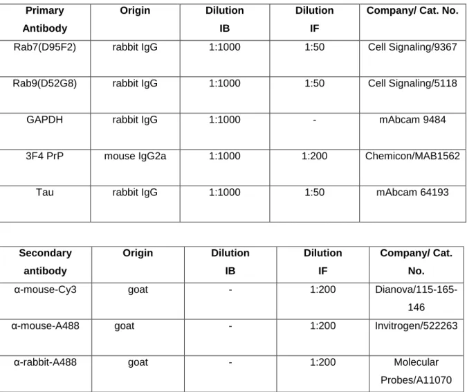
1 AntibodiesAntibodies used for immunoblotting (IB) and immunofluorescence (IF) are listed in Table 2.
Table 2 List of antibodies and their application in present study
Primary Antibody Origin Dilution IB Dilution IF
Company/ Cat. No.
Rab7(D95F2) rabbit IgG 1:1000 1:50 Cell Signaling/9367 Rab9(D52G8) rabbit IgG 1:1000 1:50 Cell Signaling/5118
GAPDH rabbit IgG 1:1000 - mAbcam 9484
3F4 PrP mouse IgG2a 1:1000 1:200 Chemicon/MAB1562
Tau rabbit IgG 1:1000 1:50 mAbcam 64193
Secondary antibody Origin Dilution IB Dilution IF Company/ Cat. No.
α-mouse-Cy3 goat - 1:200
Dianova/115-165-146
α-mouse-A488 goat - 1:200 Invitrogen/522263
α-rabbit-A488 goat - 1:200 Molecular
Probes/A11070
2 Antibiotics
Table 3 List of antibiotics
Antibiotics Company
Streptomycin Gibco
3 culture media
Table 4 List of culture media
Culture media Company
Agarose Biozym
DMEM Sigma-Aldrich
4 Chemicals
All chemicals used in this study were obtained from Amersham (Freiburg, Germany), Sigma and Fluka (Deisenhofen, Germany), Merck (Haar, Germany), Applichem (Darmstadt, Germany), Serva (Heidelberg, Germany), Roth (Karlsruhe, Germany) and BioRad (München, Germany).
5 Instruments and other materials
Table 5 List of the instruments used in this study
Appliance Model or Description Manufacture
Bio-safety Cabinet Hera safe KS Heraeus/ Osterode, Germany
Centrifuge 5415C Eppendorf/Hamburg,Germany
Centrifuge Rotina 35R Hettich/ Tuttlingen, Germany Chamber slide Lab-Tek™ II Chamber Slide,
15453
nunc/ New York, USA Culture dishes 60 mm, 351016 Becton Dickinson /NJ, USA Electro blotting apparatus Mini Trans-Blot® Bio-Rad /Munich, Germany Electrophoresis apparatus Mini-Protean® III Bio-Rad /Munich, Germany Electroporation cuvette 1mm, 748 011 Biozym/ Oldendorf, Germany
Freeze drier Alpha 1-4 LD SciQuip Ltd/ Shropshire, UK Heated magnetic stirrer iKAMAG RCT IKA-Labortechnik/ Staufen,
Germany
Ice machine - Ziegra /Isernhagen, Germany
Incubator IFE 400 Memmert/ Schwabach,
Germany Microscope Zeiss LSM 510 Meta Carl Zeiss/ Goettingen,
Germany
Power supply Power Pac 300 Bio-Rad /Munich, Germany Safe-Lock tubes 0.2, 0.5, 1.5 and 2ml Eppendorf /Hamburg,
Germany
Semi-Dry transfer Cell Transblot SD Bio-Rad/ Munchen, Germany Serological pipettes 2, 5, 10, 25ml Sarstedt /Germany
plastic tubes 15 and 50ml Sarstedt /Germany
pH meter pH 526 WTW/ Weilheim, Germany
Shakers CERTOMAT R Sartorius/ Goettingen,
Germany
Spectrophotometers EL808 Bioteck instruments/Winooski-vermont, USA Sterile filter Nalgene 0.2μm Sartorius/ Goettingen,
Germany
Sterile filter pipette tips - Biozym /Oldendorf, Germany
Thermomixer 5436 Eppendorf/ Hamburg,
German
Vortexer Genie 2™ Bender and Hobein /Zurich,
Switzerland
6 Kits
All the listed kits were used according to the manufacturer’s instructions.
Table 6 list of the kits used in this study
Name Company/ Cat. No. Application
Caspase-3 activity assay kit Promega/G7220 Apoptotic activity assay Lipofectamine 2000 Invitrogen Liposome-mediated
transfection
7 siRNA Duplex
Table 7 List of siRNA Duplex
siRNA Duplex Sequence (5’-3’) Accession/Cat. No.
siRNA-Rab7a CUGCUGCGUUCUCCUAUUU Operon
siRNA Negative control -
EUROGENTES/SR-CL000-005
siRNA-Tau CGUACACUGAGUUCGAGCU Operon
siRNA Negative control -
EUROGENTES/SR-CL000-005
8 Software
The list of scientific software used in the study is in table 8.
Table 8 List of scientific software
Program Use References
California, USA ImageJ 1.43u Densitomatric analysis National institutes of Health,
USA
ImageJ 1.43u WCIF Colocalization analysis National institutes of Health, USA
KC4 V3.4 Absorbance reader Bioteck instruments, USA LabImage 2.7.1 Densitomatric analysis Kapelan GmbH, Halle,
Germany Protein-Lynx-Global-Server v
2.1
LS MS/MS data analyzer
Micromass, Manchester, U.K Zeiss LSM 4.2.0.121 Immunofluorescence MicroImaging GmbH,
Goettingen, Germany
9 Stock solutions
Blocking solution for WB: 5% Milk Powder in TBS-T.
Cell lysis buffer I: 50 mM Tris-HCl pH 8.0, 0.5% CHAPS, 1mM EDTA, 1% triton x100.
Cell lysis buffer II: 7 M urea, 2 M thiourea, 4% CHAPS, 2% ampholytes, 1% DTT and a protease inhibitor mixture.
Electrophoresis buffer (SDS-running buffer): 192 mM glycine, 0.1% SDS, 25 mM Tris-HCl pH 8.3.
TBE: 42 mM Boric Acid, 10 mM EDTA, 50 mM Tris-HCl pH 8.0.
TBS-T: TBS and 0.1% of Tween-20.
TE: 0.01 M Tris-HCl, pH 7.4, 1 mM EDTA pH 8.0
Transblot buffer for PVDF membrane (semi dry): 192 mM Glycine, 10% methanol, 25 mM Tris-HCl pH 8.3.
IV Methods
1 Biological samples 1.1 Human brain
Frontal cortex and cerebellum samples from 30 pathologically confirmed sCJD patients (15 of each MM1 and VV2 subtypes) and 15 age-matched control cases (CON) were used in this study. All 45 samples were obtained from the Institute of Neuropathology Brain Bank (HUB-ICO-IDIBELL Biobank) and Biobank of Hospital Clinic-IDIBAPS. The mean age and gender of study cases were as described previously (Llorens et al. 2013). In brief, for sCJD western blot analysis in frontal cortex: 60 years of age in control (10M/5F), 68 sCJD MM1 (10M/5F), 63 sCJD VV2 (5M/10F). In cerebellum, the mean age was 62 in control (11M/4F), 66 sCJD MM1 (10M/5F) and 63 in sCJD VV2 (4M/10F). After post-mortem interval ranging 1h 45min. to 24h 30min., coronal sections 1cm thick were cut from one of the hemispheres. Along with selectively dissected areas of encephalon, coronal sections were rapidly frozen on metal plates over dry ice, sorted in separate bags, labeled with water-resistant ink and stored at -80oC until further use for
biochemical investigations. The other hemisphere was was immersed fixed in 4% buffered formalin for 3 weeks for morphological studies and neuropathological examination and characterization. The analysis of the codon 129 genotype of PrP gene (Met: M or Val: V) was performed after isolation of genomic DNA from blood samples according to standard methods. Western blot profile of PrPSc was
classified as type 1 or type 2 based on electrophoretic mobility after proteinase K (PK) digestion.
1.2 sCJD MM1 mice brain
Double transgenic mice overexpressing about 4-fold level of human PrPC
with methionine at codon 129 (Met129) on a murine PrP knock-out background were used, as described previously (Padilla et al. 2011). Inocula were prepared from sCJD MM1 brain tissues as 10% (w/v) homogenates. Individually identified 6-10 week-old mice were anaesthetised and inoculated with 2 mg of brain homogenate in the right parietal lobe using a 25-gauge disposable hypodermic needle (6 animals per group and time point). Mice were observed daily and the
neurological status was assessed weekly. When disease progression was evident, or at the end of lifespan, animals were euthanized, necropsy was performed, and the brain was removed. A part of the brain was fixed by immersion in 10% buffered formalin to quantify spongiform degeneration and perform immunohistological procedures. The other part was frozen at −80°C to extract protein. Survival time was calculated for each isolate and expressed as the mean of the survival day post-inoculation (DPI) of all mice scoring positive for PrPSc. Infection rate was
determined as the proportion of mice scoring positive for PrPSc from all inoculated
mice.
2 Immunoblot
Tissue lysis and immunoblotting were performed as described previously [Zafar et al., 2014]. Briefly, cells were lysed (50mM Tris-HCl, pH8, 1% Triton X-100, 0.5% CHAPS, 1mM DTT), and lysates were cleared of cell debris (1 min., 1000 xg, 4°C). Cell lysates were supplemented with protease and phosphatase inhibitors (Roche)and were separated on 12.5% 1-DE SDS-PAGE. The proteins from SDS-PAGE were transferred to a PVDF blotting membrane and were incubated with antibodies against Rab7a and Rab9 (D95F2 and D52G8, 1:1000, Cell Signalling). The primary antibody was detected by incubation of secondary antibodies of anti-rabbit in 5% milk (1:5000). Membranes were then rinsed in 1x TBS-T and incubated with the corresponding horseradish peroxidase-conjugated secondary antibody (diluted 1:2000/1:5000) for 1h at RT. Immunoreactivity was detected after immersion of the membranes into enhanced chemiluminescence (ECL) solution and exposure to ECL-Hyperfilm (Amersham Biosciences, Buckinghamshire, UK). Images were documented using the ScanMaker4 (Microtek, International), after correction for the background, and band intensities were determined by densitometry using Labimage (version 2.7.1, Kapelan GmbH, Germany) data analyzer software.
3 Immunofluorescence
Frozen cortex, cerebellum and hippocampus of wild type mice (see section III, 1.2) were embedded in cryomatrix and 5 µm thick sections were sliced using
cryostat (Leica cryostat 3050). These sections were mounted on histological glass slides, fixed in acetone for 1 minute, washed with methanol for 10 minutes and air dried. After, slides were hydrated in PBS for 10 minutes and incubated overnight with primary antibodies (anti-PrPc 3F4 (1:200) and rabbit anti-Rab7a (1:50)) diluted in PBS supplemented with 0.2% TritonX-100 at 4ºC overnight. After incubated overnight, we washed 3 times for 5 min. with PBS and incubated with secondary antibody for 1h at room temperature. The slides were washed with PBS (3x, 5 min.) and mounted with cover slip using anti fade mounting medium. Slides were visualized using a confocal laser scanning microscope.
4 Primary culture of mouse cortex
The wild type pregnant mice at embryonic day 14 under halothane (Sigma) anesthesia were sacrificed by cervical dislocation. The embryonic cerebral cortex were isolated and dissociated by mechanically dissociated and plated on polyethylenimine (1 mg/ml)-coated glass cover-slips in culture wells. For the brefeldin-A (BFA, Cell Signalling) treatment, cell cultures were first grown in Dulbecco's Modified Eagle's Medium (DMEM, Sigma-Aldrich), supplemented with 5% fetal bovine serum (FBS, Biochrom AG), 2.5% fetal calf serum (FCS, Eurobio), 2 mM glutamine, and 0.1% penicillin and streptomycin (Gibco), at a density of 7x10⁵ cell/cm2. After 3 days in vitro (DIV), the medium was replaced with N5
medium [Kawamoto & Barrett, 1986] with 180 mg/L glucose and supplemented with 5% FBS and 1% FCS. Then 3 μM cytosine arabinoside (Sigma, St Louis, MO, USA) was added to prevent astrocyte proliferation (resulting in at least 97% pure neuronal culture), and 1 μM MK-801 (Research Biochemicals International) to present excitotoxicity (Knusel et al. 1990). The medium was changed daily. On DIV 5, FCS was removed and the FBS content was reduced to 1%. Cells were cultured at 37C in humidified 5% CO2 atmosphere.
5 Liposome-mediated transient transfection
Transfection assay were performed using LIpofectamine 2000 (invitrogen) according to the manufacturer’s instructions. The primary culture of mice cortex (see section III, 4) were plated in 6-well plates at a cell density of 2-5 x 10⁵ per well
and maintained for 24h in the medium containing 10% FBS (about 70-90% confluent). After cell culture reaching approximately 70% confluency, 4 µg of each plasmid and 10µL of Lipofectamine 2000 were incubated separately in 250 µL of Opti-MEM I Reduced Serum Medium (Invitrogen). After 5 min. of incubation at room temperature, the diluted plasmids and Lipofectamine 2000 were combined and incubated for an additional 20 minutes at room temperature. The DNA-Lipofectamine 2000 complexes (5µg) were then added to each well, and the cells were incubated for 24h at 37°C in a CO2 incubator. After incubation, the
transfection medium was replaced with DMEM supplemented complete medium. Cells were collected from confluent cultures after 48h of transfection.
Small interfering RNA (siRNA) transfections were performed with 100nM siRNA using Lipofectamine 2000 (Invitrogen, Carlsbad, CA, USA) according to the manufacturer´s instructions. Rab7a siRNA duplex was a targeted with the sequence 5’-CUGCUGCGUUCUCCUAUUU-3’ and Tau siRNA with the sequence 5’-CGUACACUGAGUUCGAGCU-3’. The cells were also simultaneously transfected with Non-targeting siRNA duplex (control siRNA duplex negative control: Eurogentec) was used as a negative control. Transfected cells were cultured for 48h and were lysed (see section III, 6) for expression analysis and immunofluorescence (see section III, 3) for localization studies.
6 Cell lysis
After transfection of primary cell culture, the cells were washed with ice cold 1xPBS, scraped and centrifuged at 4ºC for 5 minutes at 400xg. The pallet was resuspended in ice cold 1x PBS and centrifuged at 4ºC for 5 min at 400xg. The washed cells were lysed in lysis buffer (50 mM Tris-HCL, pH 8, 1% Triton X-100, 0.5% CHAPS, 1mM DTT, protease and phosphatase inhibitor cocktail). Cells lysates were homogenized with ultra sonicator on ice and the lysates were centrifuged for 15 min. with 543,000xg at 4ºC. Protein concentration was estimated by Bradford essay (see section III, 7).
7 Protein concentration estimated by Bradford assay
The protein concentration was estimated by Bradford essay (Bio-Rad). Bradford reagent was prepared by diluting a 1x dye reagent (Bio-Rad) with dH2O
(1:5). To standard curve, we used different dilution of BSA in ddH2O (1000µg/L;
750µg/L; 500µg/L; 250µg/L; 100µg/L and 50µg/L) in a total volume of 20µg. Protein samples of unknown concentration were diluted in ddH2O (1:19). Protein
standards (to standard curve) and protein samples of unknown concentration were mixed with 980µl of Bradford reagent and incubated for 10 min. at room temperature. The absorbance of the samples was measured at 595 nm. The calculation of the protein concentration was done using Microsoft Office 2007 Excel software.
8 Immunofluorescence and quantification analysis
Primary cells culture (see section III, 4) and primary cell culture transfected with siRNA (see section III, 5) were plated on chambered slides (Lab-TEK II; Thermo Fisher Scientific (Nunc GmbH and Co. KG), Langenselbold Site) and fixed for 5 min. with 100% ethanol. After, the cells were permeabilised with 0.2% Triton X-100 in 1xPBS for 20 minutes. Colocalization of PrPc with Rab7a and Rab9 was
detected by applying the primary antibodies (anti-PrPc 3F4 (1:200), rabbit
anti-Rab7a (1:50) and rabbit anti-Rab9 (1:50)) for overnight at 4ºC. To Colocalization of Total-Tau with Rab7a was also detected by applying the primary antibodies (anti-Tau (1:200) and rabbit anti-Rab7a (1:50)) for over-night at 4ºC. The primary antibodies were detected by incubation the slides for 60 min. with secondary antibodies (Alexa 488 conjugated anti-rabbit (1:200) and Cy3-labeled anti-mouse secondary antibody (1:200)). Incubation with Hoechst 33342 or with TOPRO-3 iodide for 10 min. was performed to visualize nucleosomes. Finally, cover slips were placed on glass slides and mounted with Fluoromount (DAKO, Hamburg, Germany). After secondary antibody incubation all the steps were carried out in a dark humid chamber. The slides were kept dry dark at 4ºC until further microscopic evaluation.
We performed confocal laser scanning microscopy using a LSM 510 laser-scanning microscope (Zeiss, Göttingen, Germany; 488 nm Argon, 543 and 633 nm
Helium-Neon excitation wavelengths) according to the manufacturer´s instructions. Individual images of PrPc, Rab7a, Rab9 and Tau were analyzed separately by
ImageJ (WCIF plugin) software. For two-color analysis, stacks of images with a total thickness of approximately 30µm were acquired, using a dynamic range of 12 bits per pixel. Co-localization expressed as a correlation coefficient indicates the strength and direction of linear relationship between two fluorescence channels. Pearson’s linear correlation coefficient (rp) was used in this study to calculate
fluorescence channel correlation (Manders et al. 1992):
Where Ri and Gi are the red and green intensity measures, respectively.
Raver and Gaver are the average values of Ri and Gi, respectively.
The rp values can range from -1 to 1. An rp of -1 indicates a perfect negative
linear between variables, an rp of 0 indicates no linear relationship between
variables, an rp of >0 indicate a positive correlation and an rp of 1 indicates a
perfect positive linear relationship between variables. The values close to zero of the correlation coefficient are difficult to interpret when the degree of overlap is the quantity to be measure. Wherein, colocalization in the context of fluorescence microscopy were calculated according to published methodology (Manders et al. 1993):
Where Ri,coloc = Ri if Gi > 0 and Ri,coloc = 0 if Gi = 0, and where Gi,coloc = Gi if Ri > 0
and Gi,coloc = 0 if Ri = 0. The values of these coefficients, M1 and M2, ranges from
0 to 1. A value of 0 indicates that none of the signal within thresholds in that channel exists as co-localized with the other channel. A value of 1 indicates that the entire signal within thresholds in that channel exists as colocalized with the other channel. Two perfectly colocalized images will generate a scatter plot where the points fall in a line at 45° to either axis.
9 Statistical analysis
The statistical significance of the difference between control and sCJD VV1 and VV2 as also between control and siRNA in western blot was determined by student´s t-tests (GraphPad Prism 5 software). Pvalue less than 0.05 was considered statistically significant. To statistical significance of co-localization of PrPc and Rab7a and Rab9 in cell culture was determined by Pearson’s correlation
and M1 and M2 coefficients (Imagej (WCIF plugin) software.
10 Ethics Statement
Human samples from the Institute of Neuropathology Brain Bank (HUB-ICO-IDIBELL Biobank) and Biobank of Hospital Clinic-IDIBAPS were obtained following the Spanish legislation (Ley de la Investigación Biomédica 2013 and Real DecretoBiobancos, 2014) and the approval of the local ethics committees.
All animal experiments were performed in accordance with the ethical standard set by Regierungspräsidium Tübingen (Regional Council) Experimental No. FLI 231/07 file reference number 35/9185.81-2. All animal experiments have been performed in compliance with the institutional and French national guidelines, in accordance with the European Community Council Directive 86/609/EEC. The experimental protocol was approved by the INRA Toulouse/ENVT ethics committee.
IV Results
1 Intracellular levels of Rab7a and Rab9 proteins in frontal cortex and cerebellum of human brain
To know how endosomal pathway is affected in case of sCJD MM1 and VV2 disease, we analyzed the Rab7a and Rab9 proteins expression in frontal cortex of sCJDMM1 and VV2 patients and in cerebellum of sCJD MM1 and VV2 patients by western blots.
In frontal cortex, we observed a increased intracellular levels of Rab7a and a decreased intracellular levels of Rab9 in sCJD MM1 patients as compared to the control. However, patients with sCJD VV2 didn’t have any significance difference. In cerebellum, we observed an up-regulation of Rab7a and a down-regulation of Rab9 in sCJDMM1 and sCJDVV2 patients as compared to the control (Figure 11).
Figure 11. Intracellular levels of Rab7a and Rab9 proteins in frontal cortex and cerebellum human samples of sCJD MM1 and VV2 patients: Western blots of sCJD MM1(n=15), sCJD VV2
(n=15) and control (n=15) was performed separately. To observe the expression of Rab7a and Rab9 primary antibody anti-Rab7a and anti-Rab9 were used and the expression was measured by densitometry. (A) The Box Plot from western blot was used to represent the intracellular expression of Rab7 in the frontal cortex and cerebellum of sCJD MM1 and sCJD VV2 compared to the healthy age-matched control. sCJD MM1 and VV2 patients had a significant difference (P value < 0.01 (**) and P value < 0.001 (***)). (B) The Box Plot from western blot was used to represent expression of Rab9 protein in the cerebellum of sCJDMM1 and VV2 patients compared to the healthy age-matched control. Both, MM1 and VV2 subtypes of sCJD had a significant difference (P value < 0.001 (***)).
2 Intracellular levels of Rab7a and Rab9 in sCJD-Mice
To analyze the expressional regulation of Rab7a and Rab9 during disease progression, we performed immunoblotting in cortex and cerebellum from the mice with sCJD MM1 at symptomatic and symptomatic stages. In the pre-symptomatic stage, we observed low intracellular levels of Ran9 in the cortex and cerebellum and low intracellular levels of Rab7a in the cortex (figure 12A, B, C). However, we observed a high intracellular level of Rab7a in the cerebellum at pre-symptomatic stage (figure 12B).
In the symptomatic stage, we observed high intracellular levels of Rab9 in the cortex and cerebellum and high intracellular levels of Rab7a in the cortex (figure 12A, B, C). In the cerebellum, we observed low intracellular levels of Rab7a at symptomatic stage (figure 12B).
Figure 12. Intracellular levels of Rab7a and Rab9 in sCJD-Mice at pre-symptomatic and symptomatic stages: For characterization of sCJD MM1, all results of the intracellular levels of
Rab7a and Rab9 expression from mice´s cortex and cerebellum by western blot were combined and divided in two different stages of disease, pre-symptomatic and symptomatic stages. (A) Expression of Rab7a in the cortex, (B) Expression of Rab7a in the cerebellum (C) Expression of Rab9 in the cortex, (D) Expression of Rab9 in the cerebellum.
A B