Dual role of ATP on
neuromuscular
transmission in
Experimental
Autoimmune
Myasthenia Gravis
(EAMG)
Liliana Patrícia Maciel Almeida
Mestrado em Bioquímica
Departamento de Imuno-Fisiologia e Farmacologia (ICBAS-UP) Center for Drug Discovery and Innovative Medicines - MedInUP 2016
Orientador
Professora Doutora Laura Oliveira, Professora Auxiliar, ICBAS/UP
Coorientador
O Presidente do Júri,
Acknowledgments
It is utopian to think that one can achieve success alone. As such, by reading this section, I hope the reader can catch a glimpse of the appreciation I have for all the people who contributed to the conclusion of another milestone in my life, my Master Thesis.
My first acknowledgment is to Professor Laura Oliveira, my supervisor. For all the guidance, support and advice given to me, whether regarding the lab work, my professional life or my personal life. I am deeply thankful for your patience with me, your will to teach me everything you know and to push me to always give my best and also for all the confidence you placed on me. Thank you for being a mentor and, more than that, a friend in this journey. I will never forget all the enthusiasm, help and kindness you showed me through this year.
I would like to thank to Professor Paulo Correia-de-Sá, my co-supervisor, for his constant encouragement to pursue professional perfection, for always sharing his knowledge and expertise in order for me to grow as a scientist and for the help in the elaboration of written reports.
I also want to thank everyone in the group, PhD students Bruno Bragança, Isabel Silva and Mariana Certal, Doctor Cátia Vieira, Doctor Aurora Barbosa, Professor Ana Patrícia Sousa, Professor Margarida Duarte-Araújo, Professor Graça Lobo, Doctor Fátima Ferreirinha and Doctor Bernardo Noronha-Matos for welcoming me in the lab, for always being available to answer my questions. I thank you for the kindness, patience and the help you gave me.
Special thanks to Dra Teresa Magalhães-Cardoso, “HPLC master”. I will never forget all the help with the HPLC, all the knowledge and advice you shared with me. Thank you for the constant good-mood and motivation.
A big thank you also to the vivarium girls for all their assistance.
I would like to acknowledge Mrs. Belmira and Mrs. Helena for the overall help and guidance. Thank you for the funny lunch breaks and global good-mood you brought to the lab.
II FCUP/ICBAS
Acknowledgments
To Mafalda “Gonças”, Adriana “Drica”, Isabel “Escrava”, Ângela “Formigo” and Ana Filipa “Pandinha” thank you for the sharing of experiences, knowledge, advice and for welcoming me so well in the lab. Most of all, thank you for the laughs and good moments we shared. Although we’ve only known each other for a few months I feel like you gave me friends for life. A special thank to Isabel Calejo for helping me with HPLC analysis of the samples and for always being available to help me and teach me. Many thanks also to Ângela for putting up with me for 5 years now. I hope there are still many more to come “miga”.
A huge thanks to Carla “Bambi”, Diana “NeRD”, Patrícia “Tixa”, Daniela “Prazeres”, Augusto “Gusto”, Cristina “Tina”, Tânia “Anti”, Francisca “Chica” and Ana Cláudia “Tulinha” for the care, support, availability, advice and friendship. You have showed me that although miles apart, a friend can always be present in times of need.
I would also like to thank Inês Lopes and Filipa Lemos for the companionship you showed me all year long.
Thanks to all my family members, who contributed to this achievement for always having believed in me and for all the motivation through every phase.
To my brother, many thanks for all the pep talks and late night conversations that would always make me laugh even when I was not in the mood.
To my parents, who are my role-models in life. I am very grateful to have your support in every achievement in life. Thank you for always going above and beyond to provide me everything I need to pursue a career and a good life. I hope I can repay you everything you have done for me through the years and that I have made you proud.
To my boyfriend, Davide, without whom this year would have been a lot harder. I am lucky to have you as my safe-harbour. Thank you for all the love, patience and unconditional support you showed me throughout all these years and especially this last one. Thank you for always being available even if it was just to sit with me while I wrote this thesis or to listen to me while I talked for hours.
Finally to my grand-parents, my guardian-angels. Thank you for all the love and for all the valuable life-lessons you taught me while you could. Your looks of happiness
and pride with each of my accomplishments were my driving force to always do the best I could. The most honest and heartfelt thank you!
Brief-Note
Part of this work was presented at one scientific meeting as an oral communication – IJUP Meeting 2016 (9th Meeting of Young Researchers of U. Porto
(Porto, 2016):
Dual role of ATP on neuromuscular transmission in Experimental Autoimmune Myasthenia Gravis (EAMG)
L. Neves, L. Almeida, I. Silva, M.T. Magalhães-Cardoso, M.A. Timóteo, P. Correia-de-Sá and L. Oliveira
Resumo
Miastenia gravis é uma doença autoimune mediada por anticorpos e dependente de células T que se caracteriza por uma falha na contração neuromuscular. Em indivíduos miasténicos, a falha na contração é rapidamente atingida aquando de uma estimulação muscular contínua que, no caso dos músculos respiratórios pode ser fatal para o indivíduo. Além do tratamento crónico com imunossupressores, o agravamento da situação clínica é normalmente controlado com inibidores das colinesterases, cujo uso acarreta efeitos laterais significativos (revisto em Conti-Fine et al., 2006). Assim sendo, outra estratégia terapêutica valiosa pode passar pela modulação dos sistemas capazes de influenciar a libertação de acetilcolina (ACh) na junção neuromuscular (JNM), nomeadamente o sistema purinérgico.
Recentemente, o nosso grupo demonstrou que nos terminais nervosos motores de ratazanas com miastenia gravis autoimune induzida experimentalmente (EAMG) ocorre uma diminuição da atividade tónica facilitatória mediada pelos recetores pré-sináticos da adenosina A2A (A2AR) devido a uma diminuição da acumulação de
adenosina na fenda sináptica (Oliveira et al., 2015). O défice na acumulação extracelular de adenosina contrasta com a descoberta de que os animais EAMG libertam maiores quantidades de ATP após estimulação elétrica do nervo frénico (Neves, 2015, comunicação pessoal). Há muito se sabe que o ATP é co-libertado com a ACh a partir dos terminais nervosos motores (Silinsky, 1975) e que a sua ação pode ser direta, através da ativação de purinocetores P2 (Salgado et al., 2000), ou indireta, por intermédio de recetores A2AR após a sua conversão em adenosina na fenda sináptica
por uma cascata de ecto-nucleotidases (Correia-de-Sá and Ribeiro, 1996). Assim, o objetivo deste trabalho consistiu em avaliar se existia ou não algum défice na atividade das ecto-nucleotidases que pudesse explicar o paradoxo aparente entre os níveis elevados de ATP e a deficiente acumulação de adenosina na JNM de animais EAMG. Tendo em consideração a acumulação de nucleótidos de adenina na JNM dos animais miasténicos, avaliou-se o papel neuromodulador dos recetores P2 sobre a libertação de ACh em ratazanas EAMG.
A indução de miastenia gravis autoimune foi realizada em ratazanas adultas de acordo com um protocolo previamente estabelecido no Laboratório de Farmacologia e Neurobiologia do ICBAS (Oliveira et al., 2015). Após o seu isolamento, as preparações de hemidiafragma inervado de ratazana foram usadas para experiências em que se avaliou a libertação de [3H]-ACh induzida por estimulação elétrica do nervo frénico (750
pulsos de duração de 0.04 ms aplicados com a frequência de 5 Hz). Estas mesmas preparações foram usadas para estudos de cinética enzimática onde se avaliou o
VIII FCUP/ICBAS
Resumo
catabolismo extracelular de ATP e ADP e a formação dos seus metabolitos por cromatografia líquida de alta eficiência com deteção UV (HPLC-UV).
Os resultados mostram que a deficiente acumulação de adenosina na JNM de ratazanas EAMG não pode ser atribuída a quaisquer alterações no catabolismo extracelular do ATP (30 µM) e do ADP (30 µM) verificada nos animais miasténicos comparativamente com o grupo controlo. O tempo de semivida do ATP não foi significativamente (P>0.05) diferente entre animais EAMG (10 ±1 min, n=4) e controlo (injetados apenas com CFA) (10 ± 3 min, n=4). Estes tempos de semivida também não foram diferentes dos verificados em animais que não foram submetidos a qualquer tipo de tratamento (8 ± 2 min, n=8) (Magalhães-Cardoso et al., 2003). O tempo de semivida do ADP (30 µM) em animais EAMG (10±1 min, n=4) também não foi diferente dos animais controlo (10±1 min), nem tão pouco diferiu dos tempos de semivida calculados para o ATP nas mesmas condições experimentais. Estudos anteriores do grupo, demonstram que também não existe nenhuma deficiência na atividade da ecto-5’ nucleotidase que é a enzima responsável pela formação de adenosina a partir da hidrólise extracelular do AMP. Estes dados sugerem que o défice na acumulação de adenosina na JNM de animais EAMG após estimulação elétrica do nervo frénico não pode ser atribuído a alterações no metabolismo extracelular dos nucleótidos de adenina libertados.
O ATP (1 µM), aplicado por um curto período de tempo (3 minutos), reduziu a libertação de [3H]-ACh induzida por estimulação elétrica do nervo frénico em 26±10%
(n=4) e 20±6% (n=5) em preparações controlo e EAMG, respetivamente. O efeito inibitório do ATP não foi reproduzido pelo seu análogo estável, ,-imido ATP (100 µM), mas foi observado na presença de adenosina desaminase (ADA) a enzima responsável pela inativação extracelular da adenosina. Estes resultados mostram que o efeito inibitório do ATP se deve à sua pronta conversão extracelular em ADP pelas NTPDases existentes na fenda sináptica e, por conseguinte, à ativação de recetores P2 sensíveis ao ADP (P2Y1, P2Y12 e/ou P2Y13) existentes no terminal nervoso motor. O bloqueio
seletivo do recetor P2Y13 pelo MRS 2211 (10 µM) atenuou o efeito inibitório do ATP (1
µM) sobre a libertação de [3H]-ACh a partir de preparações de hemidiafragma inervado
de ratazanas controlo e EAMG. A estimulação seletiva do recetor P2Y1 com MRS 2365
(30 nM) produziu um efeito inibitório de maior magnitude que o ATP (1 µM) sobre a libertação de [3H]-ACh tanto em animais EAMG (31±4%, n=5) como em ratazanas
controlo (44±6%, n=4). Já o bloqueio seletivo deste subtipo de recetor (P2Y1) com MRS
2179 (300 nM) preveniu apenas o efeito inibitório do ATP (1 µM) em preparações isoladas de animais EAMG, mas não em ratazanas do grupo controlo. Aparentemente,
a ativação de recetores do subtipo P2Y12 parece contrariar parcialmente o efeito
inibitório do ATP (1 µM), já que o bloqueio seletivo destes recetores com AR-C 66096 (100 nM) potenciou significativamente a inibição da libertação de [3H]-ACh verificada
após a aplicação de ATP (1 µM) para 45±7% (n=4) em preparações de ratazanas EAMG.
Os resultados apresentados neste trabalho sugerem que o aumento da libertação de ATP na JNM dos animais miasténicos após estimulação elétrica do nervo frénico pode conduzir à ativação de recetores P2 sensíveis ao ADP, após a sua metabolização extracelular neste nucleotídeo pelas NTPDases existentes na fenda sináptica. Os resultados preliminares obtidos até à data sugerem o envolvimento de recetores de dois subtipos, P2Y1 e P2Y13, na inibição da libertação de [3H]-ACh após a
aplicação de ATP. A atividade inibitória destes dois recetores parece ser parcialmente contrariada pela co-ativação de recetores do subtipo P2Y12 nos animais miasténicos.
Especula-se ainda sobre os mecanismos moleculares responsáveis para interação entre os vários recetores sensíveis ao ADP responsáveis pelo controlo da libertação de ACh na JNM e qual a preponderância de cada um deles nos animais miasténicos. Apesar desta limitação, este trabalho permitiu descobrir novos alvos farmacológicos para prevenir a falência da transmissão neuromuscular em doentes miasténicos e, com isso, talvez propor novas estratégias para o tratamento desta patologia.
Abstract
Myasthenia Gravis (MG) is a T cell–dependent antibody-mediated autoimmune
disease characterized by neuromuscular contraction failure. In myasthenic individuals, contraction failure is easily achieved with continuous muscular stimulation which, in the case of respiratory muscles, could be fatal to the individual. Besides chronic immunosuppressive treatment, clinical exacerbations are usually controlled with cholinesterase inhibitors, which use has significant side effects (reviewed by Conti-Fine et al., 2006). As such, another viable therapeutic strategy might be to modulate the systems able to fine-tune control acetylcholine (ACh) release at the neuromuscular junction (NMJ), such as the purinergic system.
Recently, our group demonstrated that at motor nerve terminals of rats with experimental autoimmune Myasthenia Gravis (EAMG) there is a decrease of the facilitatory tonus mediated by presynaptic adenosine A2A receptors (A2AR) due to a
decreased accumulation of adenosine at the synaptic cleft (Oliveira et al., 2015). Deficits on adenosine accumulation contrast with the finding that EAMG animals accumulate higher amounts of ATP after electrical stimulation of the phrenic nerve (Neves, 2015, personal communication). ATP has long been known to be co-released with ACh from motor nerve terminals (Silinsky, 1975), and that its action may be exerted either directly, through the activation of P2 purinoceptors (Salgado et al., 2000), or indirectly, via A2AR
after its conversion into adenosine in the synaptic cleft by ectonucleotidases (Correia-de-Sá and Ribeiro, 1996). Therefore, we aimed at investigating if there was in fact a deficient activity of the ecto-nucleotidases responsible for the apparent paradox between increased amounts of ATP and decreased ADO accumulation at the NMJ of EAMG animals. Taking into consideration the accumulation of adenine nucleotides at the NMJ of myasthenic animals, the neuromodulatory role(s) of P2 receptors on ACh release in rats with EAMG, was also investigated.
The EAMG induction was proceeded in adult female rats as described by a previously established protocol at the Pharmacology and Neurobiology laboratory of ICBAS (Oliveira et al., 2015). After isolation, phrenic nerve hemidiaphragm preparations were used for experiments in order to evaluate [3H]-ACh release after electrical
stimulation of innervated hemidiaphragm with 5 Hz trains (750 pulses of 0.04 ms duration). Hemidiaphragm preparations were also used for enzyme kinetic experiments where the extracellular catabolism of ATP and ADP and the formation of the subsequent metabolites were evaluated by high performance liquid chromatography coupled to UV detection (HPLC-UV) system.
XII FCUP/ICBAS
Abstract
Data indicates that deficient accumulation of adenosine at motor endplates from EAMG rats cannot be attributed to changes in the kinetics of extracellular catabolism either of ATP (30 µM) nor ADP (30 µM) in myasthenic animals compared with the control group. The half-life of ATP (30 µM) was not significantly (P>0.05) different between EAMG (10 ± 1 min, n=4) and control animals (only injected with CFA) (10 ± 3 min, n=4). Neither was this kinetic parameter different from naïve animals (not submitted to any kind of treatment) (8 ± 2 min, n=8) (Magalhães-Cardoso et al., 2003). The half-life of ADP (30 µM) in EAMG animals (10 ± 1 min, n=4) was also not different from that calculated for CFA (10 ± 1 min, n=4) animals; The half-lives of ATP and ADP were also not different under the same experimental conditions. Previous studies from our group also demonstrated that there is no impairment of the activity of ecto-5’-nucleotidase that is the enzyme responsible for adenosine formation from the extracellular hydrolysis of AMP. Data suggest that the deficit in ADO accumulation at the NMJ of EAMG animals triggered by electrical stimulation cannot be attributed to changes in the extracellular metabolism of released adenine nucleotides.
Application of ATP (1 µM) during a short period of time (3 min) decreased electrically-evoked [3H]-ACh release from the phrenic nerve by 26 ± 10% (n=4) and 20 ±
6% (n=5) of control and EAMG preparations, respectively. The inhibitory effect of ATP was not reproduced by its stable analogue, adenosine 5’– imido) triphosphate (,-imido ATP, 100 µM), but it was still observed in the presence of adenosine deaminase (ADA), the enzyme responsible for the extracellular inactivation of adenosine. These results show that the inhibitory effect of ATP is most probably due to its extracellular conversion into ADP by the NTPDases present at the synaptic cleft and, consequently, to the activation of ADP-sensitive P2 receptors (P2Y1, P2Y12 and/or P2Y13) on
motoneurons. Selective blockage of P2Y13 receptor by MRS 2211 (10 µM) attenuated
the inhibitory effect of ATP (1 µM) on evoked [3H]-ACh relesae from innervated
hemidiaphragm preparations of EAMG and control rats. Selective stimulation of the P2Y1
receptor with MRS 2365 (30 nM) produced an inhibitory effect of a greater magnitude than ATP (1 µM) on the release of [3H]-ACh from both EAMG (31 ± 4%, n=5) and control
rats (44 ± 6%, n=4). The selective blockage of this receptor subtype (P2Y1) with MRS
2179 (300 nM) only prevented ATP (1 µM) transmitter release inhibition in EAMG animals preparations, but not in the control group.
Apparently, the activation of the P2Y12 receptor subtype seems to partially
counteract ATP (1 µM)-induced inhibition, since the selective blockage of these receptors with AR-C66096 (100 nM) significantly potentiated (to 45 ± 7%, n=4) the inhibitory action of ATP (1 µM) on [3H]-ACh release from EAMG animal preparations.
The results presented in this work suggest that the increased release of ATP at the NMJ of myasthenic animals after phrenic nerve electrical stimulation might lead to the activation of ADP-sensitive P2 receptors after its extracellular catabolism by NTPDases present at the synaptic cleft. Preliminary results obtained so far suggest the involvement of two receptor subtypes, P2Y1 and P2Y13, in the inhibition of [3H]-ACh
release after the application of ATP. The inhibitory activity of these two receptors seems to be partially counteracted by the co-activation of P2Y12 receptor subtype in myasthenic
animals. There is still speculation about the molecular mechanisms responsible for the interaction between the various ADP-sensitive receptors responsible for the control of ACh release at the NMJ and concerning the preponderance of each receptor subtype in myasthenic animals. Despite this limitation, this work allowed us to discover new pharmacological targets to prevent neuromuscular transmission failure in myasthenic patients, and with that, maybe, to propose new strategies for the treatment of this pathology.
Index
Brief-Note ... V Resumo ... VII Abstract ... XI Index ... XV List of Figures and Tables ... XVII List of abbreviations ... XIX
Introduction ... 1
1. Neuromuscular Junction ... 3
1.1 Presynaptic region ... 3
1.2 Postsynaptic region ... 5
1.3 ACh receptors ... 6
1.4 Neuromuscular Safety Factor ... 9
2. Myasthenia Gravis ... 10 2.1 Pathophysiology ... 10 2.2 Epidemiology ... 12 2.3 Clinical features ... 12 2.4 Diagnosis ... 13 2.5 Treatment ... 14
2.6 Animal models of Myasthenia Gravis ... 16
3. Purinergic signaling ... 18
3.1 ATP release ... 18
3.2 Metabolism of purines ... 19
3.3 Nucleoside transporters ... 22
3.4 Purinoceptor subtypes ... 23
3.4.1 P1 receptors (ADO receptors) ... 24
3.4.2 P2 receptors (ATP receptors) ... 25
3.5 Purinergic modulation of neuromuscular transmission ... 27
XVI FCUP/ICBAS
Index
Materials and Methods ... 35
1. Induction and Clinical assessment of EAMG ... 37
1.1 Evaluation of animal health conditions ... 37
2. Isolation of the phrenic nerve-hemidiaphragm ... 38
3. Kinetics of the extracellular catabolism of adenine nucleotides ... 39
4. Separation and quantification of adenine nucleotides and its metabolites by HPLC 41 4.1 Instruments ... 41
4.2 Separation process ... 41
4.3 Quantification Process ... 42
4.4 Half-life time (t1/2) determination ... 42
5. [3H]-ACh release experiments ... 43
5.1 Setting up the preparation in the organ bath ... 43
5.2 Experimental period ... 43 5.2.1 Equilibrium ... 44 5.2.2 Labeling ... 44 5.2.3 Washout ... 44 5.2.4 Release ... 44 5.2.5 Stimulation conditions ... 44
5.2.6 Released [3H]-ACh quantification ... 45
6. Drugs and Solutions ... 45
Results and Discussion ... 47
1. Kinetics of the extracellular catabolism of adenine nucleotides (ATP and ADP) in the diaphragm of control and EAMG rats. ... 49
2. Pharmacological characterization of the P2R modulating ACh release from control and myasthenic motor nerve endings ... 56
Conclusions and future work ... 63
List of Figures and Tables
Figure 1. Schematic representation of the transport, synthesis and degradative processes in a cholinergic presynaptic nerve terminal ... 4 Figure 2. Representative scheme of the NMJ. ... 5 Figure 3. Structural comparison between ionotropic and metabotropic receptors. ... 6 Figure 4. Schematic representation of the molecular organization of nAChRs in the membrane ... 8 Figure 5. Schematic representation of the muscle action potential. ... 9 Figure 6. Postsynaptic organization of muscle nAChR clusters. ... 11 Figure 7. Mechanisms of action of anti-nAChRs antibodies on the pathophysiology of MG.. ... 12 Figure 8. Schematic representations of the different EAMG induction modes. ... 16 Figure 9. The purinergic signaling cascade. ... 19 Figure 10. The graph shows the different properties of all eight members of the NTPDase family. ... 20 Figure 11. Illustrative image of all adenosine receptors and their intracellular coupling system. ... 25 Figure 12. Prototypic representation of P2XR and P2YR and their intracellular transduction pathways.. ... 26 Figure 13. Scheme of the anatomy of both left and phrenic nerves innervating the rat diaphragm. ... 39 Figure 14. Protocol used in the experiments to evaluate the kinetics of the extracellular catabolism of adenine nucleotides. ... 39 Figure 15. Phrenic nerve-hemidiaphragm preparation mounted in a horizontal organ bath where the enzymatic kinetic assays were performed. ... 40 Figure 16. RP-HPLC-UV chromatogram illustrating the separations ... 42 Figure 17. Illustration of the experimental protocol for measuring [3H]-ACh release. ... 43
Figure 18. Time-course of extracellular ATP (30 µM) catabolism in phrenic nerve hemidiaphragm preparations from control (CFA) and EAMG animals. ... 50 Figure 19. Time-course of extracellular ADP (30 µM) catabolism in phrenic nerve hemidiaphragm preparations from control (CFA) and EAMG animals. ... 52 Figure 20. Evaluation of ecto-NTPDase activity when ATP (A) and ADP (B) were used as substrates of the ectoNTPDase cascade in the rat phrenic nerve hemidiaphragm preparations from control (CFA) and EAMG animals.. ... 53 Figure 21. The effect of exogenously added ATP (1 µM) on evoked [3H]-ACh release
XVIII FCUP/ICBAS
List of Figures and Tables
[3H]-ACh release was electrically evoked twice (S1 and S2) by 750 rectangular pulses
applied with a 5Hz frequency. ... 57 Figure 22. The effect of exogenously added ATP (1 µM) in the presence or absence of P2Y13R selective antagonist, MRS 2211 (10 µM) on evoked [3H]-ACh from phrenic nerve
hemidiaphragm preparations from control (CFA) and EAMG animals [3H]-ACh release
was electrically evoked twice (S1 and S2) by 750 rectangular pulses applied with a 5Hz frequency. ... 58 Figure 23. The effect of selective blockade of P2Y12R activity with the antagonist, AR-C
66096 (100 nM), on the inhibitory action of ATP (1 µM) on evoked [3H]-ACh from EAMG
rat motor nerve terminals. ... 58 Figure 24. Evaluation of P2Y1 receptor contribution on the inhibitory action of ATP (1 µM) on evoked [3H]-ACh release from innervated hemidiaphragm preparations of control
(CFA) and EAMG rats. ... 59 Figure 25. Purinergic unbalance in the control of evoked neurotransmitter release at motor endplates of myasthenic rats (EAMG) and healthy controls (CFA).. ... 66
Table 1. Characteristics of the SLC-28 sodium coupled nucleoside transport family ... 22 Table 2. Characteristics of the SLC-29 nucleoside transport family ... 23 Table 3. Compilation of all purinergic receptors, its intracellular signaling mechanisms and principal endogenous ligands ... 24
List of abbreviations
[3H]-ACh Tritiated acetylcholine
[3H]-choline Tritiated choline
°C Centigrade degrees
µCi Microcurie
µL Microliter
µM Micromolar
ABC ATP binding cassette
Abs Antibodies
ACh Acetylcholine
AChE Acetylcholinesterase
ACR Apyrase conserved regions
ADA Adenosine deaminase
ADO Adenosine
ADP Adenosine diphosphate
AMP Adenosine monophosphate
ATP Adenosine triphosphate
Auto-Abs Auto-antibodies
cAMP Cyclic adenosine monophosphate
CAT Choline acetyl transferase
CFA Complete Freund’s adjuvant
CNS Central nervous system
CNT Concentrative nucleoside transporter
DPM Disintegrations per minute
XX FCUP/ICBAS
List of abbreviations
ENPP Ectonucleoside pyrophosphatase/phosphodiesterase
ENT Equilibrative nucleoside transporter
ENTPDase Ectonucleoside triphosphate diphosphohydrolase
EOM Extrinsic ocular muscles
EPP Endplate potential
g Gram
GDP Guanosine diphosphate
GPCR G-protein coupled receptor
GPI Glycophosphoinositol
GTP Guanosine triphosphate
h Hour
HX Hypoxanthine
Hz Hertz
IFA Incomplete Freund’s adjuvant
IMP Inosine monophosphate
INO Inosine
IP3 Inositol triphosphate
IVIg Intravenous immunoglobulin
kg Kilogram
LRP4 Low density lipoprotein receptor related protein
mA Milliampere
mAChRs Muscarinic acetylcholine receptors
MEPP Miniature endplate potential
MG Myasthenia Gravis
min Minute
mM Millimolar
mm Milimiter
MuSK Muscle specific kinase
nAChRs Nicotinic acetylcholine receptors
nM Nanomolar
NMJ Neuromuscular junction
nmol nanomol
P1R P1 receptors (Adenosine receptors)
P2R P2 receptors (ATP receptors)
PBS Phosphate buffered saline
PLC β Phospholipase β
PNP Purine nucleotide phosphorylase
RNS Repetitive nerve stimulation
SCID Severe combined immunodeficiency
SFMEG Single fiber electromyography
t1/2 Half-life time
TM Transmembrane
Tyr Tyrode
UDP Uridine diphosphate
UV Ultra-violet
VAChT Vesicular Acetylcholine Transporter
Chapter I
Introduction
2 FCUP/ICBAS
1. Neuromuscular Junction
The neuromuscular junction (NMJ) is a synapse specialized in the transmission of electrical impulses from the motor nerve terminal to the skeletal muscle, in order to generate an appropriate muscle contraction (reviewed by Fagerlund and Eriksson, 2009; Hughes et al., 2006).
The NMJ is formed by three major structural components that derive from different cell types: the presynaptic terminal originated from the motor neuron, the postsynaptic region of the skeletal muscle cell and the perisynaptic Schwann cell. All of these cells work together to ensure the success of neuromuscular transmission (reviewed by Fagerlund and Eriksson, 2009; Hughes et al., 2006).
Through the use of modern technologies, the NMJ structure has evolved from an apparent anatomical fusion of nerve and muscle, to the distinction of boundaries for muscle and nerve cells, creating the notion of a space between them called synapse. These findings suggest the existence of a soluble messenger molecule, which proved later to be acetylcholine (ACh) at least in vertebrate NMJs (reviewed by Bennet, 2000; Hughes et al., 2006).
1.1 Presynaptic region
The presynaptic terminal consists of the distal demyelinated part of the motor nerve axon, which has its cell body in the ventral horn of the spinal cord. This part of the nerve is encapsulated by a perisynaptic Schwann cell that anchors the nerve ending to the muscle membrane. These Schwann cells are reportedly not directly involved in the chemical transmission, but they have a key role in promoting motor neuronal survival (reviewed by Fagerlund and Eriksson, 2009; Hughes et al., 2006) and are responsible for synthesizing and releasing chemical neuromodulators (reviewed by Todd and Robitaille, 2006).
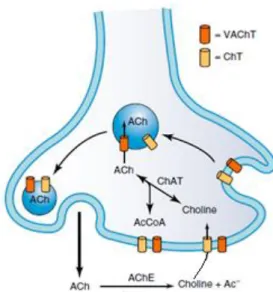
The synthesis and storage of ACh can be divided into three stages: uptake of choline into the nerve terminal resulting from the extracellular hydrolysis of released acetylcholine (ACh) by acetycholinesterase (AChE), its conversion into ACh by cytosolic choline acetyltransferase (CAT) catalyzing the transference of the acetyl group from mitochondrial acetyl coenzyme A, and packaging of ACh into synaptic vesicles through the action of the vesicular ACh transporter (VAChT) (Rang et al., 2016) (Figure 1).
4 FCUP/ICBAS
Introduction
Figure 1. Schematic representation of the transport, synthesis and degradative processes in a cholinergic presynaptic nerve terminal (adapted from Siegel et al., 2006).
Once synthetized in the cytosol of the nerve terminal, ACh accumulation in synaptic vesicles is driven by a proton-pumping ATPase called vesicular ACh transporter (VAChT) (reviewed by Eiden, 1998). These vesicles are then stored in a readily releasable pool near the active zone of release (Murthy and De Camilli, 2003; Sudhof, 2004). At this site, the amount of voltage–gated calcium channels is significantly higher compared to other regions of the presynaptic terminal (Augustine et al., 1991). As such, when an action potential reaches the nerve terminal, voltage-gated calcium channels of the P/Q type are activated, allowing high amounts of calcium ions to enter the presynaptic terminal. Calcium influx leads to the rise in the concentration of this ion near the channel pore to levels high enough to promote the fusion of synaptic vesicles containing the neurotransmitter with the presynaptic membrane (exocytosis phenomenon) releasing its content into the synaptic cleft (reviewed by Hirsch, 2007; Hughes et al., 2006). Calcium acts by neutralizing the negative charges of the presynaptic membrane allowing vesicles to approach the membrane; calcium ions also bind to specific proteins (e.g. synaptotagmin) to initiate vesicular membrane fusion through a conformational change in both vesicle and plasma membrane proteins (Sudhof, 2004). After its release, ACh has two possible fates: it can be hydrolyzed back into choline by acetylcholinesterase (AChE) or it can activate cholinergic receptors existing at the skeletal muscle fiber and at the presynaptic nerve terminal.
Under resting conditions (absence of a nerve action potential), miniature endplate potentials (MEPP) can be recorded at the NMJ as a result of the spontaneous release of a small number of ACh-containing vesicles, often termed as quantal release. When an action potential reaches the nerve terminal, several hundred vesicles are released synchronously resulting in an endplate potential (EPP) which, if large enough, is able to
evoke muscle depolarization and, subsequent, contraction (this issue will be further discussed at Post-Synaptic region chapter) (reviewed by Fagerlund and Eriksson, 2009). Acetylcholinesterase is anchored to the basal lamina (extracellular matrix that aids cell adhesion and neuromuscular signaling processes) (reviewed by Fagerlund and Eriksson, 2009) where it can catalyze the hydrolysis of ACh into choline and acetate. Originated choline can be then uptaken via high affinity-choline transporters (ChT) localized at the plasma membrane of the motor nerve terminal (Siegel et al., 2006). Choline uptake is the rate-limiting step in the biosynthesis of ACh, because treatment with hemicholinium-3 (a potent inhibitor of the high-affinity choline transporter) leads to a reduction in ACh release during prolonged nerve stimulation (reviewed by Eiden, 1998).
1.2 Postsynaptic region
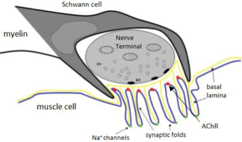
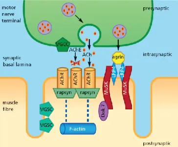
The role of the structures located on the post synaptic surface of the skeletal muscle fiber is to optimize the action of ACh to induce an EPP.
The mammalian post-synaptic membrane region can be morphologically recognized due to the presence of deep infoldings of the plasma membrane, which are called secondary synaptic folds. This feature contributes to enhance the neuromuscular transmission as it organizes ACh receptor clusters near the synapse (primary folds) and Na+ channels at the bottom of each synaptic fold (secondary folds) (reviewed by Hughes
et al., 2006) (Figure 2).
Figure 2. Representative scheme of the NMJ. The NMJ is a structure formed by three components: the perisynaptic Schwann cell, the presynaptic nerve terminal and the postsynaptic endplate region. At the presynaptic terminal, the synaptic vesicles (sv) are normally located at the active zones (az) of release. At the synapse there is a structure called basal lamina made up of laminin, agrin and collagen which provide help to neuromuscular transmission. Then, at the muscle cell region, there are multiple folds which can be identified as primary folds (shallower) where nAChRs are located (red dots) and secondary folds (deeper) where Na+ channels reside (green dots) (adapted from Hughes et al., 2006).
6 FCUP/ICBAS
Introduction
1.3 ACh receptors
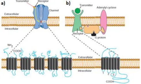
There are two types of ACh receptors: ionotropic nicotinic (nAChRs) and metabotropic muscarinic (mAChRs) receptors (Figure 3). The nicotinic receptors are found at neuromuscular junctions, autonomic ganglia and in several regions of the central nervous system (CNS), whereas muscarinic receptors were identified in the heart, smooth muscle fibers and exocrine glands, in addition to their presence in the CNS and autonomic ganglia (Rang et al., 2016).
Figure 3. Structural comparison between ionotropic and metabotropic receptors. a) Ionotropic receptors are composed by five transmembrane peptide subunits forming a pentameric pore; activation of this pore requires binding of 2 molecules of ACh to extracellular loops of two -subunits. b) Metabotropic receptors are G-protein-coupled receptors formed by a protein with seven transmembrane domains with only one binding site to ACh in the extracellular region; the third intracellular loop is essential for the interaction with a specific G-protein (adapted from Squire et al., 2008).
Like many other metabotropic receptors, the muscarinic receptor has seven transmembrane spanning domains, connected by peptide loops. The third intracellular loop has critical sequences that allow it to couple to a GTP binging protein (G-protein), explaining the GPCR designation (G-coupled protein receptors) (reviwed by Hulme et al., 1990). Once this receptor is activated, it couples to a G-protein initiating the exchange of GDP by GTP, activating the G-protein. Activated G-proteins then couple to many downstream effectors that will ultimately produce cellular effects (Squire et al., 2008). There are five subtypes of muscarinic receptors identified from M1 to M5. The
odd-numbered receptors (M1, M3) are coupled to Gq/11 proteins leading to activation of
phospholipase C-β (PLCβ) and the inositol trisphosphate (IP3) pathway. The
even-numbered receptors (M2, M4) are coupled to Gi/o proteins resulting in the inhibition of
adenylyl cyclase and in the reduction of intracellular cyclic adenosine monophosphate (cAMP) levels; these receptors may also couple to the opening of potassium channels
causing membrane hyperpolarization (Rang et al., 2016). At the NMJ, excitatory M1 and
inhibitory M2 receptors can act as presynaptic autoreceptors modulating the release of
ACh (Wessler, 1989). The balance between M1 excitation and M2 inhibition to control
neurotransmitter release at the rat motor endplate is highly dependent on the rate of neuronal activity and on the crosstalk with pre-synaptic adenosine A1 and A2A receptors
(Oliveira and Correia-de-Sá, 2005; Oliveira et al., 2002; 2009). The M1 positive feedback
control of ACh release predominates during low frequencies (e.g. 5Hz) of motor nerve stimulation. Under these conditions small amounts of adenosine are found at the neuromuscular synapse, which favors activation of inhibitory A1 receptors shutting-down
the inhibitory control of ACh operated by muscarinic M2 receptors. Upon increasing the
neuronal firing rate (to 50 Hz) adenosine accumulated at the neuromuscular junction reaches levels high enough to activate facilitatory A2A receptors, which contribute to
downmodulate M1 receptors activation favoring the negative control of the
neuromuscular transmission operated by the M2 receptor subtype (Oliveira et al., 2002).
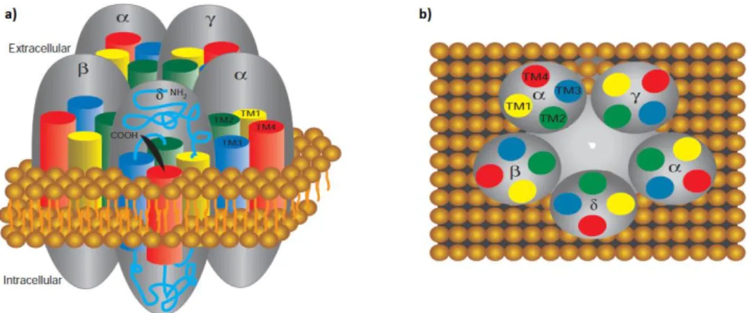
Ionotropic nicotinic ACh receptors are pentameric transmembrane protein complexes arranged around a pseudoaxis of symmetry forming an ion pore (Figure 4). At the neuromuscular junction, these receptors are present both pre- and post-synaptically and they mediate fast neurotransmission by ACh. Our group showed for the first time that the pre-synaptic nicotinic receptor responsible for ACh autofacilitation during high-frequencies of nerve stimulation contained α3β2 subunits (Faria et al., 2003). Fast desensitization of this receptor is also controlled by tonic adenosine A2A receptors
activation in order to prevent excess of transmitter release and nerve fiber damage during intense neuronal firing (Correia-de-Sá and Ribeiro, 1996; Timóteo et al., 2003).
The muscle-type nicotinic receptor is formed by two α subunits assembled together with one copy of , and δ subunits. Structural studies showed that the subunits are arranged around a central cavity that is believed to lead to the ion channel, which in the resting state is impermeable to ions; upon activation however, it opens selectively for cations. The sites for ligand binding are located toward the external perimeter of each ligand binding -subunit interface and to activate the receptor two ACh molecules are necessary.
8 FCUP/ICBAS
Introduction
Figure 4. Schematic representation of the molecular organization of nAChRs in the membrane. a) The amino and carboxy terminal extend in the extracellular space. The four membrane-spanning domains termed TM1-TM4 assume a α-helix structure to traverse the membrane. b) Top view of all five units highlighting the relative positions of all transmembrane domains, and the presence of two α subunits (adapted from Squire et al., 2008).
Binding of ACh to the nicotinic receptor triggers the influx of Na+ into the muscle
fiber to initiate depolarization of the muscle (Siegel et al., 2006). The postsynaptic total depolarization produced by ACh exocytosis triggered by a nerve action potential is the end-plate potential (EPP) (Hughes et al., 2006). This depolarization is perceived by voltage-sensitive Na+ channels (Na
v1.1), which allow the diffusion of Na+ down their
concentration gradient, spreading the depolarization phenomena throughout the muscle fibers, leading to calcium release within the muscle and the actin-myosin interaction that ultimately results in its contraction (reviewed by Conti-Fine et al., 2006; Fagerlund and Eriksson, 2009).
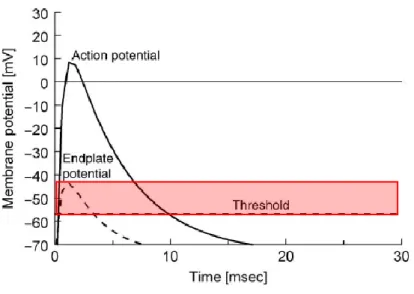
1.4 Neuromuscular Safety Factor
Neuromuscular transmission safety factor is defined as the ratio between the actual EPP and the threshold potential required to generate the muscle action potential (Figure 5). The quantal content of an impulse, the conduction properties, the architecture of synaptic folds, the density of post-synaptic nAChR and the activity of AChE in the synaptic cleft all contribute to the EPP. Normally, quantal ACh released to induce an EPP is greater than the threshold necessary to generate a muscular action potential, which accounts to a large safety factor. The current flow created after activation of nAChRs has its maximal depolarizing effect at the depths of the synaptic folds where there is a high density of voltage-gated Na+ channels. The opening of these channels
after the initial depolarization increases the effect of the transmitter release and reduces the threshold for action potential generation, thereby enhancing the safety factor. In the case of repetitive stimulation, neurotransmitter release decreases gradually in the course of a train thus reducing the amplitude of EPPs but, under normal conditions, this phenomenon is not enough to prevent action potentials generation (reviewed by Fagerlund and Eriksson, 2009; Hughes et al., 2006). However, neuromuscular transmission failure may occur in pathological conditions, such as Myasthenia Gravis, which will be further discussed in the following chapter.
Figure 5. Schematic representation of the muscle action potential. The stippled line indicates the shape of the EPP, as the full line indicates the shape of the action potential. The firing threshold of the muscle is indicated by the horizontal dashed line. The EPPs capable of surpassing the threshold potential are able to generate a muscle action potential. The safety factor is represented by the red colored rectangle and it represents the magnitude of the EPP needed to trigger muscle contraction.
10 FCUP/ICBAS
Introduction
2. Myasthenia Gravis
Myasthenia Gravis (MG) is the most common primary disorder of neuromuscular
transmission (reviewed by Sathasivam, 2014; Turner, 2007). The major characteristic feature of MG is painless, fatigable weakness (reviewed by Vincent et al., 2001). These symptoms are the result of autoantibodies (auto-Abs) attack to proteins of the post-synaptic region of NMJ which are involved in the neurotransmission phenomenon (reviewed by Berrih-Aknin et al., 2014). Among all the myasthenic patients, 85% of them are seropositive for muscle nAChR and 5% have autoreactive antibodies directed against a protein called muscle specific kinase (MuSK), which plays a central role in the clustering of nAChRs at the NMJ (reviewed by Berrih-Aknin et al., 2014; Hoch et al., 2001). Recently, the agrin receptor, low-density lipoprotein receptor-related protein 4 (LRP4), a protein that forms a complex with MuSK helping AChRs clustering, has been identified as a novel autoantigenic target (Higuchi et al., 2011; Pevzner et al., 2012) (Figure 6).
The development of those antibodies is apparently caused by the breakdown of self-tolerance in the thymus (Newsom-Davis et al., 1981) with activation of AChR-specific CD4+ T helper cells and production of pro-inflammatory cytokines, leading to the synthesis of high affinity Abs (Vincent, 2002). MG is considered a prototypical antibody-mediated autoimmune disease because it has the characteristics that fulfill all the strict criteria (reviewed by Conti-Fine et al., 2006; Vrolix et al., 2010):
a. Auto-antibodies of the blood plasma are found at the site of pathology, the NMJ in this case (Engel et al., 1977);
b. These antibodies are capable of causing the same symptoms when injected in rodents (Toyka et al., 1977);
c. Immunization of animals with muscle nAChR reproduces the disease (Patrick and Lindstrom, 1973);
d. Therapies based on the removal of blood Abs (plasma exchange) attenuates the symptoms (Newsom-Davis et al., 1978);
2.1 Pathophysiology
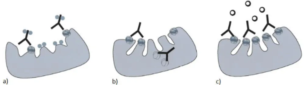
There are at least three mechanisms by which anti-nAChR antibodies may affect neuromuscular transmission (reviewed by Vrolix et al., 2010) (Figure 7):
a. Binding and activation of complement at the NMJ, which leads to focal lysis of the post-synaptic folds by the membrane attack complex (Engel et al., 1977; van der Neut et al., 2007);
b. Antigenic modulation: Abs bind and crosslink the nAChRs, inducing the endocytosis of the complex and further degradation of the nAChRs by the muscle cell (Heinemann et al., 1977; van der Neut et al., 2007);
c. Functional nAChR interference by blocking ACh binding (Almon et al., 1974).
Figure 6. Postsynaptic organization of muscle nAChR clusters. MuSK is present predominantly at the NMJ, where it is part of the receptor for agrin. Agrin is a protein synthesized by motor neurons and secreted into the basal lamina. Binding of this protein to LRP4 activates MuSK, which will trigger downstream transduction pathways culminating in the coclustering of AChRs with rapsyn. Rapsyn, is a peripheral membrane protein that attaches AChRs to the contractile F-actin protein, leading to dense AChR clustering at the primary synaptic folds.(Ruegg and Bixby, 1998; adapted from Sathasivam, 2014).
These mechanisms are, therefore, responsible for the decrease in the amount of nAChRs and for the loss of functional and anatomical structure of the NMJ. As a consequence, the EPPs induced present a lower magnitude than the threshold potential for activating the voltage-gated calcium channels responsible for the development of the muscle action potential. This is particularly evident with repetitive muscular activity since in this condition the quantal content of neurotransmitter release decreases as a function of time. In a normal situation, the neuromuscular safety factor is enough to surpass this decrease. In myasthenics, the decrease of EPP associated to the changes in the architecture of the NMJ leads to an increased probability of neuromuscular failures (reviewed by Gomez et al., 2010).
12 FCUP/ICBAS
Introduction
Figure 7. Mechanisms of action of anti-nAChRs antibodies on the pathophysiology of MG. a) Abs towards AChRs that bind complement result in destruction of the muscle endplate and a reduction in the number of available AChRs. b) Anti AChR Abs cross-link adjacent AChRs increasing their rate of internalization into muscle. c) Antibodies block the binding site for ACh at the postsynaptic nicotinic receptor (adapted from Nicolle, 2002).
2.2 Epidemiology
The incidence rate of this disease varies between 1.7 and 21.3 per million inhabitants depending on the location of the study (Carr et al., 2016). From epidemiological studies, we learned that the incidence rate of MG has a bimodal distribution, which suggests a hormonal or environmental influence on the disease onset. MG affects both sexes, at all ages and in all races (reviewed by Turner, 2007). Before the age of 50, studies show that early-onset MG is characterized by female predominance (60-70%) (Carr et al., 2016). On the other hand, between the ages of 50 and 60, there is no gender difference. After 60 years old, a very late onset MG appears and is known to be constituted mostly of male patients (Oger and Alkhawajah, 2013). Juvenile MG is variable among different races. It is uncommon in Europe and North America (10-15% of total Caucasian MG patients) and is much more frequent in Asia (50% of MG patients in China) (reviewed by Berrih-Aknin et al., 2014)
In adulthood, acquired autoimmune MG is the commonest form (reviewed by Turner, 2007). On the other hand, transient neonatal MG is caused by passive transfer of maternal Ab, especially the Anti-AChR antibodies (Béhin et al., 2008).
2.3 Clinical features
In about 2/3 of myasthenic patients, the initial symptoms are ptosis and diplopia which reflects that the extrinsic ocular muscles (EOM) and the levator palpebrae muscles are the first to show muscular weakness. The symptoms then progress in a craniocaudal direction to the facial and bulbar muscles causing reduced facial expression and speech, chewing and swallowing difficulties. Finally limb muscles are affected resulting in generalized MG (reviewed by Conti-Fine et al., 2006; Sathasivam, 2014). Weakness of the intercostal muscles and diaphragm can cause dyspnea (shortness of breath), which can evolve to severe respiratory failure. Patients with these symptoms should be
monitored for forced vital capacity and for blood gas because intubation and mechanical ventilation may be needed (reviewed by Turner, 2007).
Between different muscles, the susceptibility to MG is not the same. EOMs are particularly susceptible to show myasthenic weakness because the NMJ have less synaptic folds and, therefore, fewer postsynaptic AChRs and Na+ channels, which results
in a decreased safety factor. Also, they are more often subjected to a high frequency of neuronal firing, making them more susceptible to fatigue. Taken together, these characteristics explain why the first symptom is ocular muscle weakness. In skeletal muscles, the NMJs of the fast-twitch fibers release higher amounts of ACh, have more postsynaptic folding, and higher postsynaptic sensitivity to ACh than slow twitch fibers. These properties make fast twitch skeletal muscle fibers less prone to myasthenic failure tan slow-twitch fibers (reviewed by Conti-Fine et al., 2006).
2.4 Diagnosis
A complete medical and neurological evaluation is necessary to diagnose MG. Several number of tests may be used to establish a diagnosis of MG:
a) Tensilon test: this test consists of an intravenous injection of edrophonium, a short-acting AChE inhibitor that transiently increases the amount of ACh available, improving weakness caused by an impairment of the neuromuscular transmission. This test is sensitive in diagnosing a defect on neuromuscular transmission, but it is not specific for MG, neither it is recommended in elderly patients or patients with cardiac disease and/or pulmonary disease (reviewed by Juel and Massey, 2007).
b) Antibody tests: these tests evaluate the presence of anti-AChRs Abs in the patient’s blood. If these are not detected, anti-MuSK antibodies should be tested. The detection of serum AChR antibodies is highly specific for MG but its sensitivity is low in the presence of ocular symptoms only. Also, the absolute titer of Abs does not correlate with differences in disease severity.
c) Neurophysiology tests: these electrodiagnosis tests can be performed in two exams: repetitive nerve stimulation (RNS) and single fiber electromyography (SFMEG) (reviewed by Nicolle, 2002; Sathasivam, 2014). In RNS, the amplitude of the compound muscular action potential induced by repetitive nerve stimulation with frequencies of 3-10 Hz is measured and MG is diagnosed if a decrease in the compound muscular action potential is observed. Although it is technically easy and sensitive with the generalized disease, it is non-specific leading to false-positives (Keesey, 1989). SFEMG is the most sensitive diagnostic test and it
14 FCUP/ICBAS
Introduction
should be performed if RNS is normal and a NMJ disorder is suspected. It consists in an electromyography where action potentials of individual muscle fibers are recorded due to its 25 µm diameter recording surface (Selvan, 2011). Despite its high sensitivity, abnormalities detected are not specific for MG (Oh et al., 1992)
d) Ice pack test: An ice cube is placed over the drooping eyelid for about two minutes and, if there is ptosis improvement, a neuromuscular transmission disorder is suggested (Czaplinski et al., 2003).
2.5 Treatment
There are generally four options regarding treatment of MG which have different time courses:
a) Acetylcholinesterase inhibitors: These compounds are the first-line treatment in MG patients. They slow the hydrolysis of ACh at the NMJ, increasing the amount of ACh, which allows patients to recover from tetanic blockade and to improve muscular strength (Drachman, 1994). Pyridostigmine is the most commonly used drug, except for anti-MuSK MG patients which are insensitive to this treatment (Hatanaka et al., 2005). This is simply a symptomatic treatment, meaning that it does not retard the underlying immune attack. This treatment may have side effects due to increased muscarinic activity and a cholinergic crisis may develop if there is an excessive dosing in patients with severe MG, worsening muscle weakness (reviewed by Juel and Massey, 2007; Trouth et al., 2012).
b) Immunomodulation treatments: These treatments are used for crisis intervention, and they are seldom used for prolonged therapy (reviewed by Sieb, 2014). Two treatments belong to this category: Plasmapheresis, which consists in the direct removal of Abs against AChR from circulation (Batocchi et al., 2000) and intravenous immunoglobulin therapy (IVIg) which involves the isolation of immunoglobulins from human plasma and administration for 5 days. It’s mechanism of action is complex but it involves inhibition of complement deposition, interference with antigen recognition by sensitized Tcells and others (Samuelsson et al., 2001). These two are equal first-line treatments, however, IVIg is easier to administer and associated with less adverse events which makes it a more popular choice among physicians.(reviewed by Sathasivam, 2014). c) Chronic immunosuppression: Corticosteroids, like prednisolone are the
immunosuppressive agents most frequently used for the treatment of MG and the most effective. These may be used at low doses for years or, at high doses for
months. Anti-AChR Abs levels reduce in the first months of therapy and most patients report to have clinical benefit (reviewed by Conti-Fine et al., 2006).
i. Azathioprine is the non-steroidal immunosuppressant most frequently used. It is a purine analog that reduces nucleic acid synthesis, interfering with T and B cell proliferation (reviewed by Conti-Fine et al., 2006; Trouth et al., 2012). It usually takes up to 15 months to evaluate a clinical response, which is why it is normally used in combination with prednisone. (Palace et al., 1998).
ii. Mycophenolate mofetil blocks purine synthesis, suppressing both T and B cell proliferation and it might be used if treatment with azathioprine is not tolerated (reviewed by Trouth et al., 2012)
iii. Cyclophosphamide interferes with DNA synthesis and may preferentially suppress B lymphocytes (reviewed by Nicolle, 2002).
iv. Cyclosporine blocks the synthesis of IL-2 cytokine receptor and other key-proteins to the function of CD4+ T cells. This is the mainly used treatment in patients who do not respond/tolerate to azathioprine (reviewed by Trouth et al., 2012)
MG patients resistant to therapy have been successfully treated with cyclophosphamide in combination with bone-marrow transplant or with rituximab, a monoclonal antibody against the B-cell surface marker CD20 (reviewed by Trouth et al., 2012).
d) Surgical treatment: In early onset MG, thymic follicular hyperplasia is frequently found in women and in late onset MG, thymoma is frequently present (reviewed by Berrih-Aknin et al., 2014). As such, thymectomy is strongly recommended for patients with thymoma, because its efficacy in other situations has been questioned for lack of solid controlled prospective studies (reviewed by Trouth et al., 2012). Thymectomy may not be a viable therapeutic strategy in anti-MuSK MG because they lack the thymic characteristics that anti-AChR MG have, also suggesting different pathologic mechanisms for both forms of MG.
MG treatment should be individualized according to patient characteristics and severity of disease using the array of treatment options available. When the treatment is well fit, the majority of the patients can have largely normal lives. Even complete stable remission can be achieved in some patients (Baggi et al., 2013). Unfortunately, treatment is less effective in anti-MuSK, with a significantly increased risk of myasthenic crisis (Deymeer et al., 2007). Nonetheless, these treatments are hardly proven by controlled studies. It is largely based on serendipity and retrospective studies (reviewed by Juel
16 FCUP/ICBAS
Introduction
and Massey, 2007; Sieb, 2014) which emphasizes the need for new therapeutic strategies.
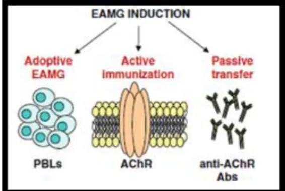
2.6 Animal models of Myasthenia Gravis
The development of animal models is crucial to the study of any disease because it allows the investigator to better understand the characteristics of the disease. Thirty years ago, Lindstrom and Patrick reported for the first time that rabbits immunized with AChRs purified from the electric organ of Electrophorus electricus develop MG-like symptoms (Patrick and Lindstrom, 1973).
Although there are other animal species suitable for experimental autoimmune
Myasthenia Gravis (EAMG) induction, rats are the most often used model (65%), with
mice accounting for 35% of the cases. Despite the fact that mice were expected to represent the most suitable model for EAMG induction, due to availability of mutant strains and to access to a huge variety of monoclonal antibodies specific for cell markers, these animals are less susceptible to EAMG induction due to a higher safety factor (high quantal amount of released ACh from the nerves) making it difficult to detect neuromuscular transmission failure than in rats (reviewed by Baggi et al., 2012). From the different rat strains, Lewis rats exhibit intermediate susceptibility, but the clinical manifestations in this strain are similar to those in human MG (Biesecker and Koffler, 1988). As such, rats have been the animal species most often used to induce EAMG, which can be achieved by active immunization, passive transfer or adoptive transfer (Figure 8):
Figure 8. Schematic representations of the different EAMG induction modes.
a) Active immunization: It is either performed by a single immunization with purified AChR in complete Freund’s adjuvant (CFA) (Lindstrom, 1980) or by immunization with a synthetic peptide which corresponds to the aminoacidic region between positions 97-116 of rat-AChR α subunit, made up in CFA followed by a second
immunization boost of the peptide in incomplete Freund’s adjuvant (IFA), 30 days after the beginning of the immunization protocol (Baggi et al., 2004). CFA and IFA are both constituted essentially by paraffin oil containing mannide mono-oleate as a surfactant. In addition, CFA contains heat-killed mycobacteria (Mycobacterium tuberculosis, in our case). These adjuvants generally act by prolonging the lifetime of injected autoantigen, by stimulating its effective delivery to the immune system, and by providing a complex set of signals to the innate compartment of the immune system (reviewed by Billiau and Matthys, 2001). The last one is the induction protocol used in this work since the anti-AChR Abs produced derive from the breakdown of self-tolerance, almost mimicking an autoimmune disease (Baggi et al., 2004).
b) Passive transfer: this induction method is basically the passive transfer of auto-Abs via daily injections of serum IgG isolated from MG patients or anti-AChR antibodies from donor animals with chronic EAMG. Also, monoclonal Abs directed to the α subunit of AChR derived from immunized animals or from cell line culture supernatants (reviewed by Baggi et al., 2012).
c) Adoptive transfer: this method is based in the transplantation of human tissues or cells in severe combined immunodeficiency (SCID) mice, lacking mature B and T cells (Schönbeck et al., 1992).
MG and EAMG have several similar characteristics: muscle weakness, fatigability, decreased response upon repetitive nerve stimulation, improvement of muscular strength following treatment with anti-cholinesterase drugs. The immunopathological features are also quite similar, such as the presence of anti-AChR Abs in serum, deposition of complement components at the NMJ, and others.
Despite their similarities, EAMG and human MG seem to have one big difference. In EAMG animals, the auto-sensitization process seems to occur only in lymph nodes apparently not affecting the thymus, yet in human MG thymic alterations are frequent (reviewed by Baggi et al., 2012; Christadoss et al., 2000).
18 FCUP/ICBAS
Introduction
3. Purinergic signaling
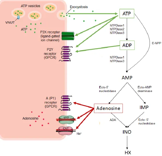
More than 30 years ago, scientists discovered that adenosine triphosphate (ATP) was a constituent of cholinergic synaptic vesicles (Dowdall et al., 1974). A year later, Eugene M Silinsky found that ATP was coreleased with ACh at the NMJ of rat phrenic nerve-hemidiaphragm upon nerve stimulation (Silinsky, 1975; Silinsky and Hubbard, 1973). These findings opened new avenues on purinergic signaling roles in the peripheral nervous system (PNS), since they make ATP a likely neurotransmitter, co-transmitter or presynaptic modulator in cholinergic (as well as many other) synapses.
Besides motor nerve endings, purines can also be released from skeletal muscle fibers (Santos et al., 2003) and Schwann cells (Liu et al., 2005; Shin et al., 2012). It is estimated that about 60% of ATP released at the motor endplate is derived from activated skeletal muscle fibers, as determined in the rat hemidiaphragm using -bungarotoxin as paralyzing agent (Santos et al., 2003). Different release sites seldom correspond to distinctive release modes. As a matter of fact, ATP can be released together with ACh by vesicle exocytosis, but the nucleotide can also be translocated to the extracellular space secondary to cell lysis and by facilitated diffusion through nucleotide transporters or electrodifusional transport through specific release channels, namely pannexin-1 hemichannels (reviewed by Yegutkin, 2008). Once in the extracellular milieu, ATP can act either directly, via P2 purinoceptors (P2R) (Salgado et al., 2000), or indirectly, via adenosine (ADO) receptors (P1 receptors, P1R) after its extracellular catabolism by ectonucleotidases (Cunha et al., 1996).
3.1 ATP release
Since ATP is a highly polarized molecule, it cannot pass freely the cell membrane. As such, ATP outflow to the extracellular space may occur by a series of mechanisms:
a) Vesicular exocytosis: As previously stated, ATP is able to be co-released with ACh from cholinergic nerve terminals. Like ACh, ATP is taken up and stored in synaptic vesicles, which can be later on released in a Ca2+-dependent manner.
b) Carrier-mediated release: Although this kind of mechanism is yet to be molecularly identified in the nervous system, specific transporters such as ATP binding cassettes (ABC) are able to translocate ATP across the plasma membrane (reviewed by Sperlagh et al., 2007; Wang et al., 1996).
c) Channels and pores: Connexin and pannexin hemichannels are also potential candidates to drive ATP across the plasma membrane in non-neuronal cells (reviewed by Sperlagh et al., 2007).
d) Cellular lysis: ATP can be released in huge amounts after cell damage or whenever the integrity of the plasma membrane is compromised (reviewed by Yegutkin, 2008).
Figure 9. The purinergic signaling cascade. ATP and adenosine release sites, metabolic enzymes and P1/P2 purinoceptors. Green arrows represent interactions with adenine nucleotides, ATP or ADP; Red arrows represent interactions with adenosine.
3.2 Metabolism of purines
The extracellular accumulation of nucleotides is regulated by several groups of ectonucleotidases located on the cell surface; some of these membrane-bound enzymes break its GPI anchor and become soluble in the interstitial medium or within body fluids. Ectonucleotidases contribute to the extracellular hydrolysis of nucleotides for which they have substrate specificity (reviewed by Robson et al., 2006; Zimmermann, 2001):
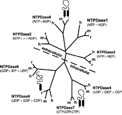
a) The E-NTPDase (ectonucleoside triphosphate diphosphohydrolase) family hydrolyzes nucleoside 5’-tri- and di-phosphates with different characteristics. They share five highly conserved sequence domains called “apyrase conserved regions” (ACRs) which are of major
20 FCUP/ICBAS
Introduction
importance for their catalytic activity. Maximal catalytic activity requires the presence of divalent cations, like calcium or magnesium, and an alkaline pH. The Km values for ATP of the purified enzymes are in the
lower micromolar range. There are 8 different NTPD genes (Figure 9). Regarding to their location, NTPDases1, 2, 3 and 8 are typical cell-surface enzymes with the catalytic site facing the extracellular medium (true E-NTPDases), NTPDases 5 and 6 are intracellularly located but after heterologous expression, they undergo secretion, and NTPDases 4 and 7 are entirely intracellularly located facing the lumen of cytoplasmic organelles such has the Golgi apparatus and lysosomal/autophagic vacuoles.
Figure 10. The graph shows the different properties of all eight members of the NTPDase family. In terms of location, there is a difference between surface-located (top) and intracellular (bottom) NTPDases. In addition, the major substrate preferences of individual subtypes and the predicted membrane topography for each group of enzymes is given (one or two transmembrane domains, indicated by barrels) (reviewed by Robson et al., 2006).
There is a considerable difference in substrate specificity between E-NTPDases: NTPDase1 (or CD39, apyrase) hydrolyzes ATP and ADP almost equally well; NTPDase2 has a 30-fold preference for the hydrolysis of ATP over ADP; NTPDase3 and 8 are functional intermediates and hydrolyze ATP approximately three times better than ADP.
b) The E-NPP (ectonucleotide pyrophophatase/phosphodiesterase) family, which comprises NPP1 to NPP3, have alkaline phosphodiesterase and