Arq Neuropsiquiatr 2008;66(1):5-7
5
Demyelinating Disease in patients
with myasthenia gravis
Denis Bernardi Bichuetti
1,2, Tatiane Martins de Barros
1,2, Enedina Maria Lobato Oliveira
1,3,5,
Marcelo Annes
1,3, Alberto Alain Gabbai
1,4Abstract – Myasthenia gravis (MG) is an autoimmune disease characterized by fluctuating muscle weakness, caused by impaired neuromuscular transmission. Patients with MG can present other autoimmune diseases in association, commonly hypo or hyperthyroidism. The association of MG to demyelinating disease is rare and has been described before. We report on three Brazilian patients with MG that presented distinct demyelinating diseases, two monophasic and one recurrent neuromyelitis optica, several years after the diagnosis of MG, and discuss their clinical courses.
Key Words: myasthenia gravis, demyelinating disease, association.
Doenças desmielinizantes em pacientes com miastenia gravis
Resumo – Miastenia gravis (MG) é doença autoimune caracterizada por episódios de fraqueza muscular alternados com melhora, causada por bloqueio da junção neuromuscular. Pacientes com MG podem apresentar outras doenças autoimunes, comumente hipo ou hipertiroidismo, e a associação de MG com doenças desmielinizantes é raramente descrita. relatamos três pacientes brasileiros com MG que desenvolveram doenças desmielinizantes, dois monofásicos e um neuromielite óptica recorrente, vários anos após o diagnóstico de MG e discutimos seus cursos clínicos.
PAlAvrAs-chAve: miastenia gravis, doenças desmielinizante, associação.
Federal University of são Paulo, são Paulo sP, Brazil: 1Neurology and Neurosurgery department; 2Post-Graduate student of Neurology; 3Assistent
Phisi-cian; 4Full Professor, discipline of Neurology; 5Neurologist consultant for Bayer schering Pharma do Brasil.
received 3 August 2007, received in inal form 14 November 2007. Accepted 10 december 2007.
Dr. Denis Bernardi Bichuetti – Rua Pedro de Toledo 377 - 04039-000 São Paulo SP - Brasil. E-mail: denisbichuetti@globo.com
Myasthenia gravis (MG) is an autoimmune disease characterized by fluctuating muscle weakness, caused by impaired neuromuscular transmission. Autoantibod-ies speciic for the human nicotinic acetylcholine recep-tor are present in 70 to 80% of the patients, and the re-maining cases can be associated with antibodies targeted at muscle speciic kinase (MusK) and other proteins in the post-synaptic membrane1,2. MG has a bimodal incidence: (1) 20 to 40 years-old, predominated by women, and (2) 60 to 80 years-old, predominated by men2.
Patients with MG commonly have thyroid disease and can present with nonspeciic immune system abnormal-ities, such as positive auto antibodies and alopecia area-ta3-5. rarely, MG can be part of multiple autoimmune syn-dromes, including autoimmune diabetes mellitus, throm-botic thrombocytopenic purpura, sjögren syndrome, sys-temic lupus erythematosus, vitiligo, among other rare dis-eases5. In the past 20 years there has been an increasing number of reports on patients with MG presenting de-myelinating diseases (dd)6-15, including multiple sclerosis,
neuromyelitis optica (NMo), transverse myelitis and op-tic neuritis. however, it is not known whether this associ-ation is also part of unspeciic immune activassoci-ation, genet-ic susceptibility or if it just happens by random.
We herein describe three Brazilian patients with MG that presented different dd and discuss their clinical courses.
methoD
Arq Neuropsiquiatr 2008;66(1)
6
Myasthenia gravis; demyelinating disease Bichuetti et al.
results
We found three patients with MG that developed dd during follow-up; their clinical, electrophysiological and autoantibodies status are described in Tables 1 and 2. Briely, patient 1 was diagnosed with generalized MG at the age of 26. Anti-thyroid antibodies were present (anti-thyroid peroxidase and anti-tireoglobulin), but she nev-er developed thyroid disease symptoms. she undnev-erwent thymectomy one year later and evolved asymptomatic on pyridostigmine treatment, thymus pathology disclosed lymphoid hyperplasia. At age 32, she presented mild trun-cal and gait ataxia associated to bilateral horizontal nys-tagmus. Brain magnetic resonance imaging (MrI) disclosed pons and cerebellum FlAIr and T2 hyperintense signal and mild gadolinium enhancement compatible with de-myelination (Figure), cerebro-spinal luid was normal. she was treated with 3g Iv methylprednisolone and symptoms resolved completely. MrI follow-up images disclosed res-olution of the preview lesion and no new T2 or FlAIr
ab-normalities. she is currently on pyridostigmine for symp-tomatic myasthenia control.
Patient 2 had intermittent diplopia since the age of 27 and was further diagnosed with ocular myasthenia. he was initially treated with prednisone and pyridostigmine with mild response to treatment. Prednisone was switched to azathioprine due to hypertension, but symptoms only re-solved when cyclosporine was started. At age 45 he devel-oped pain, low visual acuity and ptosis in the left eye. Fun-doscopic examination revealed a swollen disc on the left eye. MrI of the brain disclosed small unspeciic periven-tricular and subcortical white matter changes on T2 and FlAIr sequences. cyclosporine dose was raised and de-lazacort introduced for symptomatic treatment of pto-sis. symptoms resolved completely and his visual acuity returned to normal 2 months later.
Patient 3 was diagnosed with generalized MG at the age of 27 and underwent thymectomy one year after di-agnosis. Four years later she presented with left side par-esthesias and her spinal cord MrI disclosed a demyelin-ating lesion from c5 to c7, absent cerebrospinal luid oli-goclonal bands and normal brain MrI. symptoms resolved after 3g Iv methylprednisolone. Two months later she de-veloped bilateral acute visual loss that resolved within 30 days. on irst evaluation she had an atrophic optic disc on the left side, mild spastic gait and increased muscle stretch relexes. A clinical diagnosis of neuromyelitis op-tica was made based on the Wingerchuck criteria16 and she was started on azathioprine plus prednisone. during the next 2 years she presented with 1 myelitis and 2 optic neuritis relapses, treated with Iv methylprednisolone. An-cillary test revealed an anti-nuclear-antibody (ANA) titer of 1/320. she recently became pregnant and stopped her medication, but her neurological exam discloses only mild
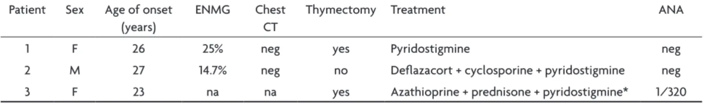
Table 1. Myasthenia gravis clinical data.
Patient sex Age of onset (years)
eNMG chest cT
Thymectomy Treatment ANA
1 F 26 25% neg yes Pyridostigmine neg 2 M 27 14.7% neg no delazacort + cyclosporine + pyridostigmine neg 3 F 23 na na yes Azathioprine + prednisone + pyridostigmine* 1/320
na, not available; neg, negative; ANA, antinuclear antibodies; eNMG, electroneuromyography; *patient 3 is currently under no medication due to pregnancy.
Table 2. Demyelinating disease clinical data.
Patient Age of onset (years)
Time from MG to dd (years)
Presentation Follow-up MrI
1 32 6 Ataxia, vertigo and INo resoled with 3g methylprednisolone Brainstem 2 45 18 oN resolved spontaneously Unspeciic 3 27 14 Myelitis evolved to recurrent NMo Myelitis c5-c7
MG, myasthenia gravis; dd, demyelinating disease; INo, internuclear ophtalmoplegia; NMo, neuromyelitis optica; oN, optic neuritis.
Arq Neuropsiquiatr 2008;66(1)
7
Myasthenia gravis; demyelinating disease Bichuetti et al.
low visual acuity without fatigue. data from her thymec-tomy was unavailable from the other hospital.
Discussion
We report on a series of Brazilian patients that pre-sented distinct dd 6-18 years after the diagnosis of MG. Two of them presented a monophasic course (patients 1 and 2), one evolved to recurrent neuromyelitis optica (pa-tient 3). The occurrence of dd in association to MG have been reported before, and are described as monophasic events (myelitis, acute disseminated encephalomyelitis and optic neuritis)10-13 and recurrent diseases (multiple sclerosis, recurrent transverse myelitis and NMo)7,9-12,14. some authors state that this association may not happen by chance, as the incidence of dd in patients with MG is much higher than expected in the general population6,15, and both may be part of multiple autoimmune syndromes or genetic predisposition to autoimmunity5.
recently, two reports have focused on this association. Gotkine et al.10 reported on 5 patients that presented dd after the diagnosis of MG, three had a monophasic event and two had a recurrent illness. The authors suggest that the association might be caused by subclinical systemic lupus erythematosus in three of them due to the presence of antinuclear antibodies, including both cases with recur-rent dd, although this has been contested due to the fact that two of their patients could actually have NMo16-18. Kister et al.9 report on 4 patients that presented recurrent NMo years after undergoing thymectomy for MG. Two of these patients were positive for NMo-Ig antibodies in their serum18 and ANA and anti-GAd were also present in 3 patients, disclosing systemic immune abnormalities. In both series, as in other cases reported before7,12-15 patients developed dd years after undergoing thymectomy, and the authors suggest that thymectomy might have induced immune dysregulation. Indeed, a long-term study of thy-mectomized patients showed that a signiicant number of patients presented different autoantibodies in their serum years after thymectomy, 43% developed ANA positivity, 12.5% of them developed autoimmune diseases, and more than 60% had at least 1 expansion within the cd8 and cd4 T-cell repertoire, compared with non-thymectomized pa-tients and healthy control subjects19.
Two of our patients have undergone thymectomy and only one presented positive ANA (patient 3); interestingly, this was the only one that evolved to a recurrent dd. This patient resembles those reported by Kister et al.9, who suggested that unspeciic immune dysregulation, either due to thymectomy or genetic susceptibility might inlu-ence the development of a second autoimmune disease. Although all our patients achieved a good control of my-asthenia symptoms, we could not determine the inluence
of developing dd on MG control due to the small num-ber of patients in this series. Furthermore, in our service only 3 out of 630 patients with MG developed dd (0,5%), much less than the rate observed by Gotkine et al.10. While we evaluated 630 ambulatory patients, Gotkine et al sur-veyed 214 patients that were admitted to the hospital, which does not relect the whole population of patients with MG in a tertiary care center. This methodological difference may have left to selection bias of more severe patients, among them those with dd.
In conclusion, demyelinating diseases are rare among patients with MG and may be part of an autoimmune syndrome spectra or genetic predisposition to autoim-munity. In this series of patients, only one developed a relapsing-remitting disease with positive low titer ANA and the others presented a monophasic course. As this is a small retrospective series, it was not possible to de-termine whether dd was related to thymectomy and its potential impact on myasthenia gravis clinical control.
references
1. Vincent A, Palace J, Hilton-Jones D. Myasthenia gravis. Lancet 2001; 357:2122-2128.
2. Romi F, Gilhus NE, Aarli JA. Myasthenia gravis: clinical, immunolog-ical, and therapeutic advances. Acta Neurol Scand 2005;111:134-141.
3. Kiessling WR, Plughaupt KW, Ricker K, Haubitz I, Mertens HG. Thy
-roid function and circulating antithy-roid antibodies in myasthenia gra -vis. Neurology 1981;31:771-774.
4. Suzuki S, Shimoda M, Kawamura M, et al. Myasthenia gravis accom
-panied by alopecia areata: clinical and immunogenetic aspects. Eur J
Neurol 2005;12:566-570.
5. Meyer O. [Immunogenetics in the understanding of multiple autoim
-mune syndromes]. Ann Med Interne (Paris) 1988;139:155-158. 6. Keesey JC. Does myasthenia gravis affect the brain? J Neurol Sci 1999;
170:77-89.
7. Furukawa Y, Yoshikawa H, Yachie A, Yamada M. Neuromyelitis opti
-ca associated with myasthenia gravis: characteristic phenotype in Jap -anese population. Eur J Neurol 2006;13:655-658.
8. Achari AN, Trontelj JV, Campos DJ. Multiple sclerosis and myasthe
-nia gravis: a case report with single iber electromyography. Neurolo -gy 1976;26:544-546.
9. Kister I, Gulati S, Boz C, et al. Neuromyelitis optica in patients with myas
-thenia gravis who underwent thymectomy. Arch Neurol 2006;63:851-856. 10. Gotkine M, Fellig Y, Abramsky O. Occurrence of CNS demyelinating
disease in patients with myasthenia gravis. Neurology 2006;67:881-883.
11. Aita JF, Snyder DH, Reichl W. Myasthenia gravis and multiple
sclero-sis: an unusual combination of diseases. Neurology 1974;24:72-75. 12. Lindsey JW, Albers GW, Steinman L. Recurrent transverse myelitis, my
-asthenia gravis, and autoantibodies. Ann Neurol 1992;32:407-409.
13. Goldman M, Herode A, Borenstein S, Zanen A. Optic neuritis, trans
-verse myelitis, and anti-DNA antibodies nine years after thymectomy
for myasthenia gravis. Arthritis Rheum 1984;27:701-703.
14. Mapelli G, De Palma P, Franco F, Fini M. Myasthenia gravis and
re-current retrobulbar optic neuritis: an unusual combination of diseases.
Ophthalmologica 1986;192:234-237.
15. Isbister CM, Mackenzie PJ, Anderson D, Wade NK, Oger J. Co-occur
-rence of multiple sclerosis and myasthenia gravis in British Columbia.
Mult Scler 2003;9:550-553.
16. Ikeda K, Araki Y, Iwasaki Y. Occurrence of CNS demyelinating disease in patients with myasthenia gravis. Neurology 2007;68:1326. 17. Kister I, Herbert J, Swerdlow ML, Bergamaschi R, Piccolo G, Oger J.
Occurrence of CNS demyelinating disease in patients with myasthe
-nia gravis. Neurology 2007;68:1326-1327.
18. Weinshenker BG, Jacob A. Occurrence of CNS demyelinating disease in patients with myasthenia gravis. Neurology 2007;68:1326.
19. Gerli R, Paganelli R, Cossarizza A, et al. Long-term immunologic ef