INSTITUTO DE INVESTIGAÇÃO E FORMAÇÃO AVANÇADA ÉVORA, JULHO 2019 ORIENTADOR (A/ES):Ana Teresa Caldeira João Paulo Prates Ramalho António Manuel Deométrio Rodrigues Lourenço Pereira
Tese apresentada à Universidade de Évora para obtenção do Grau de Doutor em Bioquímica
Su Yin Ooi
LABELLING OF PROTEINACEOUS
3 INSTITUTO DE INVESTIGAÇÃO E FORMAÇÃO AVANÇADA
President of the jury
Name António José Estevão Grande Candeias
Email [email protected] Department Chemistry department
Professional category Associate Professor with Aggregation
Vowels
Name Maria do Rosário Caeiro Martins (Member)
Email [email protected] Department Chemistry department
Professional category Assistant professor
Name Alfredo Jorge Palace Carvalho (Member)
Email [email protected] Department Chemistry department
Professional category Assistant professor
Name Rui Miguel Azevedo Bordalo (Member)
Email [email protected]
Department Catholic University of Porto
Professional category Investigator
Name Luís Miguel Santos Loura (Member)
Email [email protected] Department Coimbra University
Professional category Associate Professor
Name Paula Cristina de Sério Branco (Member)
Email [email protected]
Department New University of Lisbon - Faculty of Science and Technology
Professional category Assistant Professor w / aggregation
Name João Paulo Cristovão Almeida Prates Ramalho (Advisor)
Email [email protected] Department Chemistry department
4 INSTITUTO DE INVESTIGAÇÃO E FORMAÇÃO AVANÇADA
Dedicated to my beloved family Para a minha familia
5 INSTITUTO DE INVESTIGAÇÃO E FORMAÇÃO AVANÇADA
ABSTRACT
Easel paintings are important Cultural Heritage assets with significant historic and cultural value. They usually possess a multi-tiered structure, composed of different layers, some of which may present protein binders. Proteins have been commonly used as paintings medium, adhesives and coating layers in easel paintings. Hence, their recognition is a crucial step for easel painting’s conservation and restoration processes. The present work presents a novel fluorescent labelling methodology, using a coumarin derivative chromophore, C392STP (sodium (E/Z)-4-(4-(2-(6,7-dimethoxycoumarin-3-yl)vinyl)benzoyl)-2,3,5,6-tetrafluorobenzenesulfonate) as a fluorophore probe to bond proteinaceous binders used in paintings. The method was developed and optimized using commercial proteins and proteins extracted from hen’s egg yolk and white, bovine milk, and rabbit skin. In order to mimic the real conditions, paint models of easel paintings have been prepared by mixing proteins such as ovalbumin, casein and rabbit glue with different pigments (lead white, chrome yellow and black bone) and the fluorescent labelling method was miniaturized and tested. The results revealed that proteins in concentration as low as 6.0 μg/ml could be detected.
Finally, for validation methodology, real micro samples of easel paintings were analyzed. The extracted proteins were submitted to the fluorescent labelling method developed and clearly identified in electrophoretic profiles. The results evidence the applicability of this methodology as an effective and useful analytical tool for the identification of protein binders obtained from easel paintings and, possibly in other art work.
Additionally, theoretical quantum chemical calculations based on the Density Functional Theory (DFT) and Time Dependent Density Functional Theory (TD-DFT) have been performed in the C392STP coumarin and in a related coumarin derivative ((E/Z)-4-(2-(6,7- dimethoxycoumarin-3-yl)vinyl)-N-propylbenzamide), that mimics the coumarin bonded to lysine. The calculations confirm the experimental trends in absorption wavelengths and are in good agreement with the experimental absorption spectra, providing a comprehensive characterization of the main spectral features of the studied compounds.
6 INSTITUTO DE INVESTIGAÇÃO E FORMAÇÃO AVANÇADA
KEYWORDS
7 INSTITUTO DE INVESTIGAÇÃO E FORMAÇÃO AVANÇADA
RESUMO
Identificação de ligantes proteicos em arte
As pinturas de cavalete são um componente importante do Património Cultural, com um significativo valor histórico e cultural. Geralmente possuem uma estrutura composta por diferentes camadas, algumas das quais podem apresentar ligantes proteicos. As proteínas surgem geralmente em pinturas de cavalete como meio de suporte da pintura, adesivos e camadas de revestimento. A sua identificação é, portanto, um passo crucial para os processos de conservação e restauração da pintura de cavalete. O presente trabalho apresenta uma nova metodologia de marcação fluorescente, utilizando um cromóforo derivado da cumarina, C392STP ((E/Z)-4-(2-(6,7- dimetoxicoumarin-3-yl)vinil)-N-propilbenzamida) como sonda fluorescente para marcar os ligantes proteicos usados em pinturas. O método foi desenvolvido e otimizado utilizando proteínas comerciais e proteínas extraídas da gema e clara de ovo de galinha, de leite de bovino e de pele de coelho. Para simular as condições reais, foram preparados modelos de pintura de pinturas de cavalete, misturando-se proteínas como ovalbumina, caseína e cola de coelho, com diferentes pigmentos (branco de chumbo, amarelo de crómio e negro de osso) e o método de marcação fluorescente foi miniaturizado e testado. Com base nos resultados obtidos, o método revelou-se capaz de detetar proteínas a concentração tão baixa quanto 6,0 μg / ml.
Finalmente, para validação do método, foram analisadas micro amostras reais de pinturas de cavalete. As proteínas extraídas foram submetidas ao método de marcação fluorescente desenvolvido, tendo sido claramente identificadas em perfis eletroforéticos. Os resultados evidenciam a aplicabilidade desta metodologia como uma ferramenta analítica eficaz e útil para a identificação de ligantes proteicos extraídos de pinturas de cavalete e, possivelmente, de outras obras de arte.
Adicionalmente, foram realizados cálculos quânticos baseados na Teoria Funcional da Densidade (DFT) e na Teoria do Funcional da Densidade Dependente do Tempo (TD-DFT) da cumarina C392STP de um derivado desta cumarina,
((E/Z)-N-propyl-4-(2-(6,7-dimethoxy-2-8 INSTITUTO DE INVESTIGAÇÃO E FORMAÇÃO AVANÇADA
oxo-2H-chromen-3-yl)vinyl)benzamide)), que modela a cumarina ligada a lisina. Os cálculos confirmam as tendências experimentais observadas nos comprimentos de onda de absorção e estão de acordo com os espectros de absorção experimentais, fornecendo uma caracterização abrangente das principais características espectrais dos compostos estudados.
PALAVRAS-CHAVE
9 INSTITUTO DE INVESTIGAÇÃO E FORMAÇÃO AVANÇADA
ACKNOWLEDGEMENT
There have been many people who have helped me during the research. I am grateful to the Erasmus Mundus Programme for the financial support for a duration of 27 months through the gLINK project. I would like to thank them especially my supervisors, Professor Ana Teresa Caldeira for helping me a lot during the research especially in the application on paint models and easel paintings. Specially thanks to Professor João Paulo Prates Ramalho, who has allocated a lot of times with me to do the theoretical quantum chemical calculations. Much appreciation from me to Professor António Manuel Deométrio Rodrigues Lourenço Pereira who taught me a lot on the lab work in fluorescent labelling. With your guidance, the research has been progressing more smoothly. I would also like to thank Glink project for the financial support. A big thank you especially to Miss Cátia Salvador who have helped me in operating some procedures in the laboratory also all the colleagues in the laboratory who provided helps when I needed and special thanks to my dearest friend, Miss Reaksa, who has helped me in reading the thesis. Finally, special thanks to my family members, my parents: Cheng Huat and Ah Lik, siblings: Su Min, Su Li, Yin Pin who support me throughout the journey unconditionally.
10 INSTITUTO DE INVESTIGAÇÃO E FORMAÇÃO AVANÇADA
TABLE OF CONTENTS
ABSTRACT ... 5 RESUMO ... 7 ACKNOWLEDGEMENT ... 9 TABLE OF CONTENTS ... 10 LIST OF TABLES ... 13 LIST OF FIGURES ... 14 LIST OF ABBREVIATIONS ... 18 Chapter 1 INTRODUCTION ... 201.1 Scope and objectives ... 22
1.2 Easel painting ... 24
1.3 Organic materials in easel paintings ... 26
1.4 Proteinaceous binder detection ... 27
1.5 Use of coumarin derivatives ... 35
1.6 Theoretical quantum calculation ... 38
Chapter 2 THEORETICAL QUANTUM CHEMICAL CALCULATIONS ... 56
2.0 Overview ... 57
2.1 Introduction ... 57
2.2 Theoretical quantum chemical calculation ... 60
2.3 Results and discussions ... 61
2.3.1 Study of the free coumarin derivative chromophore C392STP ... 61
2.3.2 Study of (E/Z)-4-(2-(6,7- dimethoxycoumarin-3-yl)vinyl)-N-propylbenzamide ... 69
2.4 Conclusion ... 74
Chapter 3 FLUORESCENT LABELLING METHODOLOGY DEVELOPMENT ... 78
3.0 Overview ... 79
3.1 Introduction ... 79
3.2 Materials and methodology ... 83
3.2.1 Materials ... 83
11 INSTITUTO DE INVESTIGAÇÃO E FORMAÇÃO AVANÇADA
3.2.3 Test of the optimized method by using the proteins extracted from hen’s egg,
bovine milk and animal glue ... 84
3.3 Results and discussions ... 87
3.3.1 Spectroscopic characteristics ... 88
3.3.3 Electrophoretic profiles ... 96
Chapter 4 APPLICATION OF FLUORESCENT LABELLING METHODOLOGY ON PAINT MODELS ... 104
4.0 Overview ... 105
4.1 Introduction ... 105
4.2 Methodology ... 107
4.2.1 Materials ... 107
4.2.2 Protein content ... 107
4.2.3 Mimitize real conditions using paint models of Easel paintings ... 108
4.3 Results and discussions ... 110
4.3.1 Fluorescent labelling ... 110
4.3.2 Electrophoretic profiles of paint models with one pigment... 113
4.3.3 Electrophoretic profiles of paint models with three pigments ... 115
4.4 Conclusion ... 117
Chapter 5 APPLICATION OF FLUORESCENT LABELLING METHODOLOGY ON REAL SAMPLES ... 121
5.0 Overview ... 122
5.1 Introduction ... 122
5.2 Methodology ... 125
5.2.1 Microsamples collections ... 125
5.2.2 Protein binders’ identification ... 126
5.3 Results and discussions ... 127
5.4 Conclusion ... 130
Chapter 6 FINAL REMARKS ... 134
6.1 General conclusion ... 135
6.2 Recommendations for future research ... 138
13 INSTITUTO DE INVESTIGAÇÃO E FORMAÇÃO AVANÇADA
LIST OF TABLES
Table 1.1 Examples of research using different methodologies to detect proteins in art. ... 32
Table 1.2 Key references of theoretical quantum chemical calculations on coumarin derivatives. ... 39
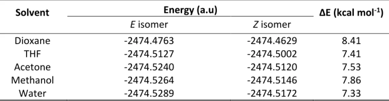
Table 2.1 Calculated energies for the E-392STP and Z-C392STP isomers in different solvents at the B3LYP/6-31+G(d) theory level. ... 62
Table 2.2 Calculated properties of the C392STP coumarin in different solvents at the PBE0/311+G(2d,p) level of theory. ... 64
Table 2.3 Calculated absorption data of the lowest energy transition for E and Z isomers in different solvents. ... 66
Table 2.4 Experimental and calculated spectral properties of the coumarin isomers at hybrid Pbe0 functional with 6-311+g (2d, p) theory level. ... 73
Table 3.1 Optimization process of fluorescent labelling methodology. ... 84
Table 3.2 Composition of concentration and resolution gels in PAGE. ... 86
Table 3.3 Molecular weight (kDa) of the commercial proteins displayed in PAGE profiles. ... 99
Table 3.4 Molecular weight (kDa) of the extracted proteins displayed in PAGE profiles. ... 101
Table 4.1 Constitution of paint models. ... 108
Table 4.2 Fluorescents labelling between paint models and C392STP. ... 112
14 INSTITUTO DE INVESTIGAÇÃO E FORMAÇÃO AVANÇADA
LIST OF FIGURES
Figure 1.1 PhD research roadmap. ... 23
Figure 1.2 Structure of an easel painting [10]. ... 24
Figure 1.3 Proteinaceous binders commonly used. ... 26
Figure 1.4 Factors contributing to the degradation of easel paintings. ... 27
Figure 1.5 Example of fluorescent labelling targeting primary amines of proteins (adapted from Skelley et al., 2003) [54]. ... 31
Figure 1.6 Chemical structure of coumarin. ... 35
Figure 1.7 Model of C392STP labelling a protein (ovalbumin). ... 37
Figure 2.1 Structural formulas of the C392STP, sodium (E/Z)-4-(4-(2-(6,7-dimethoxy-coumarin-3-yl) vin(E/Z)-4-(4-(2-(6,7-dimethoxy-coumarin-3-yl) benzo(E/Z)-4-(4-(2-(6,7-dimethoxy-coumarin-3-yl)-2,3,5,6-tetrafluorobenzenesulfonate. ... 58
Figure 2.2 Reaction between C392STP and a protein side chain amino acid amine group. ... 59
Figure 2.3 Chemical structure of (E/Z)-4-(2-(6,7- dimethoxycoumarin-3-yl)vinyl)-N-propylbenzamide. ... 59
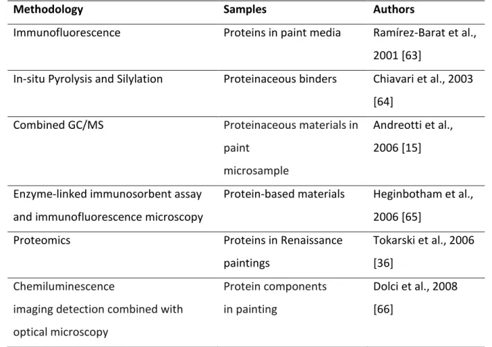
Figure 2.4 Optimized molecular geometry for E-C392STP and Z-C392STP in THF at the B3LYP/6-31+G(d) level and the HOMO and LUMO orbitals. ... 61
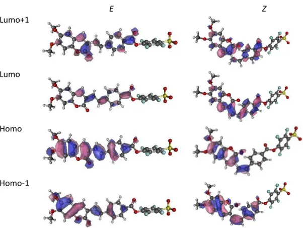
Figure 2.5 Schematic drawings of the frontier molecular orbitals of both isomers involved in the most important transitions for E- and Z-C392STP in acetone. ... 65
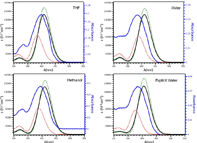
Figure 2.6 Calculated UV-Vis spectra for the Z and E isomers of C392STP (green and red, respectively) and for their mixture (E/Z 84:16) (black) and comparison with the experimental (blue) spectra in THF, methanol and water. Adapted from González-Pérez et al. [24]. ... 66
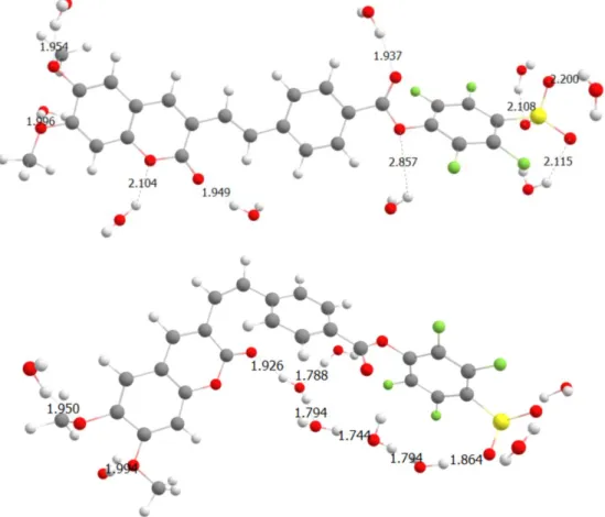
Figure 2.7 Optimized molecular geometry for E-C392STP (up) and Z-C392STP (down) in the PCM/water explicit model, at the B3LYP/6-31+G(d) level. ... 67
Figure 2.8 Theoretical and experimental IR spectra of sodium (E/Z)-4-(4-(2-(6,7-dimethoxy-coumarin-3-yl) vinyl) benzoyl)-2,3,5,6-tetrafluorobenzenesulfonate. ... 68
15 INSTITUTO DE INVESTIGAÇÃO E FORMAÇÃO AVANÇADA
Figure 2.9 Reaction of C392STP coumarin and propylamine to produce (E)-4-(2-(6,7-
dimethoxycoumarin-3-yl)vinyl)-N-propylbenzamide ... 69
Figure 2.10 Optimized molecular geometry of the E and Z conformers of the coumarin derivative
in acetonitrile at B3LYP/6-31G(d,p) level. ... 69
Figure 2.11 Schematic drawings of the frontier molecular orbitals of the both isomers involved in
the most important transitions. ... 71
Figure 2.12 Comparison between the experimental (blue line) and the calculated absorption
spectra of the coumarin E (black) and Z (red) isomers. ... 72
Figure 2.13 Experimental and calculated IR spectra of (E)-4-(2-(6,7-
dimethoxycoumarin-3-yl)vinyl)-N-propylbenzamide. ... 74
Figure 3.1 Synthesis of sodium
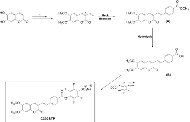
(E/Z)-4-(4-(2-(6,7-dimethoxycoumarin-3-yl)vinyl)-benzoyl)-2,3,5,6-tetrafluoro-benzenesulfonate (C392STP). ... 81
Figure 3.2 Reaction of
(E/Z)-4-(4-(2-(6,7-dimethoxycoumarin-3-yl)vinyl)benzoyl)-2,3,5,6-tetrafluorobenzenesulfonate and propylamine to produce (E)-4-(2-(6,7- dimethoxycoumarin-3-yl)vinyl)-N-propylbenzamide. ... 87
Figure 3.3 UV-Vis spectra of C392STP in acetonitrile. ... 89 Figure 3.4 UV-Vis spectra of BSA (black) and Fluorescent Labelled BSA (red) in sodium bicarbonate
buffer. ... 90
Figure 3.5 UV-Vis spectra of Ovalbumin (black) and Fluorescent Labelled Ovalbumin (red) in
sodium bicarbonate buffer. ... 90
Figure 3.6 UV-Vis spectra of Casein (black) and Fluorescent Labelled Casein (red) in sodium
bicarbonate buffer. ... 91
Figure 3.7 UV-Vis spectra of the Collagen (black) and Fluorescent Labelled Collgen (red), was
recorded in a mixture of glacial acetic acid (1mL) and sodium bicarbonate buffer solution (4mL), due to its low solubility. ... 91
Figure 3.8 UV-Vis spectra of Fish Gelatin (black) and Fluorescent Labelled Fish Gelatin (red) in
sodium bicarbonate buffer. ... 92
Figure 3.9 UV spectra of the fluorescent labelled BSA (red), ovalbumin (black), casein (green),
16 INSTITUTO DE INVESTIGAÇÃO E FORMAÇÃO AVANÇADA
Figure 3.10 FTIR spectra of BSA (blue) and Fluorescent Labelled BSA (red). ... 94
Figure 3.11 FTIR spectra of Ovalbumin (blue) and Fluorescent Labelled Ovalbumin (red). ... 94
Figure 3.12 FTIR spectra of Casein (blue) and Fluorescent Labelled Casein (red). ... 95
Figure 3.13 FTIR spectra of Collagen (blue) and Fluorescent Labelled Collagen (red). ... 95
Figure 3.14 FTIR spectra of Fish Gelatin (blue) and Fluorescent Labelled Fish Gelatin (red)... 96
Figure 3.15 Electrophorectogram of commercial BSA, casein, ovalbumin, collagen and fish gelatin without labelling and after fluorescent labelling. ... 98
Figure 3.16 Electrophorectogram of extracted proteins [ovalbumin (egg yolk and egg white), casein (milk) and rabbit skin glue]. ... 101
Figure 4.1 Easel painting models prepared using (a) hen’s egg, (b) bovine milk, and (c) rabbit skin glue, as binder with different pigments, lead white, yellow ochre and black bone. 109 Figure 4.2 Easel painting models prepared using (a) hen’s egg, (b) bovine milk, and (c) rabbit skin glue, as binder with three different pigments, lead white, chrome yellow and black bone. ... 109
Figure 4.3 Fluorescent proteins with different concentrations of chromophore. ... 111
Figure 4.4 Protein content of paint models prepared from (a) hen’s egg, (b) bovine milk, and (c) rabbit skin glue. The labels are the median ± SD of the 3 replicates. ... 114
Figure 4.5 Electrophoretic profile of proteins extracted from the paint models labelled with fluorescent coumarin 392 TFP ester. ... 115
Figure 4.6 Protein content of the paint models prepared from hen’s egg (PMO), bovine milk (PMC) and rabbit skin glue (PMRG). The labels are the median ± SD of 3 replicates. ... 116
Figure 4.7 Electrophoretic profiles of (a) PMO, (b) PMC and (c) PMRG. ... 117
Figure 5.1 Portraits by Giorgio Marini (a) A portrait of Frei Manuel do Cenáculo, 1887, (ME1281), Museum of Évora (Évora, Portugal); (b) portrait of a bearded gentleman, 1897, and (c) portrait of a lady, 1886, private collection (Évora, Portugal). ... 125
Figure 5.2 FTIR spectra of the microsamples from the portrait of Frei Manuel do Cenáculo. .. 127
Figure 5.3 FTIR spectra of the microsamples from the portrait of a bearded gentleman. ... 128
Figure 5.4 FTIR spectra of the microsamples from the portrait of a lady. ... 128
17 INSTITUTO DE INVESTIGAÇÃO E FORMAÇÃO AVANÇADA
Figure 5.6 Electrophoresis profiles of microsamples extracted from paint models (A) and
extracted from easel paintings (B). M2- portrait of Frei Manuel do Cenáculo, 1887, (ME1281), Museum of Évora (Évora, Portugal); Mb- portrait of a bearded gentleman, 1897, and Md- portrait of a lady, 1886, private collection (Évora, Portugal). ... 129
Figure 6.1 Scheme of fluorescent labelling procedure. ... 135 Figure 6.2 Time used for protein detection. ... 136
18 INSTITUTO DE INVESTIGAÇÃO E FORMAÇÃO AVANÇADA
LIST OF ABBREVIATIONS
C392STP Coumarin 392 4-sulfotetrafluorophenyl coumarin ester DFT Density functional theory
TD-DFT Time dependent density functional theory PAGE Plyacrylamide gel electrophoresis
BNP Biblioteca nacional de portugal XRF X-ray fluorescence
SEM-EDX Scanning electron microscope with energy dispersive X-ray spectroscopy
GC-MS Gas chromatography-mass spectrometry LC-MS Liquid chromatography-mass spectrometry ELISA Enzyme-linked immunosorbent assay SERS Surface enhanced raman scattering UV ultraviolet
MM Molecular mechanics IR infrared
HOMO Highest occupied molecular orbital LUMO Lowest unoccupied molecular orbital
g gram mmol milimoles equiv equivalent nm Nano-meter kcal kilocalorie THF tetrahydrofuran BSA Bovine serum albumin R.T. Room temperature ml millilitre
19 INSTITUTO DE INVESTIGAÇÃO E FORMAÇÃO AVANÇADA
rpm Rotation per minute HCl Hydrochloric acid μg Microgram μl microliter MW Molecular weight kDa kilodalton mg miligram μm Micrometer PM Paint model h hour
20 INSTITUTO DE INVESTIGAÇÃO E FORMAÇÃO AVANÇADA
Chapter 1 INTRODUCTION
21 INSTITUTO DE INVESTIGAÇÃO E FORMAÇÃO AVANÇADA
Easel paintings are extremely important Cultural Heritage assets with significant historic and cultural value. It emerged in the Middle Ages and since then have become one of the most important art expressions. Possessing multilayered structures composed of different layers, these artworks contain a diversity of organic materials, namely proteic compounds commonly produced from egg, milk or animal skin and bones. Proteins in paintings can be found in painting binders, adhesives, and additives in coating layers. The wide range of organic and inorganic materials mixtures in the painting’s matrices makes the detection of the different protein materials a difficult task and contribute to the complexity of the materials identification [1]. The degradation of the materials in the paintings due to aging and improper storage conditions also complicate the protein materials detection [2, 3]. Furthermore, improper restoration practices like repainting, coatings application and over cleaning that can cause alteration of the original painting materials can interfere with the protein identification too [2, 4, 5]. Besides that, strategies commonly used to detect proteins, such as chromatographic, spectroscopic and proteomic techniques are useful but costly because the instruments involved are expensive, while immunological methodologies involve expensive commercially manufactured antibodies. It is then of paramount importance to develop analytical, low invasive, approaches in protein identification in order to design appropriate restoration and conservation methods or even to acquire deeper insights into a particular artist’s technique.
22 INSTITUTO DE INVESTIGAÇÃO E FORMAÇÃO AVANÇADA
1.1 Scope and objectives
The main objective of this PhD research was to develop a new fluorescent labelling method to identify different proteinaceous materials usually used in art. In order to perform the fluorescent labelling, a coumarin chromophore has been used. Initially, computational studies on the coumarin chromophore and of a related compound intended to mimic the chromophore-amino acid complex were done to explore its spectral features. Following the theoretical quantum chemical calculations, experimental works using C392STP [6] to fluorescent label the protein binders followed by electrophoretic separation and identification with protein patterns by PAGE (Polyacrylamide gel electrophoresis), that allows its detection and identification, were performed. Firstly, we have tested and optimized the protocols to bond C392STP to commercial proteins such as BSA (A2153), ovalbumin (A5378), casein (C3400), collagen (C9879) and fish gelatin (G7765). The optimized method was then used on proteins extracted from hens’ egg yolk and white, bovine milk and rabbit skin using the previously optimized protocol [7]. To improve the fluorescent labelling methodology, by taking into account the complexity of the painting matrices, namely the presence of pigments and the aging processes, we have studied the method on laboratory made paint models. The proteinaceous content was extracted from the paint models and the extracted protein was used to bind with C392STP. Finally, the method was applied on real easel paintings samples. Figure 1.1 shows the PhD research roadmap of the work developed on this thesis. From the studies of C392STP using theoretical methods to the construction of the protocols for fluorescent labelling using C392STP and continuing to applications, this roadmap can provide the reader with a brief overview of this research route.
23 INSTITUTO DE INVESTIGAÇÃO E FORMAÇÃO AVANÇADA
24 INSTITUTO DE INVESTIGAÇÃO E FORMAÇÃO AVANÇADA
1.2 Easel painting
Generally, easel means a frame with legs which is made from wood. It is used to hold either a picture, a painting or a drawing [8, 9]. Easel painting refers to a midsize painting that an artist has painted on an easel. Easel paintings are composed of a few layers as shown in Figure 1.2. Usually, the materials used for the supports are either wood, canvas or metal.
Figure 1.2 Structure of an easel painting [10].
The first layer applied on the support is the ground layer/preparation layer which can either be gesso (animal glue and calcium sulfate or animal glue with calcium carbonate), oil (drying oil and white lead pigment) or bole (red clay, normally applied on gesso layer). After producing a smooth surface, chromatic layers/paint layers (a mixture of organic binders and pigments) are applied. Several binders used include egg tempera (egg yolk and pigment), drying oil (linseed/walnut/poppyseed), distemper (animal glue or casein paint) and ecaustic (wax). Lastly, a surface coating (semitransparent glazes and transparent varnish) is applied for the aesthetic presentation as well as for protective purposes [8–12]. Easel paintings emerged in the Middle Ages and since then have been one of the most important art expressions, constituting today’s relevant Cultural Heritage assets with important historical and cultural values. Paintings as earlier as 15th century are exhibited in the museums.
25 INSTITUTO DE INVESTIGAÇÃO E FORMAÇÃO AVANÇADA
In the history of art, artists used a variety of materials to paint. The materials and techniques used may vary among the artists, studios or guilds [11]. Since the period of Renaissance, artists chose natural binding media which can produce the desired effects [12]. Another interesting point to consider is the techniques to draw the underdrawing which also differed among the artists and workshops [13]. The compositions of the easel paintings produced in different countries might be different, depending on the availability of the materials and the difference in the practice of the artists. For an example, the painting “Lady of the Rose” in the National Museum of Machado de Castro, Coimbra, dating from the first half of the 15th century, exhibits as main characteristics the use of vibrant colors, the lack of
perspective, and larger scale of the sacred figures, with the use of tempera and painted on chestnut panel. This information evidence that it is a work from the workshop of Coimbra [14]. The artist frequently adapted his skills to the resource available at the time. An example is the famous Portuguese-Flemish painter, Frei Carlos, who has produced his paintings adapting the concepts of the Portuguese painters.
The main objective of research on easel painting is to understand the structure of the easel paintings in order to plan a conservation strategy, according to the information gathered. Normally, the first step is to identify the painting materials used. The characterization of the materials allows us to understand the structure of the paintings, as well as the cause of the changes, happened on the paintings. Besides, material studies may also reveal previous restoration works done [3, 15]. After gathering enough information on the paintings, a strategy to preserve the paintings, such as the selection of materials suitable for restoration [3], can be decided. Another point to be considered is the creation of an environment suitable to display or store the artwork [5]. The presence of the proteins can be the nutritional source for the growth of microorganism that contributes to the deterioration of the paintings. For instance, the mixture of ovalbumin, collagen, and casein found in the paintings was suggested to be the cause of microbial contamination [11].
26 INSTITUTO DE INVESTIGAÇÃO E FORMAÇÃO AVANÇADA
1.3 Organic materials in easel paintings
A diversity of organic materials, especially proteinaceous compounds (Figure 1.3), has been used as binders, adhesives and additives in coating layers in easel paintings. Egg ovalbumin, milk casein and animal glue collagen produced from animals’ bones, cartilages and skins are among the most commonly found proteins in the artworks [4, 16, 17]. These proteic compounds sometimes used together with siccative oils is known as tempera [4, 18].
Figure 1.3 Proteinaceous binders commonly used.
Unfortunately, those organic materials are particularly susceptible to the environmental conditions [5] as shown in Figure 1.4. Exposure to the environment cause changes in the organic materials which contribute to composition changes. The surface of the painting, particularly, can degrade under different conditions of lighting, humidity, and temperature [5, 19]. Another factor responsible for structural damages are the degradation compounds produced by pollution [4, 20].
In this way, materials identification, particularly proteins, from paint samples are challenging because:
i. The detection of the different protein materials in these complexes matrices is a difficult task; wide ranges of organic and inorganic materials mixture in the paintings contribute to the complexity of the materials identification [1, 17, 21].
ii. Protein alterated/ degradated [1, 17]. VARNISH
Egg white
MEDIUM LAYER/PAINT LAYER
Egg tempera/ protein- based
paint
GROUND LAYER
27 INSTITUTO DE INVESTIGAÇÃO E FORMAÇÃO AVANÇADA
iii. Limited amount of sample; destruction on the artworks must be limited to a minimum [17, 21].
iv. Low protein concentration in the sample [22]. v. Low solubility [4].
Figure 1.4 Factors contributing to the degradation of easel paintings.
Due to the high importance of the preservation of these artworks, the correct identification of proteinaceous binders is a crucial step for an understanding of the techniques used by the artist, and to provide relevant information for conservation and restoration processes [19]. It is then of paramount importance to develop low invasive analytical methodologies in protein identification in artwork materials that are suitable for protein identification in painting samples.
1.4 Proteinaceous binder detection
The ubiquitous presence of proteins in artworks as binders, adhesives and additives in coating layers makes their identification an important step for characterizing the artist technique and for the development of appropriate conservation and restoration treatments. The pioneer work was initiated by Ostwald [23] in 1936, by using biological dyes such as iodesine or acid
Degradation of Easel paintings Daylight radiation - visible - infrared - UV Humidity Pollution Temperature variations
28 INSTITUTO DE INVESTIGAÇÃO E FORMAÇÃO AVANÇADA
green to stain proteins. The use of iodesine/acid green to stain proteins allowed the identification of some binders for the first time. Solutions of ammonium hydroxide containing iodesine stained tempera and glue red while solutions of methyl violet stained tempera and glue violet. Acid green could stain gelatin and casein and vanillin were also able to stain tempera red or violet [24]. The study of binders in paintings using staining tests were also reported by other studies [25]. Nile blue could stain drying oil effectively while acid fuchsin stained animal glue and egg tempera. Acid fuchsin has already been found to be effective in staining protein through the reaction with the ammonium group of proteins since the early 20th century [24, 25].
The use of other classical colorimetric reactions such as the ninhydrin reaction, the Biuret reaction, the Millon reaction, and the Sakaguchi reaction, among others, have been used for the detection of proteins in paintings [26]. These methods based on the production of visible stains presented limited sensitivity and, depending on the pigment present in the painting sample, produced visible stains that could be difficult to distinguish. Nowadays analytical methods such as high-performance liquid chromatography (HPLC), gas chromatography (GC), combined with mass spectrometric (MS) detection, and thin-layer chromatography infrared spectrometry methods are commonly used in the identification of proteinaceous binders in paintings being capable to distinguish between egg, animal glue and milk proteins [15, 22, 27–33].
Proteomic is used in a broad range of studies like clinical medicine, forensic, food analysis and the origins of life [34, 35]. In the early 2000, the use of proteomic methods was suggested in cultural heritage studies to analyze proteins in artworks including archeological samples [17, 34–37]. Proteomics techniques were adapted to the scientific analysis of archeological microsamples and research work was developed [35, 36] focused on the optimization of the protocols to address the problem of the small quantity of the aged and deteriorated microsamples.
A new technique using Matrix-Assisted Laser Desorption/Ionization-Time of Flight Mass Spectrometry (MALDI-TOF-MS) was used to detect protein components in painting. Painting models were formulated using egg white, lead white pigment, and linseed oil while another
29 INSTITUTO DE INVESTIGAÇÃO E FORMAÇÃO AVANÇADA
with whole egg, lead white pigment and linseed oil [33]. Application of new protocol of MALDI analysis has also been tried on paint models produced from a mixture of lead white pigment and egg yolk, whole egg, linseed oil or both egg and oil [38] while the further simplification of MALDI protocol was tested on a mixture of inorganic pigments and egg, milk and collagen applied on glass slide [39]. Another study by Romero-Pastor and colleagues [18] tried for the first time the use of PCA on MALDI-TOF-MS-data in cultural heritage using paint models prepared with rabbit glue and rabbit glue with cinnabar or azurite in glue tempera production. Three painting models were prepared based on old medieval recipes.
Nonetheless, there are a few limitations of those methods that need to be considered. Firstly, chromatographic and proteomics techniques involve laborious sample treatment [39] which only experts can implement [4, 40]. It is important to take note that chromatographic methods involve the hydrolysis of the protein in the sample and this can reduce the amount of information (origins of protein, degradation level and interaction between pigment and binder) one can get from the sample [30]. Secondly, expensive instruments are required in performing those techniques [40]. Thirdly, the analysis of the sample using chromatographic and spectroscopic methodology is largely dependent on the characteristics of the sample such as the composition of the sample, level of degradation and contamination [41, 42]. Furthermore, the interpretation of the analysis using chromatographic techniques is difficult if the sample is a complex mixture of organic and inorganics materials [43, 44] and the results have lower specificity [33] when compared with immunological methods.
More recently immunological techniques inspired from biological methods such as Enzyme-Linked ImmunoSorbent Assay (ELISA) [41] or Surface Enhanced Raman Scattering (SERS) nanotags has been successfully used to localize/identify protein binders. The ELISA technique is particularly sensitive and specific in protein identification using antigen-antibody reactions [5, 21, 40, 42]. ELISA is a potential complementary analysis when the information of biological sources of proteins is needed from a sample. The emerging immunological techniques were tested to be used in cultural heritage studies. In the optimization of indirect ELISA to identify proteinaceous binders usedin art work, tests have been made [21, 45] on paint models constructed based on old recipes that mimic easel painting/mural painting.
30 INSTITUTO DE INVESTIGAÇÃO E FORMAÇÃO AVANÇADA
Layers of paint models were prepared by using gypsum and animal glue applied on wood panels. Then, the protein binders like whole egg/egg yolk/milk/bovine glue/rabbit glue were mixed with pigments in the ratio of pigment to binder=3:1. In order to test the dot-ELISA test, Potenza and colleagues [46] have prepared model paintings using egg and rabbit glue mixed with red ochre pigment for tempera layer and applied it on mortar surfaces. Gambino et al. (2013) [47] further study the application of non-competitive dot-blot immunoassay and addressed pigment and aging effects using microsamples of paint replicas prepared in the laboratory; egg white and pigments were mixed and applied on glass slides. In the study by Sciutto et al. (2011) [48], the use of chemiluminescent immunochemical microscope imaging has been tested on ovalbumin and casein followed by testing on collagen [44]. Testing have been done on paint models prepared according to ancient painting recipes in which gypsum and rabbit glue (weight ratio of 19:1) has been applied as ground layer and a mixture of inorganic pigments and rabbit glue (blue smalt: rabbit glue=2:1)/ egg (lead white: egg=20:13)/ casein (red ochre: casein=10:7) as paint layers.
In order to address the challenge of doing on site analysis, one recent study by Zangheri et al (216) [40] tried a newly developed portable analytical device on paint models which were prepared according to ancient painting recipes; gypsum and rabbit glue were applied on wood panel followed by inorganic pigments and egg (egg white: yolk: water= 1:1:1). The combination of immunological techniques and chemiluminescence detection reported the possibility of ovalbumin identification in the paint models as well as from samples of canvas painting, wall painting and painted wood panels. Nevertheless, the main limitation of this method is that it can only detect proteins which are part of the assay [5, 21, 40, 42, 45]. Therefore, negative results from ELISA assays cannot confirm the absence of other proteins. Furthermore, ELISA test is time-consuming [49]. The analysis are also highly costly as materials such as expensive commercially manufactured antibodies specifically tailored for cultural heritage study [41] are needed. Both chromatographic techniques as well as immunological techniques involve expensive equipment and require specialized personnel [5].
31 INSTITUTO DE INVESTIGAÇÃO E FORMAÇÃO AVANÇADA
In recent years, protein detection based on fluorescence techniques using fluorescent organic compounds has received much attention. Its use for protein detection in paintings was proposed in the end of the 1980s [50]. Thereafter, several dyes like fluorescamine, LISSA, fluorescein isothiocyanate (FITC) and cycloheptaamylose-dansyl chloride complex (DC-C7A) fluorochrome have been introduced [50, 51]. More recently, a ruthenium complex commercial dye, SYPRO Ruby, has also been used for detection of proteins in the works of art, including paintings. These fluorescent dyes interact with different functional groups in the proteins [52, 53] producing fluorescent products. Both fluorescamine and LISSA reacts with primary amines of proteins during fluorescent labelling [24, 54]; the reaction involving primary amines is shown in Figure 1.5.
Figure 1.5 Example of fluorescent labelling targeting primary amines of proteins (adapted
from Skelley et al., 2003) [54].
In the reaction between FITC and proteins, the sulfhydryl group can also be involved. The same happens with DC-C7A in which the sulfonyl chlorides/isothiocyanates of DC-C7A react with the primary amines and thiols of the proteins [24, 55]. Sypro Ruby is a noncovalent stain adapted from the biomedical field for protein identification through protein mapping, using gel electrophoresis [24, 56]. Staining using Sypro Ruby have been tried on paints [56], a polychromy section of an altarpiece [37, 57], sculptures [58] and on easel painting samples [52]; proteins commonly used in art work (egg, animal glue, fish glue) could be successfully
32 INSTITUTO DE INVESTIGAÇÃO E FORMAÇÃO AVANÇADA
detected. These fluorescent dye, however, are very expensive and must be used with parsimony [59].
The applicability of a particular molecule as a fluorescent dye is highly dependent on its photophysical and photochemical properties like UV−vis absorption and fluorescence spectra, molar extinction coefficients, quantum efficiencies, Stokes shifts, pH and thermal stabilities among others [60]. Many coumarins (benzopyranones) and coumarin derivatives fit these conditions, which make it particularly adequate for use in fluorescent labelling [61]. The use of coumarin derivatives as fluorescent dyes will be discussed in the following section. The identification of proteinaceous components in paintings remains a challenging task and much effort have been made to develop simpler and less time consuming approaches [39, 40, 46, 62]. Table 1.1 shows a list of some recent research on current methods used in protein detection in art.
Table 1.1 Examples of research using different methodologies to detect proteins in art.
Methodology Samples Authors
Immunofluorescence Proteins in paint media Ramírez-Barat et al., 2001 [63]
In-situ Pyrolysis and Silylation Proteinaceous binders Chiavari et al., 2003 [64]
Combined GC/MS Proteinaceous materials in paint
microsample
Andreotti et al., 2006 [15]
Enzyme-linked immunosorbent assay and immunofluorescence microscopy
Protein-based materials Heginbotham et al., 2006 [65]
Proteomics Proteins in Renaissance paintings
Tokarski et al., 2006 [36]
Chemiluminescence
imaging detection combined with optical microscopy
Protein components in painting
Dolci et al., 2008 [66]
33 INSTITUTO DE INVESTIGAÇÃO E FORMAÇÃO AVANÇADA
Immunofluorescence microscopy Proteins in painting Vagnini et al., 2008 [1]
Gas chromatography/mass spectrometry
Organic paint media Colombini et al., 2010 [22]
Proteomic strategies Proteinaceous binders in paintings
Leo et al., 2009 [17]
FT-NIR spectroscopy Organic components in
painting materials
Vagnini et al., 2009 [1]
Enzyme-linked immunosorbent assay (ELISA) and immuno-fluorescence microscopy (IFM) techniques
Proteins in ancient paint media
Cartechini et al., 2010 [41]
Liquid chromatography–tandem mass spectrometry Protein binders in historical paints Fremout et al., 2010 [30] GC/MS Proteinaceous materials from paint microsample
Lluveras et al., 2010 [43]
Surface enhanced Raman scattering (SERS) nanotags
Avian egg, animal glue, or casein binders
Arslanoglu et al. ,2011 [67] Enzyme-linked immunosorbent assay
(ELISA)
Bovine milk (or casein) and chicken albumen
Palmieri et al., 2011 [45]
Indirect Enzyme-Linked
Immunosorbent Assay (ELISA) method
Proteinaceous binding media and adhesives
Schultz and
Petersen, 2011 [42]
Multiplexed chemiluminescent immunochemical imaging technique
Organic components in the complex stratigraphy of paintings Sciutto et al., 2011 [48]
Radiographs and technical photographs, x-ray fluorescence
Materials in the portraits executed between
Soares et al. 2012 [3]
34 INSTITUTO DE INVESTIGAÇÃO E FORMAÇÃO AVANÇADA
the 16th and 19th centuries
from National Library of Portugal (BNP)
Dot-blot immunoassay Egg white Gambino et al. 2013
[47] Enzyme-linked immunosorbent assay
(ELISA)
Animal glue and hen-egg yolk
Palmieri et al. 2013 [21]
Chemiluminescent imaging detection
Animal glues Sciutto et al., 2013 [44]
MALDI-MS Lipid- and protein-based
binders
Calvano et al. 2015 [39]
XRF, optical microscopy, Raman spectroscopy,
and SEM-EDX
Pigments and fillers on the paintings by
Giorgio Marini
Bordalo et al. 2016 [68]
Combined surface analysis and microanalytical techniques Materials of the underdrawings of the Flemish-Portuguese easel paintings Valadas et al. 2016 [13] Chemiluminescent immunochemical contact imaging
Chicken ovalbumin Zangheri et al. 2016 [40]
Scanning electron microscopy analyses,
Energy-dispersive X-ray spectroscopy, μ-X-ray diffraction, μ-Raman, μ-FTIR and optical microscopy, immunological assays
Painting materials of the easel paintings by Giorgio Marini
Salvador et al. 2017 [11]
35 INSTITUTO DE INVESTIGAÇÃO E FORMAÇÃO AVANÇADA
1.5 Use of coumarin derivatives
Coumarins/benzo-α-pyrones/2H-chromen-2-one/1-benzopyran-2-one are large family of compounds, consisting of a fusion of a pyrone and a benzene ring, with the pyrone carbonyl at position 2 (Figure 1.6) [69].
Figure 1.6 Chemical structure of coumarin.
Coumarins possess a variety of biological activity including antibacterial, anticancer, anticoagulant, antifungal, antihelmintic, anti-HIV, anti-inflammatory, antimicrobial, antioxidant, antiviral, estrogenic, dermal photosensitising, vasodilator, molluscucidal, sedative and hypnotic, analgesic and hypothermic activity [70–78]. Studies reported the used of coumarin derivatives in different fields like biology, chemistry, medicine, and pharmacology [79]. It has been applied as anesthetic in laboratory experiments, for fixing odors in perfume, in flavoring and in synthetic vanilla production, as a constituent of lavender oil, and as a natural source of essential antioxidants [80, 81].
Coumarin derivatives represent one of the most important chemical classes of fluorescent organic compounds, being one of the most extensively investigated and commercially significant groups of organic fluorescent materials [6, 70, 71, 82–84]. Some of its substituted derivatives can emit strong fluorescent light [84, 85]. It has been applied in fields such as biological science, environmental monitoring, clinical chemistry, DNA sequencing and genetic analysis by fluorescence in situ hybridization (FISH) [86]. Due to the outstanding photophysical properties of coumarin, it is used in:
i. High-performance liquid chromatography
36 INSTITUTO DE INVESTIGAÇÃO E FORMAÇÃO AVANÇADA
iii. Generating fluorescent derivatives
iv. Probing proteins [69, 86]
Coumarin derivatives provide some of the most important commercial fluorescent brightening agents and appropriately substituted compounds are also used as highly effective fluorescent dyes on synthetic fibers and in daylight fluorescent pigments, conveying a vivid brilliance to a range of paint and printing ink applications. In addition, fluorescent coumarins may be used in a range of applications which specifically exploit their light emission properties, including non-destructive flaw detection, tunable dye lasers, emission layers in organic light-emitting diodes (OLED) and solar energy collectors. The most commonly-encountered fluorescent coumarins either absorb in the UV region emitting blue light (FBAs) or are yellow dyes emitting a green fluorescence [70, 87, 88]. Although several derivatives that both absorb and emit at long wavelengths are known, there is much interest in the molecular design and synthesis of new coumarins derivatives which would extend the available range of long-wavelength emitting fluorescent materials [87, 88].
In particular, coumarins that react with target biomolecules, metals or reactive groups have been extensively exploited as fluorescent labels [89–94]. Examples include the coumarins that have amine reactive moieties, tetrafluorophenyl (TFP) or N-hydroxysuccinimide (NHS) esters, which are effective dyes for biolabelling of molecules possessing primary amine groups [60, 95–97].
It is known that the 4-sulfotetrafluorophenyl coumarin esters, like coumarin 392 STP ester, bonds covalently with amino acids [98], particularly with the lysine side chain amine, providing an efficient labelling. Coumarin392STP, the coumarin derivative fluorophore that was studied in this work, possesses very interesting physicochemical characteristics as a large Stokes shift, pH-independence of absorbance and emission and excellent photo-stability [99]. Other properties of this coumarin derivative include a high fluorescent quantum yield, and easiness to synthesize, and its possession of photophysical and spectroscopic properties which can be easily tailored according to the desired application [6, 70]. These properties,
37 INSTITUTO DE INVESTIGAÇÃO E FORMAÇÃO AVANÇADA
together with its low cost, point to the possibility of C392TFP to become a fluorescent dye with a wide range of applications in bioimaging and biolabelling.
This work proposes using the coumarin derivative chromophore (Coumarin 392 4-sulfotetrafluorophenyl coumarin ester) [6] to develop a new simple, fast and affordable protocol to detect and identify protein binders used in easel paintings. Figure 1.7 shows a model of C392STP labelling a protein. The proteinaceous extracted from the paints are made react with the coumarin chromophore that binds to the proteins and its fluorescent properties allow an easy detection and identification of the proteins separated by gel electrophoresis. Furthermore, the step of electrophoresis gel staining is not needed in the identification process.
38 INSTITUTO DE INVESTIGAÇÃO E FORMAÇÃO AVANÇADA
1.6 Theoretical quantum calculation
Quantum chemistry is based on the application on chemistry of methods derived from the laws of quantum mechanics [100]. A few years after the introduction of the Schrödinger equation, the Hartree-Fock approach was introduced and it has been widely used in the quantum chemistry research [101]. Not long after, post-Hartree Fock ab initio method, based on the wave function, has been introduced to address the question on the correlation between electrons [101].
Parallelly, Density Functional Theory (DFT) which is based on Hohenberg-Kohn [102], and Kohn and Sham [103] theorems were also proposed [101, 104, 105]. It is also known as static DFT and formally has the form of an effective one-particle Schrödinger equation [104]. Since then, DFT has been used widely in modeling the ground states of molecules [106, 107]. DFT methods have an increasing popularity in first principles quantum chemical calculations that explore the electronic structure [84, 108–110]. Less CPU time is required when compared with conventional ab initio calculations, it computes the results with greater accuracy than the Hartree-Fock Theory and solvent effect can be taken into account [84, 108, 110]. That makes nowadays DFT as the leading method in theoretical quantum chemical calculation for electronic structure exploration [109].
A few years later, time dependent DFT (TD-DFT), an extension of DFT, has been proposed by Runge and Gross [111]. The TD-DFT method has been proposed for the exploration of electronic excited-states energies [84, 112]. It is formulated in the form of an effective two-particle equation [104] which can be used to calculate the transition energies, dipole moments and emitting geometries [113]. It is now established that TD-DFT theory is an accurate method for analyzing structural, thermodynamic, kinetic and spectroscopic properties [84, 114–120]providing good results with a lower computational cost [101, 105, 121] when compared with other approaches. Parac and Grimme (2002) [122] have shown the applicability of TD-DFT on calculating excitation energies of large molecules.
39 INSTITUTO DE INVESTIGAÇÃO E FORMAÇÃO AVANÇADA
There are in the literature many examples of DFT and TD-DFT calculations on coumarin and coumarin derivatives, a few of them reported in Table 1.2. In one study 30 coumarin derivatives were studied to investigate molecular properties such as the energy of the highest occupied molecular orbital (HOMO) and the lowest unoccupied molecular orbital energy (LUMO), the energy gap and the dipole moment, by using DFT together with the B3LYP functional and the 6-31G* basis set [81]. Also, a recent work has produced benchmarking studies that explored the spectroscopic characteristic of the absorption spectra of 25 coumarin derivatives [123, 124].
In the study, DFT and TDDFT methods with different functionals and basis set have been used to study the absorption spectra of a diversity of simple coumarins and furanocoumarins derivatives. The calculated spectra were well estimated when comparing to the experimental spectra. The properties of solvated coumarins have also been studied using TDDFT calculations [125–127]. The combination of the functionals and the polarizable continuum model (PCM) was found useful in the study of solvent effects. It is known that PCM is able to give a good estimation of solvent effects [128] with solvatochromic and Stokes’ shift calculated presenting good agreement with experimental values [126]. The choice of the functional is largely dependent on the type of the coumarin, the accuracy required, and the computational cost allocated [125, 128].
Table 1.2 Key references of theoretical quantum chemical calculations on coumarin
derivatives.
Subjects Methodology Authors
Coumarins 151 and 120 Time-Dependent Density Functional Theory (TDDFT) calculations against CASSCF, CASPT2 (both single and multistate versions), CIS, and ZINDO
Cave 2002 [129]
Coumarin derivatives Density Functional Theory framework (DFT) at Becke–Lee–Yang–Parr functional (B3LYP)/6-311+G(2d,2p) basis set
Preat et al., 2005 [120]
40 INSTITUTO DE INVESTIGAÇÃO E FORMAÇÃO AVANÇADA
Acetyl coumarin DFT and Hartree- Fock (HF) at 6-31G* and 6-311++G** basis sets
Bahgat 2006 [130]
Coumarin derivatives PBE0/6-31+G(d) Jacquemin et al. 2006 [84] Coumarin based dyes TDDFT with the Baer, Neuhauser, and
Livshits BNL RSH functional,
Stein et al. 2009 [131]
7-acetoxy-4-methyl coumarin
Density functional theory at B3LYP/6-311+G** basis set
Arivazhagan et al. 2010 [80] Coumarin–thiourea
conjugate
Ab initio molecular orbital calculations Shiraisi et al. 2010 [118]
3-cyano-4-methylcoumarin
Density functional theory (DFT) at 6-31G(d,p) basis sets
Chaitanya et al. 2012 [79] 3,4-dihydrocoumarin
and 3-methylcoumarin
SQM force field method based on ab initio and DFT calculation at 6-311++G(d,p) basis set Arivazhagan et al. 2014 [132] 7-Acetoxy-4-(Bromomethyl)Coumarin DFT calculation at B3LYP/6-311++G(d,p) basis set Erdogdu et al. 2015 [86] coumarin 151 TDDFT/EFP1 Ramegowda et
al. 2015 [127] V-shaped bis-coumarins DFT and TDDFT
with the M06-2X hybrid
exchange–correlation functional
Šimon Budzák et al. 2016
Coumarin derivatives Density functional theory (DFT) methods at B3LYP functional and a 6-31G* basis set
Hmamouchi et al. 2016 [81] Simple coumarins and
Furanocoumarins derivatives
DFT functional CAM-B3LYP, WB97XD, HSEH1PBE, MPW1PW91 and TD-B3LYP with 6-31 + G (d,p) basis set
Irfan et al., 2017 [124]
41 INSTITUTO DE INVESTIGAÇÃO E FORMAÇÃO AVANÇADA
The literature also shows several studies focused on a specific type of coumarins. For instance, properties such as conductivity, solvatochromism, gas-phase spectroscopy, solution-phase spectroscopy and electroabsorption spectroscopy of coumarin 151 [129, 133– 135] and coumarin 120 [129, 135, 136] were thoroughly studied. The properties of coumarin 120 and coumarin 151 were investigated using TDDFT calculations founding close agreement with the experimental S1S0 excitation energies [129]. It is also reported that the PBE0 and the MPW1PW91 hybrid functionals gave the better results presenting results nearer to the experimental values [114, 129]. Barone and colleagues presented the UV-Vis absorption spectrum of 7-amino-coumarin based on TDDFT calculations [126, 137]. According to the calculations, the B3LYP functional provided the more reliable geometry the 7-aminocoumarin, among those functionals tested.
In this work, theoretical quantum chemical calculations based on the DFT and TDDFT have been performed on both the E and the Z isomers of C392STP coumarin, the fluorophore label molecule that we used as a probe to bond proteinaceous binders used in paintings. Calculations were also done on a related compound that mimics the fluorescent coumarin bonded with a protein amino acid amine side chain.
42 INSTITUTO DE INVESTIGAÇÃO E FORMAÇÃO AVANÇADA
Bibliography
[1] M. Vagnini, L. Pitzurra, L. Cartechini, C. Miliani, B. G. Brunetti, and A. Sgamellotti, “Identification of proteins in painting cross-sections by immunofluorescence microscopy,”
Anal. Bioanal. Chem., vol. 392, no. 1–2, pp. 57–64, 2008.
[2] A. J. Cruz, “Em busca da imagem original: Luciano Freire e a teoria e a prática do restauro de pintura em Portugal cerca 1900,” Conserv. Património, vol. 5, pp. 67–83, 2007.
[3] C. M. Soares, R. M. Rodrigues, A. J. Cruz, and C. Rêgo, “Historical and material approach to the paintings at the Portugal National Library: contributions to the history of conservation and restoration of easel painting in the 19th century,” Int. J. Herit. Digit. era, vol. 1, no. supplement 1, pp. 283–288, 2012.
[4] M. P. Colombini and F. Modugno, “Characterisation of proteinaceous binders in artistic paintings by chromatographic techniques,” J. Sep. Sci., vol. 27, no. 3, pp. 147–160, 2004. [5] J. Arslanoglu, J. Schultz, J. Loike, and K. Peterson, “Immunology and art: Using
antibody-based techniques to identify proteins and gums in artworks,” J. Biosci., vol. 35, no. 1, pp. 3–10, 2010.
[6] S. M. Martins, P. C. Branco, and A. M. D. L. Pereira, “An Efficient Methodology for the Synthesis of 3-Styryl Coumarins,” J. Braz. Chem. Soc., vol. 23, no. 4, pp. 688–693, 2012. [7] C. Salvador, A. Branco, A. Candeias, and A. T. Caldeira, “Innovative approaches for
immunodetection of proteic binders in art,” E-Conservation J., no. 5, pp. 1–10, 2017. [8] “Cambridge Dictionary Online,” Cambridge University Press, 2008. [Online]. Available:
https://dictionary.cambridge.org/dictionary/english/easel. [Accessed: 16-Apr-2018]. [9] “Oxford Dictionary of English,” Oxford University Press, 2015. [Online]. Available:
https://en.oxforddictionaries.com/definition/easel. [Accessed: 16-Apr-2018].
[10] J. R. Allred, “Characterization of Hidden Paint Layer Topography Using a Stereographic XRF Approach,” Delft University of Technology, 2017.
[11] C. Salvador, R. Bordalo, M. Silva, T. Rosado, A. Candeias, and A. T. Caldeira, “On the conservation of easel paintings: evaluation of microbial contamination and artists materials,” Appl. Phys. A Mater. Sci. Process., vol. 123, no. 1, p. 80, 2017.
43 INSTITUTO DE INVESTIGAÇÃO E FORMAÇÃO AVANÇADA
the biological origin of animal glues used in paintings through mitochondrial DNA analysis,”
Anal. Bioanal. Chem., vol. 399, no. 9, pp. 2987–2995, 2011.
[13] S. Valadas, R. Freire, A. Cardoso, J. Mirao, P. Vandenabeele, O. J. Caetano, and A. Candeias, “New insight on the underdrawing of 16th Flemish-Portuguese easel paintings by combined surface analysis and microanalytical techniques,” Micron, vol. 85, pp. 15–25, 2016.
[14] “Museu Nacional Machado de Castro, Coimbra,” 2019. [Online]. Available: http://www.museumachadocastro.gov.pt/en-GB/4
coleccoes/painting/ContentDetail.aspx?id=438.
[15] A. Andreotti, M. Bonaduce, M. P. Colombini, G. Gautier, F. Modugno, and E. Ribechini, “Combined GC/MS analytical procedure for the characterization of glycerolipid, waxy, resinous, and proteinaceous materials in a unique paint microsample,” Anal. Chem., vol. 78, no. 13, pp. 4490–4500, 2006.
[16] W. Fremout, S. Kuckova, M. Crhova, J. Sanyoya, S. Saverwyns, R. Hynek,…, and L. Moens, “Classification of protein binders in artist’s paints by matrix-assisted laser desorption/ionisation time-of-flight mass spectrometry: An evaluation of principal component analysis (PCA) and soft independent modelling of class analogy (SIMCA),”
Rapid Commun. Mass Spectrom., vol. 25, no. 11, pp. 1631–1640, 2011.
[17] G. Leo, L. Cartechini, P. Pucci, A. Sgamellotti, G. Marino, and L. Birolo, “Proteomic strategies for the identification of proteinaceous binders in paintings,” Anal. Bioanal.
Chem., vol. 395, no. 7, pp. 2269–2280, 2009.
[18] J. Romero-Pastor, N. Navas, S. Kuckova, A. Rodríguez-Navarro, and C. Cardell, “Collagen-based proteinaceous binder-pigment interaction study under UV ageing conditions by MALDI-TOF-MS and principal component analysis,” J. Mass Spectrom., vol. 47, no. 3, pp. 322–330, 2012.
[19] M. Elias, N. Mas, and P. Cotte, “Review of several optical non-destructive analyses of an easel painting. Complementarity and crosschecking of the results,” J. Cult. Herit., vol. 12, no. 4, pp. 335–345, 2011.
44 INSTITUTO DE INVESTIGAÇÃO E FORMAÇÃO AVANÇADA
in Islamic Art Museum , Cairo,” J. Cult., vol. 12, pp. 412–419, 2011.
[21] M. Palmieri, M. Vagnini, L. Pitzurra, B. G. Brunetti, and L. Cartechini, “Identification of animal glue and hen-egg yolk in paintings by use of enzyme-linked immunosorbent assay (ELISA),” Anal Bioanal Chem, vol. 405, pp. 6365–6371, 2013.
[22] M. P. Colombini, A. Andreotti, I. Bonaduce, F. Modugno, and E. Ribechini, “Analytical strategies for characterizing organic paint media using gas chromatography/mass spectrometry,” Acc. Chem. Res., vol. 43, no. 6, pp. 715–727, 2010.
[23] W. Ostwald, “Iconoscopic studies I: Microscopic identification of homogenous binding mediums,” Tech. Stud. F. Fine Art, vol. 4, no. 3, pp. 135–44, 1936.
[24] S. Dallongeville, N. Garnier, C. Rolando, and C. Tokarski, “Proteins in Art, Archaeology, and Paleontology: From Detection to Identification,” Chem. Rev., vol. 116, no. 1, pp. 2–79, 2016. [25] J. Plesters, “Cross-sections and Chemical Analysis of Paint Samples,” Stud. Conserv., vol. 2,
no. 3, pp. 110–157, 1956.
[26] M. C. Gay, “Essais D’Identification Et De Localisation Des Liants Picturaux Par Des Colorations Specifique Sur Coupe Mince,” Ann. du Lab. Rech. des Museés Fr., pp. 8–24, 1970.
[27] A. Lluveras, I. Bonaduce, A. Andreotti, and M.P. Colombini, “GC/MS Analytical Procedure for the Characterization of Glycerolipids, Natural Waxes, Terpenoid Resins, Proteinaceous and Polysaccharide Materials in the Same Paint Microsample Avoiding Interferences from Inorganic Media,” Anal. Chem., vol. 82, no. 1, pp. 376–386, 2010.
[28] M. T. Doménech-Carbó, “Novel analytical methods for characterising binding media and protective coatings in artworks,” Anal. Chim. Acta, vol. 621, no. 2, pp. 109–139, 2008. [29] R. Checa-Moreno, E. Manzano, G. Mirón, and L. F. Capitan-Vallvey “Comparison between
traditional strategies and classification technique (SIMCA) in the identification of old proteinaceous binders,” Talanta., vol. 75, no. 3, pp. 697-704, 2008.
[30] W. Fremout, M. Dhaenens, S. Saverwyns, J. Sanyova, P. Vandenabeele, D. Deforce, and L. Moens, “Tryptic peptide analysis of protein binders in works of art by liquid chromatography-tandem mass spectrometry,” Anal. Chim. Acta, vol. 658, no. 2, pp. 156– 162, 2010.
45 INSTITUTO DE INVESTIGAÇÃO E FORMAÇÃO AVANÇADA
[31] S. Kuckova, M. Crhova, L. Vankova, A. Hnizda, R. Hynek, and M. Kodicek, “Towards proteomic analysis of milk proteins in historical building materials,” Int. J. Mass Spectrom., vol. 284, no. 1–3, pp. 42–46, 2009.
[32] A. Nevin, D. Comelli, G. Valentini, D. Anglos, A. Burnstock, S. Cather, and R. Cubeddu, “Time-resolved fluorescence spectroscopy and imaging of proteinaceous binders used in paintings,” Anal. Bioanal. Chem., vol. 388, no. 8, pp. 1897–1905, 2007.
[33] C. Tokarski, E. Martin, C. Rolando, and C. Cren-Olivé, “Identification of Proteins in Renaissance Paintings by Proteomics,” Anal. Chem., vol. 78, no. 5, pp. 1494–1502, 2006. [34] S. Have, Y. Ahmad, and A. I. Lamond, “Analytical & Bioanalytical Proteomics – Current
Novelties and Future Directions,” no. 3, pp. 1–6, 2011.
[35] R. Vinciguerra, A. De Chiaro, P. Pucci, G. Marino, and L. Birolo, “Proteomic strategies for cultural heritage : From bones to paintings,” Microchem. J., vol. 126, pp. 341–348, 2016. [36] C. Tokarski, E. Martin, C. Rolando, M. Post-traductionnelles, and A. Cedex, “Identification
of Proteins in Renaissance Paintings by Proteomics John the Baptist , St . Sebastian ( XVth century ), and,” Analysis, vol. 78, no. 5, pp. 1494–1502, 2006.
[37] S. Dallongeville, M. Richter, S. Schäfer, M. Kühlenthal, N. Garnier, C. Rolanda, and, C. Tokarski, “Proteomics applied to the authentication of fish glue: application to a 17th century artwork sample,” Analyst, vol. 138, no. 18, p. 5357, 2013.
[38] C. D. Calvano, I. D. Van Der Werf, and F. Palmisano, “Fingerprinting of egg and oil binders in painted artworks by matrix-assisted laser desorption ionization time-of-flight mass spectrometry analysis of lipid oxidation by-products,” Anal Bioanal Chem, vol. 400, no. 7, pp. 2229–2240, 2011.
[39] C. D. Calvano, I. D. Van Der Werf, F. Palmisano, and L. Sabbatini, “Identification of lipid- and protein-based binders in paintings by direct on-plate wet chemistry and matrix-assisted laser desorption ionization mass spectrometry,” Anal. Bioanal. Chem., vol. 407, no. 3, pp. 1015–1022, 2015.
[40] M. Zangheri, G. Sciutto, M. Mirasoli, S. Prati, R. Mazzeo, A. Roda, and M. Guardigli, “A portable device for on site detection of chicken ovalbumin in artworks by chemiluminescent immunochemical contact imaging,” Microchem. J., vol. 124, pp. 247–
46 INSTITUTO DE INVESTIGAÇÃO E FORMAÇÃO AVANÇADA
255, 2016.
[41] L. Cartechini, M. Vagnini, M. Palmieri, L.Pitzurra, T. Mello, J. Mazurek, and G. Chiari, “Immunodetection of proteins in ancient paint media,” Acc. Chem. Res., vol. 43, no. 6, pp. 867–876, 2010.
[42] J. Schultz and K. Petersen, “Antibody-Based Techniques to Distinguish Proteins and Identify Sturgeon Glue in Works of Art,” in Proceedings of Symposium 2011 – Adhesives and
Consolidants for Conservation, 2011, pp. 1–13.
[43] A. Lluveras, I. Bonaduce, A. Andreotti, and M. P. Columbini, “GC/MS analytical procedure for the characterization of glycerolipids, natural waxes, terpenoid resins, proteinaceous and polysaccharide materials in the same paint microsample avoiding interferences from inorganic media,” Anal. Chem., vol. 82, no. 1, pp. 376–386, 2010.
[44] G. Sciutto, L.S. Dolci, M. Guardigli, M. Zangheri, S. Prati, R. Mazzeo, and A. Roda, “Single and multiplexed immunoassays for the chemiluminescent imaging detection of animal glues in historical paint cross-sections,” Anal. Bioanal. Chem., vol. 405, no. 2–3, pp. 933– 940, 2013.
[45] M. Palmieri, M. Vagnini, L. Pitzurra, P. Rocchi, B.G. Brunetti, A. Sgamellotti, and L. Cartechini, “Development of an analytical protocol for a fast, sensitive and specific protein recognition in paintings by enzyme-linked immunosorbent assay (ELISA),” Anal. Bioanal.
Chem., vol. 399, no. 9, pp. 3011–3023, 2011.
[46] M. Potenza, G. Sabatino, F. Giambi, L. Rosi, A. M. Papini, and L. Dei, “Analysis of egg-based model wall paintings by use of an innovative combined dot-ELISA and UPLC-based approach,” Anal. Bioanal. Chem., vol. 405, no. 2–3, pp. 691–701, 2013.
[47] M. Gambino, F. Cappitelli, C. Cattò, A. Carpen, P. Principi, L. Ghezzi, … and F. Villa, “A simple and reliable methodology to detect egg white in art samples,” J. Biosci., vol. 38, no. 2, pp. 397–408, 2013.
[48] G. Sciutto, L.S. Dolci, A. Buragina, S. Prati, M. Guardigli, R. Mazzeo, and A. Roda, “Development of a multiplexed chemiluminescent immunochemical imaging technique for the simultaneous localization of different proteins in painting micro cross-sections,”
![Figure 1.5 Example of fluorescent labelling targeting primary amines of proteins (adapted from Skelley et al., 2003) [54]](https://thumb-eu.123doks.com/thumbv2/123dok_br/15696282.1066450/30.918.150.798.465.706/figure-example-fluorescent-labelling-targeting-primary-proteins-skelley.webp)