Printed version ISSN 0001-3765 / Online version ISSN 1678-2690 http://dx.doi.org/10.1590/0001-3765201820170514
www.scielo.br/aabc | www.fb.com/aabcjournal
Advances in enzyme bioelectrochemistry
ANDRESSA R. PEREIRA1
,GRAZIELA C. SEDENHO1
, JOÃO C. P. DE SOUZA1,2
and FRANK N. CRESPILHO1
1
São Carlos Institute of Chemistry, University of São Paulo, Av. Trabalhador São-carlense, 400, 13560-970 São Carlos, SP, Brazil 2
Goiano Federal Institute, Rodovia Sul Goiana, Km 1, 75901-970 Rio Verde, GO, Brazil
Manuscript received on July 5, 2017; accepted for publication on October 11, 2017
ABSTRACT
Bioelectrochemistry can be defined as a branch of Chemical Science concerned with electron-proton transfer and transport involving biomolecules, as well as electrode reactions of redox enzymes. The bioelectrochemical reactions and system have direct impact in biotechnological development, in medical devices designing, in the behavior of DNA-protein complexes, in green-energy and bioenergy concepts, and make it possible an understanding of metabolism of all living organisms (e.g. humans) where biomolecules are integral to health and proper functioning. In the last years, many researchers have dedicated itself to study different redox enzymes by using electrochemistry, aiming to understand their mechanisms and to develop promising bioanodes and biocathodes for biofuel cells as well as to develop biosensors and implantable bioelectronics devices. Inside this scope, this review try to introduce and contemplate some relevant topics for enzyme bioelectrochemistry, such as the immobilization of the enzymes at electrode surfaces, the electron transfer, the bioelectrocatalysis, and new techniques conjugated with electrochemistry vising understand the kinetics and thermodynamics of redox proteins. Furthermore, examples of recent approaches in designing biosensors and biofuel developed are presented.
Key words: bioelectrocatalysis, biofuel cells, bioelectrochemistry, immobilization, proteelectrode in-teractions, redox enzymes.
Correspondence to: Frank Nelson Crespilho E-mail: [email protected]
* Contribution to the centenary of the Brazilian Academy of Sciences.
INTRODUCTION
The term “bioelectrochemistry” can be defined as the area of the science that utilizes electrochemical principles and techniques to investigate processes of biological relevance (Guidelli et al. 2001), in particular, focusing on the electrochemical properties of biological molecules. Since 1933, when Brdicka (Brdicka 1933) discovered the
catalytic properties of proteins, scientists have studied their bioelectrochemistry, and the investigation of the fundamental features of electron transfer (ET) in proteins has aroused great interest for the development of devices such as biosensors and biofuel cell for medical applications.
electrochemistry was performed in solution, but good results were not obtained because of the adsorption and denaturation of the enzymes on the electrode surfaces and the highly irreversible electrode reactions that are related to electrode fouling (Armstrong 1990). At the same time, electrochemical enzyme biosensors were developed using enzymes immobilized in films on electrodes (Kauffmann and Guilbault 1992, Guilbault 1984), and it was demonstrated that enzymes immobilized in films retain high catalytic activity, even though mediators must be used to shuttle electrons between the enzymes and the electrode surfaces instead of direct electron transfer (DET). Although bioelectrochemistry and bioelectrocatalysis have been investigated since 1933, Table I summarizes the books that were published in this field for the last 20 years. Besides proteins, DNA is another biomolecule that has been studied recently by electrochemistry (Bartels et al. 2017); however, this topic is out of the scope of this review.
Here, we will summarize some important topics in the field of bioelectrochemistry related to enzymes, such as the enzymes that have been studied to
develop different types of biosensors and the bioanodes and biocathodes developed for biofuel cells. In addition, we will discuss how ET occurs between enzymes and the electrode surfaces. Moreover, we will discuss how modification of the protein-electrode interface could improve the ET because this a key parameter for improving the communication between the protein and the electrode surface. Lastly, some examples of the applications of this area are given, such as the development of biosensors and biofuel cells.
REDOX ENZYMES
Enzymes are divided into six main classes: oxidoreductases, transferases, hydrolases, lyases, isomerases, and ligases, and this division is based on the type of reaction catalyzed by the enzyme (Nelson and Cox 2005). Here, we are interested in the oxidoreductases, which are responsible to catalyze biological oxidation and reduction reactions. The oxidoreductases can be divided into dehydrogenases, oxygenases, and oxidases (May and Padgette 1983), where the dehydrogenases are
TABLE I
Books published in the field of bioelectrochemistry in the last 20 years.
Title Year Reference
Encyclopedia of electrochemistry: bioelectrochemistry: volume 9 2002 Bard et al. 2002
Bioelectrochemistry of membranes 2004 Walz et al. 2004
Bioinorganic electrochemistry 2007 Hammerich and Ulstrup 2007
Bioelectrochemistry research developments 2008 Bernstein 2008
Bioelectrochemistry: fundamentals, experimental techniques and applications 2008 Bartlett 2008 Bioelectrochemical systems: from extracellular electron transfer to biotechnological
application
2009 Rabaey et al. 2009 Bioelectrochemistry: fundamentals, applications and recent developments 2011 Alkire et al. 2011
Biological electrochemistry 2012 Dryhurst 2012
Nanobioelectrochemistry: from implantable biosensors to green power generation 2013 Crespilho 2013 Implantable bioelectronics: devices, materials and applications 2014 Katz 2014
Biofilms in bioelectrochemical systems: from laboratory practice to data interpretation 2015 Beyenal and Babauta 2015
Electrochemical biosensors 2015 Cosnier 2015
considered the largest type of this class of enzyme. Dehydrogenases can be sub-divided by their cofactor and coenzymes requirements, for example, nicotinamide adenine dinucleotide phosphate (NAD(P))-dependent or flavin coenzyme-dependent, and they are used in the development of bioanodes of biofuel cells. For example, glucose dehydrogenase (GDh) and alcohol dehydrogenase (ADH) are two enzymes that have been utilized to develop biodevices. GDh catalyzes the oxidation of glucose to gluconolactone, according to Equation 1 and ADH catalyzes the reversible interconversion of alcohols to aldehydes or ketones, where Equation 2 is an example of ethanol oxidation utilizing an ADH β-nicotinamide adenine dinucleotide (NAD+)-dependent enzyme.
glucose+GDh(FAD)→gluconolactone+GDh(FADH2) (1)
CH3 CH2 OH+NAD+ CH3 CHO+NADH+H+ (2)
Oxygenases are known to incorporate molecular oxygen directly into organic substrates and exhibit very high efficiency and selectivity. They convert alkanes to alcohols, olefins to epoxides, sulfides to sulfoxides, and cleave aromatic rings or oxidize their substituents. The oxygenases are divided into two classes: dioxygenases and monooxygenases. The first type incorporates both atoms of an oxygen molecule into the organic substrate, while the second incorporates only one atom of molecular oxygen into the substrate, and the other oxygen atom is reduced to water at the expense of a reductant, such as NAD(P)H (May and Padgette 1983).
The last type of oxidoreductases is the oxidases, which include flavoprotein oxidases, metalloflavoprotein oxidases, and hemeprotein oxidases. In this case, the most used enzyme for electrochemical studies is glucose oxidase (GOx). This enzyme is responsible for the catalytic oxidation of the glucose to gluconolactone using
molecular oxygen as an electron acceptor, and its product is non-enzymatically hydrolyzed to gluconic acid and hydrogen peroxide (Bankar et al. 2009).
All enzymes have a polypeptide backbone arranged in secondary and tertiary structures and feature a redox cofactor that might be metal complexes or an organic molecule bound to a specific site. Thus, the most common redox cofactors, so-called redox-active centers, are quinones, flavins, NAD(P)H, hemes, iron-sulfur clusters, and copper centers. The quinones are two-electron, two-proton redox centers and are known to be hydrogen atom carriers, which implies that their redox reactions vary with pH. For this species, the intermediate semiquinone radical is accessible and often stable, allowing sequential one-electron oxidation or reduction reactions (Bartlett 2008). Flavins are divided into flavin adenine dinucleotide (FAD), and flavin mononucleotide (FMN), and they are also two-electron, two-proton redox centers (Walsh 1980). As for quinones, their redox potentials are pH-dependent.
NAD+ and NADP+ are two-electron, one-proton redox couples. In this case, the intermediate radicals are not accessible, and they are considered hydride carriers in biological systems. The difference between them is the presence of an additional phosphate on the ribose ring of the adenosine; however, their redox potentials are the same (Bartlett 2008).
redox centers that can pick up or release one electron at a time (Beinert et al. 1997). Lastly, there are copper centers, where copper acts as a one-electron center, changing between the Cu+ and Cu2+ states.
DIRECT ELECTRON TRANSFER AND MEDIATED ELECTRON TRANSFER IN PROTEINS
An enzyme reaction at an electrode surface can proceed in two ways. The first approach is mediated electron transfer (Figure 1a), which is based on the utilization of redox mediators and, in this case, the enzyme catalyzes the oxidation or reduction of the mediator (Cardosi and Turner 1987, Bartlett et al. 1991). In this type of system, the catalytic process involves the enzymatic transformation of the analyte and the mediator. In the second, in contrast, direct (mediatorless) electron transfer occurs (Figure 1b) (Tarasevich 1985). In this case, the electron is directly transferred from the active center of the enzyme to the electrode surface, which provides important information about the thermodynamics and kinetics of the biological redox process.
Because many proteins have their redox sites buried deeply in their structure, the redox center is isolated from the environment; thus, DET with bulk electrodes is hindered. In this case, the electrical communication between the enzyme and the electrode surface can be established by using charge-carriers, so-called ET mediators. These agents are artificial electron acceptor or donor molecules able to shuttle electrons from the redox center of the enzyme to the electrode and vice versa (Katz et al. 2007).
An ideal redox mediator should provide a rapid reaction with the enzyme, exhibit reversible electrochemistry (large rate constant for the interfacial ET at the electrode surface), be stable in the oxidized and reduced forms under the working conditions, have a low overpotential for regeneration, and do not participate in side reactions
during ET. Furthermore, the redox potential of the mediator should be more positive for oxidative biocatalysis and more negative for reductive biocatalysis, compared to the redox potential of the enzyme active site (Chaubey and Malhotra 2002).
There are several redox mediators ranging from organic to inorganic molecules, including methylene blue, methyl violet, Prussian blue, thionin, toluidine blue, quinone derivates, ferrocene and its derivates, and inorganic redox ions such as ferri/ferrocyanide (Kavanagh and Leech 2013). For the selection of a suitable mediator, some factors must be considered, such as the redox potential, stability, and solubility under the working conditions, and the properties of the enzyme and the mediator (Kavanagh and Leech 2013). This is because the hydrophobic/hydrophilic properties of the mediator and the enzyme and the size and shape of the mediator affect the penetration of the mediator close to the enzyme redox site (Katz et al. 2007).
of devices with mediated ET processes is quite complicated because the compartmentalization of the device using membranes is necessary.
Nowadays, many researchers are interested in achieving DET between an electrode and the active center of an enzyme, and this is very important for the development of next-generation enzyme biosensors and biofuel cells (Willner 2002). In addition, non-mediated bioelectrochemistry at solid electrodes has been developed as a potentially powerful method for mechanistic studies of redox proteins (Frew and Hill 1988). DET has been observed in redox proteins where the redox center is close to the surface of the protein, such as cytochrome c (Eddowes and Hill 1977) and ferredoxin (Armstrong et al. 1982). However, for proteins such as GOx where the prosthetic group, FAD, is deeply embedded within a protective protein shell, it is difficult to observe this type of charge transfer.
An immobilized enzyme capable of DET will allow the electrochemical measurement of the enzyme substrate without the addition of any mediator to analyze the ET process (Zhao et al. 1992). In addition, it has been suggested that DET may proceed most easily to or from electrode surfaces when the environment is similar to the native environment of the redox protein (Zhao et al. 1992). Thus, obtaining DET between enzymes and electrode surfaces is important, once this process could be applied to the study of enzyme-catalyzed reactions in biological systems and in the investigation of the mechanisms of redox reactions at enzymes molecules (Cai and Chen 2004).
The first reports of DET with a redox-active protein were published in 1977 independently by Eddowes and Hill (Eddowes and Hill 1977) and by Yeh and Kuwana (Yeh and Kuwana 1977). They showed the reversible electrochemistry, using cyclic voltammetry (CV), of cytochrome-c on bipyridyl-modified gold and tin-doped indium oxide electrodes. Subsequently, in 1978/1979, Figure 1 - Schematic representation
of (a) mediated electron transfer, where Mr corresponds to the redox mediator, and (b) direct electron transfer.
2016). These systems produce high currents because the enzyme and mediator are present in high concentration at the electrode surface. An alternative approach to the mediated system is the use of soluble enzymes functionalized with electron mediators. For example, GOx has been covalently modified with ferrocene, osmium, and ruthenium complexes by the formation of bonds with lysine or histidine residues (Katz et al. 2007).
Russian scientists reported indirect evidence that DET was also possible for larger redox proteins with enzyme activity. In this case, it was shown that laccase-modified (Berezin et al. 1978, Tarasevich et al. 1979) and peroxidase-modified (Yaropolov et al. 1979) carbon electrodes exhibit DET in the presence of their substrates.
Many redox proteins have demonstrated efficient DET reactions; however, these proteins have no intrinsic catalytic activity but act as ET components in biochemical pathways, e.g., ferredoxins and azurin (Guo and Hill 1991). On the other hand, efficient DET reactions with electrodes have been reported for few redox enzymes, e.g., GOx (Martins et al. 2014) and bilirubin oxidase (Shleev et al. 2005b). In principle, two experimental approaches could establish if DET occurs between the enzyme and the electrode surface: indirect evidence based on the catalytic response current in the presence of the substrate and direct evidence from the independent electrochemical activity of the redox cofactor in the absence of the substrate.
Many enzymes with known DET properties contain a metallocenter at the active site, e.g., heme, iron-sulfur cluster, and copper (Gorton et al. 1999), as exemplified in Figure 2a. However, there are some enzymes with DET properties that contain only an organic cofactor, such as flavin (Figure 2b) (Wilson and Turner 1992).
For DET occurrence in the redox proteins, there are some prerequisites. According to Marcus theory (Marcus 1956), the DET rate between two redox sites will depend on three factors: the reorganization energy, which is divided into the inner and outer contributions, where the first is related to the energy necessary to modify bond distances and the second is related to the energy necessary to reorganize the solvent; the potential difference between the redox centers; and the distance between the redox sites (Carter et al. 1995). Thus, ET between large redox proteins and the electrode surface is usually slow and sometimes
difficult to achieve (Heller and Degani 1998) because the redox center is deeply embedded in the protein structure. As mentioned above, in many cases, a direct enzymatic electrochemical reaction is difficult because of factors such as the way in which the enzyme is adsorbed on the electrode surface, which could result in the denaturation and loss of electrochemical activity and bioactivity. Moreover, the large size of the enzyme results in the inaccessibility of the redox center, making it difficult to obtain DET (Cai and Chen 2004).
As cited above, electron tunneling from the enzymatic redox center to the electrode surface and vice versa can be described by the Marcus-Hush-Chidsey formalism. Initially, the Marcus model was developed for homogeneous ET (Marcus 1956, Zwolinski et al. 1955) but, at the same time, Hush contributed similar ideas concerning heterogeneous ET (Hush 1958). Subsequently, Chidsey showed Figure 2 - Structural representation of (a) bilirubin oxidase from Myrothecium verrucaria (PDB: 2XLL) emphasizing the metallocenter, and (b) glucose oxidase from Aspergillus niger
the dependence of the ET rates on distance at the electrode, and the dependence of the ET with the temperature at the metal-electrolyte interface (Chidsey 1991). Applying the Marcus-Hush-Chidsey model to bioelectrochemistry, it is possible to conclude that the ET depends upon the enzyme structure, the position of the redox center inside the protein structure, the enzyme orientation, and the ET distance, which varies exponentially (Cooney et al. 2008). The semi-classical Marcus theory affirms that the ET rate (ket) is governed by the Gibbs free energy (ΔG°), the reorganization energy (λ), and electronic coupling (HAD) between the electron donor (D) and acceptor (A) at the transition state. Equation 3 describes a non-adiabatic ET, which occurs for the most protein processes (Luz et al. 2014), according to Marcus theory:
2 2 0 2
4 ( )
4 4 DA ET b b H G k exp k T
h k T
− ∆ +
=
π λ
λ
πλ (3)
Here, h is the Planck constant, kB is the Boltzmann constant, and T is the temperature.
For the calculation of the ET rate constants (koxi/red) of heterogeneous systems, it is necessary to consider the overpotential. Moreover, koxi/red depends on the Fermi level of the electrode, and the weight of each energy state is calculated by Fermi-Dirac statistics; thus, Equation 4 describes ET in heterogeneous systems (Chidsey 1991).
[ [
(
( ))
( )
0' 2
/
exp / ] / 4 ]
exp 1 4 / RT
max red oxi
F E E RT x RT k k dx x − − ± − − = + ∫ ∞ ∞ λ λ
πλ (4)
Here, kmax is the asymptotic value of the rate constant at high overpotential, which is given by Equation 5.
(
)
2 2 4 expo max A V k r N hRT π β= − (5)
Here, V0 represents the degree of electronic coupling between the donor and the acceptor, β is the decay coefficient, and r is the distance between redox centers.
Thus, one possibility to improve the DET between the electrode surface and the enzyme is to shorten the distance between the active center and the electrode by modifying the electrode surface or the protein structure, as will be described in Section Protein-electrode interfaces. Another approach that has been utilized is the incorporation of nanoparticles to the electrodes (Zhang et al. 2004, Gan et al. 2004, Hilliard et al. 2002, Pereira et al. 2011). This approach is promising because nanoparticles have high specific surface areas and excellent biocompatibility and conductivity. For example, gold nanoparticles can adsorb redox enzymes without loss of the enzyme activity (Hayat 1989), and the nanoparticles act as conduction centers, facilitating the transfer of electrons. Zhao et al. 2006 showed the DET of GOx immobilized on gold nanoparticles by a Nafion film; moreover, they showed that GOx retains its electrocatalytic behavior for the oxidation of glucose.
In addition, some materials present on the electrode surface can facilitate DET, as described by Cai and Chen (Cai and Chen 2004). They found that carbon nanotubes (CNT) improve the DET of hemoglobin. This could be attributed to the oxygen-containing groups (Musameh et al. 2002) present on the CNT surface, its small size, electronic structure, and electrical conductivity.
Thus, both mediated and direct ET have advantages and disadvantages, and it is necessary to analyze the goal of the study and its applications to choose the most suitable method.
PROTEIN-ELECTRODE INTERFACES
bioelectrocatalysis of some enzymes, it is necessary to modify the electrode or protein structure. Nanoparticles, carbon nanotubes, graphene, among others have been used to modify electrode surfaces successfully. Regarding the modification in enzyme structure, deglycosylation and oligomerization procedures have been employed to improve the ET and bioelectrocatalysis.
In the case of electrode modification, “smart” materials, such as nanomaterials, have been incorporated on electrode surfaces to improve the ET. For instance, nanoparticles (Katz and Willner 2004, Xiao et al. 2003, Crespilho 2006a, b, 2008, 2009), carbon nanotubes (Azamian et al. 2002, Zhao et al. 2009), and graphene (Gao and Duan 2015, Kuila et al. 2011) have been used. Concerning nanoparticles, gold nanoparticles (AuNPs) can be functionalized with organic molecules containing thiol groups (Xiao et al. 2003, Luz and Crespilho 2016, Willner et al. 2006, 2007), and these molecules can provide wiring between the enzyme and electrode with a better protein orientation, which improves the ET. Figure 3 shows a simple method that uses the self-assembly of AuNPs, cysteine (Cys), poly(allylamine hydrochloride) (PAH), and cytochrome-c (Cyt-c) (Luz and Crespilho 2016). In this study, a AuNP-modified electrode and an unAuNP-modified electrode were studied, and it was observed that the ET was facilitated by the incorporation of the AuNPs at the protein/electrode interface. Therefore, the presence of AuNPs decreases the effective ET distance between the redox center of Cyt-c and the electrode surface.
Carbon nanotubes are interesting materials for bioelectrodes because of their attractive properties, such as their inherently high surface area, tubular structure, and electrocatalytic properties (Guiseppi-Elie et al. 2002), which allow for effective communication between the CNTs and redox proteins. In addition, carbon nanotubes approximate a redox active center, as for cytochrome-c (Davis et
al. 1997, Wang et al. 2002a, b), and they also can be deeply embedded within a glycoprotein such as GOx (Guiseppi-Elie et al. 2002, Zhao et al. 2002). Another attractive property of carbon nanotubes is the possibility of aligning CNT assemblies on the electrode surface, wherein the length and the density of the assemblies can be controlled (Gooding et al. 2003). Graphene is another carbon nanomaterial that has been applied to enzymatic ET studies, and it has extraordinary electron transport properties (Li et al. 2008b, Zhang et al. 2005) and a very high surface area (Li et al. 2008a). Graphene can promote ET in a matrix and facilitate the DET Figure 3 - Stepwise schematic illustration showing the fabrication of the Au/Cys/AuNP-PAH/Cys/Cyt-c electrode and photographs of the cysteine, AuNP-PAH, and Cyt-c solutions used to modify the electrodes. Inset: interaction sites between Cys, AuNP-PAH, and Cyt-c. Region I represents the electrostatic interactions between the carboxylic groups of cysteine and amine groups of PAH. In region II, AuNP-PAH interacts with cysteine mainly via a S–Au bond. Region III represents the immobilization of Cyt-c, whose interactions with Cys are dominated by electrostatic forces between the carboxylic acid groups and NH3+
process between the redox center of protein and the electrode (Shan et al. 2009, Kang et al. 2009).
Other strategies to improve the ET have been used, such as the modification of surface with polymers, the functionalization of the electrode surface, the use of redox mediators, and the use of mesoporous materials and composites. Conducting and redox polymers have been applied to bioelectrodes since the 1980s (Degani and Heller 1989, Foulds and Lowe 1986, 1988, Pandey 1988) to improve the communication between the enzyme and electrode, thus resulting in fast ET. Redox mediators are used to promote effective ET because some oxidoreductases enzymes are not able to transfer electrons by themselves (Moehlenbrock and Minteer 2008). Furthermore, many low-molecular-weight redox active compounds have been used (Chaubey and Malhotra 2002), such as methylene blue (McCord and Fridovich 1970), toluidine blue O (Boguslavsky et al. 1995), and ferrocene (Jonsson et al. 1989). Some mesoporous materials have also been used to study ET and develop biosensors (Dai et al. 2004). Composites have been studied as a platform to enhance the performance of bioelectrodes, particularly nanocomposites have been utilized to improve the amperometric response of bio-relevant molecules such as dopamine, hydrogen peroxide, or NADH (Le Goff et al. 2011).
The modification and functionalization of electrodes can improve the ET because these strategies result in the excellent adsorption of the enzyme on the electrode surface. In this context, the modification and functionalization of flexible carbon fibers (FCF) has been studied to prepare an optimal electrode for the investigation of bioelectrochemical processes (Martins et al. 2014, de Souza et al. 2016, Pereira et al. 2016, Pereira et al. 2017a, Olyveira et al. 2012b). A high-performance GDh bioanode was developed by modifying an FCF array using acid treatment with H2SO4/HNO3, which resulted in the formation of
nitrated carbon nanoblisters on the FCF surface, promoting the modification and functionalization of the electrode (de Souza et al. 2016), as shown in Figure 4. This modification provided a bioelectrode with unprecedented electrocatalytic performance.
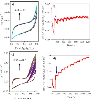
Another reported modification of FCF is their functionalization with quinone-like groups by chemical treatment with KMnO4/H2SO4 (Pereira et al. 2017a). The quinones are well-known for their ability to catalyze the oxidation of NADH (Gorton and Dominguez 2002, Katz et al. 1994, Abdellaoui et al. 2016), allowing the improvement of the bioelectrocatalysis of NAD-dependent enzymes. ADH was immobilized on the pristine FCF and on the FCF functionalized with quinone-like groups, and the performances of the electrodes were compared using electrochemical measurements in the presence of several ethanol concentrations, as shown in Figure 5.
As shown, after the modification of the FCF surface, there is an improvement in the bioelectrocatalysis, and the current densities increase around 10 times. This indicates that the
Figure 4 - (a) Pristine FCF agglomerates. (b) and (c) show the
modification of electrode surfaces could enhance the communication between the electrode and the enzyme.
Besides the modification of the electrode surface, another possibility to improve the charge transfer is the modification of the protein structure. Mano and coauthors (Courjean et al. 2009, Prevoteau et al. 2010) showed that the deglycosylation of glycoproteins could be utilized to decrease the distance between the active center of an enzyme and the electrode surface, improving the DET. Deglycosylation corresponds to the cleavage of the glycans of the protein structure without changing the protein core (Courjean et al. 2009). The glycans are bonded to the protein
backbone by glycosidic and amidic bonds, which could be cleaved by enzymatic or chemical routes. Thus, after this procedure, the enzyme could be utilized as the biocatalysts in solid electrodes with the active center closer to the surface, resulting in an increased faradaic current and improving the performance of the bioanodes in biofuel cells.
Enzymatic procedures (Courjean et al. 2009) for deglycosylation usually involve exoglycosidases and endoglycosidases. The last can be divided into three main groups: endo-D, H, and F, which are responsible for the portions linked by asparagine; endo-β-D-galactosidase, responsible for the hydrolysis of galactosidic bonds in some prosthetic groups; and the endoglycopeptidases, which are divided into N-glycosidases and O-glycosidases, which hydrolyze N-acetylglycosamineasparagine and N-acetylgalatosamineserine/threonine, respectively. Enzymatic routes allow for mild conditions, but they are specific for certain glycans. On the other hand, chemical methods (Patel et al. 1993, Dwek et al. 1993, Edge et al. 1981) are not specific with respect to how the glycan is bonded to the protein. Moreover, they can remove all the glycans under appropriate reaction conditions. In this case, the reactants cleave the glycosidic bonds involving mainly neutral sugars. Trifluoromethanesulfonic acid (TFMS) is a strong acid that has been utilized for the chemical deglycosylation of glycoproteins. This acid cleaves the glycosidic bonds, without changing the protein core, maintaining the enzyme activity (Edge et al. 1981). For example, for the deglycosylated horseradish peroxidase (HRP), a recombinant enzyme, it was showed a much higher rate of heterogeneous DET than for native one. In addition, the percentage of adsorbed enzyme molecules oriented for DET was increased compared to the wild-type HRP. The glycosylation could be considered as the reason for the absence of any electrochemical response of laccase from Coriolopsis fulvocinerea under anaerobic Figure 5 - (a) Cyclic voltammograms of pristine FCF-ADH
and (b) the amperometric response in the presence of several ethanol concentrations using the same electrode. (c) Cyclic
voltammograms of quinone modified FCF-ADH, and (d) the
amperometric response for increasing ethanol concentration
of the quinone-modified FCF-ADH. All measurements were
carried out in N2-saturated, 0.1 mol L-1
sodium phosphate
buffer (pH 7.5) containing 0.6 mmol L-1 NAD+
at 25 ºC. For the cyclic voltammograms, the scan rate was 50 mVs-1
conditions; that is, it increases the distance of the electron tunneling between the laccase and conducting carbon (Shleev et al. 2005a). For GOx, a monolayer of the deglycosylated enzyme was
immobilized on a vitreous carbon electrode, and it was observed that the electrooxidation of glucose started at –490 mV versus Ag/AgCl (Courjean et al.
2009). As shown in Figure 6a, the faradaic currents relative to the FAD/FADH2 cofactors inside both enzymes is much higher for the deglycosylated
enzyme. By using the Laviron formalism (Laviron 1979), the rate of electron transfer obtained for GOx was found to be 0.2 s-1, while this parameter
corresponds to 1.58 s-1 for the deglycosylated enzyme, indicating that, after deglycosylation, the DET is improved.
Another way to change the enzyme structure is to promote enzyme oligomerization because oligomers are more hydrophobic than native
species, which improves the interaction of the enzyme with carbon electrode surfaces. Protein oligomerization can be considered as a type of protein aggregation, and it is dependent on several
factors related to protein structure levels and the protein environment (Wang et al. 2010). Because the environmental conditions can influence protein oligomerization, the pH and hydrophobicity are considered important parameters in determining protein aggregation rates. For instance, GOx has
been oligomerized using a Brønsted acid (TFMS in this case), where the pH of the reaction mixture was drastically reduced, exposing the hydrophobic
chains of the enzyme and stabilizing its structure; consequently, the catalytic activity was retained (Pereira et al. 2017b). This oligomerized enzyme
is more hydrophobic than native GOx, which improves the adsorption of the enzyme on carbon surfaces, promoting an enhancement of DET, as
shown in Figure 6b.
ENZYME IMMOBILIZATION
In 1971, the term “enzyme immobilization” was coined by E. Katchalski-Katzi to designate “enzymes physically confined or localized in a certain defined region of space that retain their catalytic activity and can be used repeatedly and continuously” (Katchalskikatzir 1993). The use of immobilized enzymes in industrial processes has been of great interest since the 1960’s because the anchoring of enzymes on a support facilitates the handling of the enzyme, solves the solubility
Figure 6 - (a) Cyclic voltammograms of GOx (dotted line) and deglycosylated GOx (dGOx) (solid line) adsorbed on glassy carbon electrodes (20 mM
phosphate buffer, pH 7.4, 37 °C, scan rate: 20 mV s-1 , an argon atmosphere). Reprinted from (Courjean et al. 2009) with permission of John Wiley and Sons; (b) Cyclic voltammograms of FCF-GOx (dotted line) and FCF-Ol-GOx (solid line) bioelectrodes (0.10 M
sodium phosphate buffer, pH 7.5, 25 °C, scan rate:
100 mV s-1
problem of some enzymes, and minimizes or eliminates protein contamination of the product. In addition, the immobilization allows the recovery and reuse of the enzyme because of the facile separation of the biocatalyst from the product and the improvement in the enzyme stability (Klibanov 1979).
The development of biodevices, such as biosensors, implantable biodevices, and biofuel cells, requires the immobilization of enzymes on electrode surfaces. Immobilized enzymes for bioelectrochemistry have several advantages, as in industrial applications. However, some drawbacks of this approach need to be considered, for example, difficult reproducibility, greater cost, and lower catalytic activity because of limitations in the mass transfer or conformational changes.
The way that the protein is immobilized significantly affects the interactions between the electrode surface and the enzyme and the electrical communication with the redox site of the protein. Therefore, the performance of the immobilized enzyme depends on the enzyme and the method of anchoring.
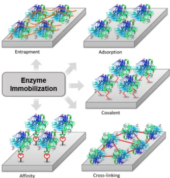
The most frequently used methods for enzyme immobilization are divided into five types: non-covalent adsorption, non-covalent bonding, entrapment, cross-linking, and affinity (Figure 7) (Sassolas et al. 2012, Guisán 2006). The characteristics, advantages, and drawbacks are detailed in the following sections.
ADSORPTION
Immobilization by adsorption is based on physical interactions, such as van der Waals forces, hydrogen bonds, and electrostatic interactions between the support and the biomolecule (Jesionowski et al. 2014). The adsorption method involves placing the electrode in contact with the enzyme solution for a period or depositing the enzyme solution onto the electrode surface until the solvent evaporates. In
both cases, the unadsorbed molecules are removed by washing the electrode with a buffer solution. The presence of defects on the electrode surface can improve the adsorption of enzymes, for example, defects can be easily obtained on FCF electrodes by chemical treatment with permanganate ions in sulfuric acid solution, which results in the exfoliation of the surface (Pereira et al. 2016, 2017a, Martins et al. 2014) or the treatment with sulfuric acid and nitric acid, which promotes the formation of nanoblisters on the FCF electrode surface (de Souza et al. 2016).
by the LbL technique, the electrode is immersed in sequence into aqueous solutions containing the positively and negatively charged materials to be immobilized. This procedure is repeated until the desired number of layers is achieved. In contrast, the LB technique employs monolayers insoluble in water, and these LB films are transferred from the air/water interface onto electrode surface by vertical dipping into the aqueous solution. This procedure can be repeated several times to form multilayers (Iost et al. 2011b). The LbL and LB techniques have been employed for the construction of nanostructured films containing enzymes for different electrochemical applications, such as biosensing and bioelectronic devices (Iost and Crespilho 2012, Siqueira et al. 2010, Crespilho et al. 2006a, c, 2008, Iost et al. 2011b, Caseli et al. 2008).
The adsorption method is the simplest, easiest, and the most inexpensive method of enzyme immobilization onto solid surfaces. Furthermore, this method, generally, does not affect the active site of the enzyme, which contributes to the preservation of its electrocatalytic activity. However, because of the weak bonds involved, the enzyme molecules tend to leach from the electrode surface. The interactions between enzyme and electrode are destroyed by desorption forces, such as high ionic strength and pH. Another disadvantage is that this method is non-specific; thus, it results in the immobilization of other proteins and substrates (Cao and Schmid 2005).
COVALENT BONDING
An important method of enzyme immobilization is covalent attachment because this kind of bond usually provides the strongest attachment between the enzyme and support compared to other types of enzyme immobilization, such as non-covalent adsorption. In this method, enzyme molecules are anchored to the electrode material by multiple
covalent bonds between functional groups. To maintain the enzyme activity, the active site must not participate in covalent bonding.
The reactive amino acid residues should be located on the enzyme surface because access to buried residues is restricted. The most common enzyme functional groups used for covalent immobilization are the amino groups of N-terminal amino acids and (ε)-amino groups of lysine residue (Cao and Schmid 2005); γ- and β-carboxyl groups of glutamic acid and aspartic acid residues and C-terminal carboxyl groups; the guanidinyl groups of arginine residues; the sulfhydryl groups of cysteine residues; the imidazolyl group of histidine residue; the thioether moiety of methionine residue. In addition, in the case of glycosylated enzymes, covalent bonds may be formed by the sugar residues. In particular, enzymes containing cysteine residue can be directly immobilized on gold electrodes by thiol bonds.
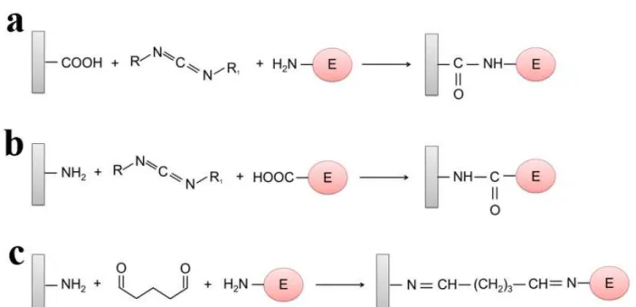
The presence and the types of functional groups on the electrode surface are also important for covalent immobilization, and the most common functional groups are carboxylic acid groups and amino groups. The functional groups are activated by multifunctional reagents, for example, carbodiimide and glutaraldehyde. Carbodiimide can provide the linkage between amino groups of the protein and carboxylic acid groups of the electrode surface and vice versa (Figures 8a and 8b). Glutaraldehyde can be used to bond the amino groups of the support and the enzyme (Figure 8c) (Sassolas et al. 2012).
and extreme pH (Mateo et al. 2007). However, the disadvantage of this immobilization method is that matrix cannot be re-used because of the irreversible attachment of the enzyme.
ENTRAPMENT
Enzymes can be easily immobilized on
electrodes
via
their inclusion in a polymer
network, such as organic or inorganic polymeric
matrices. Some examples of materials used
for the entrapment of enzymes are Nafion,
polyacrylamide, polypyrrole,
chitosan, agarose,
polyaniline, and silica sol-gel (Sassolas et al.
2012, Klotzbach et al. 2008).
The method of enzyme entrapment varies according to the polymer. In the case of Nafion, two methodologies have been successfully employed: (a) the casting of a Nafion suspension on enzymes anchored on the electrode surface, thereby trapping the enzyme between the electrode and the Nafion membrane after solvent evaporation (Pereira et al. 2017a, de Souza et al. 2016) and (b) the casting of a mixture containing Nafion and enzyme on the electrode surface (Klotzbach
gas bubbles, mechanical stirring, and hydrophobic solvents. On the other hand, the main drawbacks of the entrapment immobilization method are enzyme leakage and mass transfer limitations caused by the polymer barrier (Sassolas et al. 2012, Sheldon 2007).
CROSS-LINKING
Enzyme immobilization can also be performed by cross-linking protein molecules using a bifunctional chemical cross-linker (Sassolas et al. 2012). The most used reagent for this purpose is glutaraldehyde (Barbosa et al. 2014). This dialdehyde reacts mainly with the primary amino groups of proteins; thus, the cross-linking of enzymes, either to a solid support or between protein molecules, generally implies the ε-amino group of lysine residues or N-terminal group of the protein chain. Eventually, the reaction proceeds with the nucleophilic functional groups of amino acid side-chains, such as other amines, thiol, phenol, and imidazole (Habeeb and Hiramoro 1968). Co-reticulation of the target enzyme with a functionally inert protein with a high density of superficial lysine residues, such as bovine serum albumin, is quite common for the development of bioelectrodes (Olyveira et al. 2012b, Crespilho et al. 2009a).
The reaction mechanism of glutaraldehyde with proteins can proceed by several mechanisms (Figure 9). This is because glutaraldehyde assumes different monomeric and polymeric conformations in aqueous solution, and each structure can react at different points of the protein chain (Barbosa et al. 2014).
This immobilization method is simple and provides a strong interaction between the biomolecules. The cross-linking method can be combined with other enzyme immobilization methods, for example, entrapment (Olyveira et al. 2012b, Crespilho et al. 2009b). However, cross-linking has some disadvantages, such as enzyme
activity losses because of distortions in the protein conformation, poor reproducibility, and low mechanical stability (Sassolas et al. 2012, Sheldon 2007).
AFFINITY
Enzymes can be immobilized on the electrode surface by (bio)affinity bonds between a specific group of the protein and the support. For this, the electrode surface must be activated, for example with lectin, avidin, or metal chelates; alternatively, the protein must be conjugated with a compound with an affinity for the support (Guisán 2006). This method has the advantage that it provides controlled and oriented immobilization of the enzyme. However, in some cases, genetic engineering may be required for the production of tagged enzymes (Sassolas et al. 2012, Datta et al. 2013).
with histidine at specific positions. Another strategy to immobilize enzymes by affinity bonds is to exploit the strong interaction between biotin and avidin or streptavidin. For this, the biotin is bound to the enzyme through lysine residues by the reaction with biotin-ester reagents; alternatively, the enzyme can be genetically biotinylated (Guisán 2006). In addition, carbohydrates present in the surface of glycoproteins can be employed in the immobilization. Carbohydrates have a high affinity for lectins, such as concanavalin A, which can be easily immobilized on the electrode surface. This kind of immobilization is reversible and has the great advantage of using tags that are naturally present in the enzyme and located in areas of easy accessibility and are not essential for biological activity (Sassolas et al. 2012, Andreescu and Marty 2006).
BIOELECTROCATALYSIS
Electrocatalyst accelerates the rate of chemical reaction, but, in this case, the reaction takes place on the surface of an electrode (Masa and Schuhmann 2016). Electrocatalysis can also be defined as the enhancement in the electrochemical reaction rate provided by a species that is not consumed in the reaction, i.e., the electrocatalyst (Grubb 1963). When the electrode material has sites that can adsorb the reagent, the probability of achieving an energetic situation that favors ET increases significantly. Thus, materials that have this property are called electrocatalysts (Bard and Faulkner 1980). Therefore, the performance of an electrocatalyst depends on the electronic structure of the atoms on the electrode surface (Hammer and Norskov 1995, Hammer et al. 2000, Tersoff and Falicov 1981), as well the chemical nature of the surface (Chen and McCreery 1996), its morphology, and crystal structure (Lebedeva et al. 2002). An electrocatalytic cycle can be summarized by three typical steps (Masa and Schuhmann 2016): i) substrate transportation from the electrolyte bulk
to the active site, ii) electrocatalytic reaction, and iii) product transportation from the catalyst surface. In step (ii), sub-reactions must be considered, which includes the substrate adsorption, ET, and product desorption.
In bioelectrocatalysis, the electrocatalysts are biomolecules, e.g., whole cells or enzymes, in particular, the enzymes of the oxidoreductase group, which catalyze redox reactions (Ghindilis et al. 1997, May 1999), and the fundamental principles are the same as for electrocatalysis. Although there are different types of oxidoreductases, the oxidases and hydrogenases are worthy of attention because they catalyze the same kind of reaction, i.e., the catalytic reactions of these enzymes start from the same substrate and finish with the same product via distinct mechanisms (Ferri et al. 2011). For instance, both GDh and GOx produce gluconolactone, as shown by Equations 1 and 6, respectively. The oxidases transfer an electron to oxygen, while the dehydrogenases transfer electrons directly to an electron acceptor molecule (a coenzyme).
glucose+GOx(FAD)→gluconolactone+GOx(FADH2) (6a)
GOx(FADH2)+O2→ GOx(FAD)+H2O2 (6b)
For enzyme-modified electrodes, ET between the electrode surface and the redox center of the protein is dependent on the enzyme orientation, and of the localization of the enzyme active center, which must be located at a short distance from the electrode to allow the tunneling of the electron (Heller 1992, Falk et al. 2012). Thus, the greatest challenge in bioelectrocatalysis is the development of an electrochemical interface that establishes electrical communication between the enzyme and the electrode surface (Bartlett 2008).
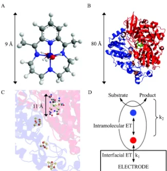
presence of non-protein cofactors, such as metals for metalloenzymes or organic molecules as coenzymes (McCall et al. 2000), is required. In bioelectrocatalysis, the cofactors can serve as redox centers that exchange charge between the enzyme and electrode. Usually, the cofactors are located inside the protein structure and they have high selectivity and specificity (Masa and Schuhmann 2016). Enzymes are a special group of molecular electrocatalysts (Hexter et al. 2014), and they can be attached to the electrode by numerous weak interactions. Therefore, ET between the substrate and the electrode is possible if the enzyme is suitably orientated and the enzyme active site is located a short distance from the electrode, allowing electron tunneling (Heller 1992, Falk et al. 2012). In electron tunneling in a biological process, the electrons can travel up to 14 Å between redox centers through the protein medium under physiological conditions (Page et al. 1999); consequently, it is important that the redox centers be close to the electrode surface. In general, electrocatalysts are small molecules, such as the cobalt complex that catalyzes the hydrogen evolution reaction (Figure 10a) (McCrory et al. 2012); however, the enzymes usually utilized as biocatalysts have diameters around 100 Å (Figure 10b) (Hexter et al. 2014). Thus, in most cases, there is a large distance between the enzyme’s catalytic site and the protein surface, and at least one electron relay center is needed to ensure rapid intramolecular ET (as shown in Figure 10c). Fast interfacial ET between the electrode and the relay system is possible only if the enzyme makes good electronic contact with the electrode surface (Hexter et al. 2014). Figure 10d shows a pictorial model of bioelectrocatalysis, where the interfacial ET between the enzyme and the electrode is separated from the catalytic events. In other words, the bioelectrocatalysis process occurs in two steps.
In comparison to conventional catalysts, enzymes have well-defined active sites, because they are formed of a transition metal ion as a part
of the complex protein matrix. In this case, the electron densities of the redox centers are altered by the peptides and residual chemical groups present in the protein backbone to values matching the energy of substrates, allowing a faster conversion of the substrates (Masa and Schuhmann 2016).
UTILIZATION OF ELECTROCHEMISTRY TO STUDY PROTEIN REDOX REACTIONS
VOLTAMMETRIC TECHNIQUES
bioelectrochemistry has focused on the mechanism of ET and how it is linked to other physiological functions. For small biochemical systems, the voltammetric method is a common choice, providing insight into the kinetics and thermodynamics of ET reactions. Moreover, these methods provide valuable information concerning the mechanisms and interactions of such biochemical systems (Gulaboski et al. 2012). However, for proteins, the utilization of voltammetry to understand their redox reactions is difficult because of the protein size and the presence of large lipophilic tails that prevent the transfer of electrons between the protein and the electrode.
Because the active center are often bound to internal buried sites in the secondary structure of the enzymes, access by the electrodes is difficult, which implies a slow electron exchange with the electrodes (Armstrong 1990). Consequently, early biosensors and biofuel cells utilized mediators for electrons flow. However, mediation is not the ideal approach to study fundamentals of catalytic enzyme reactions because the influence of the enzyme and the enzyme substrate on the electrochemistry of the mediator must be considered. Thus, to facilitate DET between the enzyme and the electrode surface, some types of protein thin films at the electrode surfaces have been developed (Rusling and Zhang 2001). A possible approach is so-called protein-film voltammetry (PFV) (Armstrong et al. 1997), which avoids mediation and allows the direct observation of the enzymatic ET, as well as its catalytic reaction. The concept of the PFV was developed by Fraser Armstrong and provides an important way to investigate how ET in proteins is coupled to chemical reactions, for instance, in catalysis. PFV involves co-adsorbing proteins with aminocyclitols and polymixins to give monolayers with highly reversible voltammetry utilizing an edge plane pyrolytic graphite electrode (Armstrong et al. 1997). This method solves problems such as the protein diffusion because the protein under
investigation is adsorbed on the electrode, forming a stable film of enzyme molecules.
The PFV approach represents a method for studying the fundamental electrochemical of enzyme redox chemistry. In this methodology, the protein is deposited on the electrode surface mainly by self-assembly from the aqueous electrolyte in which the protein is dissolved. Thus, the redox features of the adsorbed protein can be monitored by applying a controlled potential to the protein-modified electrode, that is, by using different voltammetric techniques (Gulaboski et al. 2012). Learning how to adsorb a protein in a native and active configuration on the electrode surface and understanding the voltammetric results on both a quantitative and qualitative level are two intrinsic challenges of PFV. Some factors make PFV an excellent technique to study the electrochemistry of different proteins. For example, the sample economy: the amount of sample required to form a monolayer is around 10-11 mol cm-2; the sensitivity of the method: the investigation of the reactions requires a small amount of the sample; and the rate of the reaction: the voltammetric waveform and current are not limited by diffusion (Armstrong 2002).
a pair of compact reduction and oxidation peaks, and the average value of these peaks gives the reduction potential, while their changes in shape and separation as the scan rate gives information on the ET kinetics (Armstrong 2002).
As cited above, CV is the most popular method for the study of thin protein film electrochemistry. For reversible electrochemical reactions, the interconversions between oxidized and reduced forms of the enzyme are fast, considering the time scale of the voltammogram. The ideal cyclic voltammogram must have symmetric oxidation and reduction peaks of equal heights and no oxidation-reduction peak separation (Bard and Faulkner 1980) The integration of the CV surface area provides the charge (Q), in coulombs involved in the process, allowing the determination of the total surface concentration (ΓT), in other words, the amount of the enzyme in the film (Equation 7).
T
Q
=
nFA
Γ
(7)Here, n is the number of electrons transferred in the reaction, F is the Faraday constant, A is the electrode area in cm2, and Γ
T is the total surface concentration of electroactive protein in mol cm-2. The ideal reversible peak current (Ip) for a reversible thin electroactive film on an electrode is:
2 2
4
T p
n F A I
RT
ν
Γ
= (8)
where R is the gas constant, T is the temperature in Kelvin, and ν corresponds to the scan rate. This model where the Ip increases linearly with increasing scan rate is ideal and is known as ideal thin-layer voltammetry (Rusling and Zhang 2001). However, the cyclic voltammograms of enzyme films are usually significantly different from predictions of an ideal thin layer model (Rusling and Zhang 2001, Rusling 2003), and these deviations depend on the enzyme and film properties. For example, the shape of the cyclic voltammogram can be unsymmetrical
when only partial electrolysis of the redox sites in the films occurs during the scan.
For films where the cyclic voltammograms are controlled by diffusion, the integral under the peak is not proportional to the surface concentration of electroactive centers in the film, and this occurs because only a fraction of the protein has been electrolyzed. In this case, the peak current for an n-electron reaction in a film is:
(
5)
3/ 2 1/ 2 1/ 22.69 10
p ct f
I = × n AD ν C (9)
where the concentration of electroactive species Cf is equal to ΓT/d, d is the film thickness, and Dct is the charge transport diffusion coefficient (Murray 1984).
As described previously, PFV has been utilized to study systems in the absence of a substrate. However, this approach could also be used for mechanistic studies in the presence of substrate. In this case, rotating-disc voltammetry (RDV) can be used to obtain KM and kcat, and the limiting current (IL) of the enzyme films in solution with the substrate is determined by the Koutecky-Levich approximation (Shaked and Whitesides 1980) (Equations 10 and 11).
1 1 1
L cat Lev
I = I +I (10)
2/3 1/6 1/ 2
0.62
LevI
=
nFAD Cv
−ω
(11)Here, C is the bulk concentration of the substrate, D is the diffusion coefficient of the substrate, v is the kinematic viscosity of the solution, and ω is the electrode rotation rate.
Icat in Equation 10 corresponds to the catalytic current for the enzyme reaction with the substrate, and the electrochemical form of the Michaelis-Menten Equation is (Sucheta et al. 1993):
(
cat)
cat
M nFA k C I
C K
Γ =
+
where Γ corresponds to the surface coverage of enzyme that can be measured by CV, and KM and kcat are the apparent Michaelis-Menten parameters.
Pulsed voltammetry can provide better sensitivity and resolution when compared to other voltammetric methods. For thin enzyme films, the square-wave voltammograms give a reversible ET with symmetric peaks resulting from subtraction of the currents measured at the end of each forward and reverse pulse. As CV, forward-reverse SWVs peaks are valuable for mechanistic analysis. However, using this method, there is no direct relation between the electroactive surface area and the integral under the curve; consequently, this parameter is better determined using CV.
Marcus theory provides a more realistic description of enzyme thin-film voltammetry; however, using CV, the analysis methods are less accessible than that explained above. On the other hand, using SWV, Marcus theory can be applied. In this case, non-linear regression analysis of SWV data allows the estimation of reorganization energies and the ET rate constants (Rusling et al. 2008). In addition, SWV with large amplitude pulses could be used to study more complex ET processes in multicenter enzymes with multiple redox centers (Jeuken et al. 2002).
IN SITU TECHNIQUES
Bioelectrochemical methods combined with spectroscopic techniques can provide detailed information about the activity and rate of an enzymatic process and gain direct structural insight into functionally relevant states (Ash and Vincent 2016). Vincent’s group have studied metalloenzymes under direct electrochemical control by infrared (IR) spectroscopy (Healy et al. 2011, Grabarczyk et al. 2014, Ash et al. 2015). This in situ spectroelectrochemistry with IR radiation is shown in Figure 11 and is based on attenuated total reflectance (ATR) measurements, where the working
electrode, modified with the protein of interest, is placed above the ATR prism (Ash and Vincent 2016, Hidalgo et al. 2015). This makes it possible to carry spectroscopic analysis under precise electrochemical control. This technique has been used in the investigation of hydrogenases that have metallic active sites, such as [NiFe] and [FeFe] hydrogenases (Ash, et al. 2017a, b, Paengnakorn et al. 2017, Healy et al. 2013). In these cases, the shifts in the ν(CO) or ν(CN) vibrational bands are monitored, allowing the identification of short-lived intermediates, the diagnosis of redox-coupled structural changes, and the monitoring of side reactions. Furthermore, some potential-dependent changes can be observed in bands associated with dehydrogenase active site ligands. This spectroelectrochemical method has also been used to study a [MoFe] nitrogenase (Paengnakorn et al. 2017), demonstrating the versatility of this approach.
et al. 2010). This approach is based on redox-dependent absorbance changes, which can be monitored in the fluorescence domain by means of a Förster resonance energy transfer (FRET) donor– acceptor pair, whereby the redox site is the energy acceptor and an externally linked dye label is the fluorescent donor (Krzeminski et al. 2011, Salverda et al. 2010). Lastly, another in situ technique is based on magnetic spectroscopy, where magnetic circular dichroism is used for the evaluation of the proteins with in situ control of electrochemical potential (Marritt et al. 2006). This methodology was utilized in the analysis of the redox behavior of the cytochrome-c, a hemoprotein. The technique was demonstrated to be a robust analytical tool for the determination of heme properties in multiheme enzymes (Marritt et al. 2006).
The most recent techniques for studying bioelectrochemical processes, by using in situ techniques, are the differential electrochemical mass spectrometry (DEMS) (de Souza et al. In
Press) and potentiometric titrations combined with electron paramagnetic resonance (Artz et al. In Press). The DEMS technique was applied in the study of the bioelectro-oxidation of ethanol by ADH, and allows the concomitantly detection of the two substrates (NADH and acetaldehyde) by using electrochemical and mass spectrometric techniques on-line. This new technique can be useful for other redox enzymes when their products are gaseous or volatile. The scheme of DEMS setup and the bioelectrohemical cell are represented in figure 12a and 12b. This system is composed by a conventional electrochemical cell connected in a mass spectrometer and the bioelectrochemical cell had an FCF with immobilized ADH as working electrode, the electrical contact was provided by a gold ring and wire, and the working electrode is supported over a polytetrafluoroethylene (PTFE) membrane that allows only the passage of gaseous and volatile compounds.
Figure 11 - Schematic representation of the spectroelectrochemical ATR-IR cell designed for protein
film infrared electrochemistry experiments, showing the relative location of electrodes and the direction of solution flow. The enzyme, Hyd-1, is adsorbed on carbon beads which are cast directly onto the Si internal reflection element. Adapted and reprinted from (Hidalgo et al. 2015) with permission of John
APPLICATIONS: BIOSENSORS AND BIOFUEL CELLS
In addition to answer fundamental questions about the ET processes in biological systems involving macromolecules, whole cells, and membranes, bioelectrochemistry has a wide range of practical applications, such as in biosensors, immunoassays, energy conversion, wastewater treatment, and bioelectrosynthesis (Bartlett 2008, Olyveira et al. 2012a). As the present review addresses the bioelectrochemistry of enzymes, we will describe the main applications of enzymatic devices, such as biosensors and biofuel cells (BFCs). These biodevices have been successfully used in numerous areas, ranging from environmental monitoring to in vivo energy harvesting.
ENZYMATIC BIOSENSORS
Enzymatic biosensors are analytical devices that use enzymes to detect and/or quantify specific chemicals. The selective analysis provided by these devices is based on biochemical molecular
recognition. Because of their specificity, portability, fast response, and low cost, enzymatic biosensors are present in many different fields, such as food quality control, the monitoring of industrial processes and pollutants, and biomedical analyses (Iost et al. 2011a, Oliveira et al. 2014, Luz et al. 2013). A major application of biosensors is in blood glucose monitoring for the management of diabetes. Glucose biosensors are commercially available and account approximately 85% of the world market for biosensors. Most personal blood glucose monitors are based on disposable, screen-printed enzyme electrode strips. Each strip contains printed working and reference electrodes, where the working electrode coated with the necessary reagents (enzyme, mediator, stabilizer, surfactant, linking, and binding agents) are deposited in a dry form (Turner 2013).
The first glucose enzyme electrode was reported in 1962 by Clark and Lyons. They used GOx, and the measurements were based on monitoring the consumed oxygen. As discussed, GOx catalyzes Figure 12 - (a) DEMS setup: (1) electrochemical half-cell, (2) connection between electrochemical half-cell and mass spectrometer, (3) pre-vacuum chamber, (4) turbomolecular vacuum pumps, (5) quadrupole, and (6) controller. The red spheres represent volatile compounds, and the blue and yellow ones are the ionized fragments. (b) Zoomed area between 1 and 2, where CE is the counter-electrode, WE is the working counter-electrode, the black region is FCF with the immobilized ADH, the yellow portion is a gold electrical connection, and RE is the reference electrode (Ag/AgCl/Cl
the oxidation of glucose to gluconolactone. In the presence of O2, the natural GOx electron acceptor, the oxidized form of the enzyme, GOx-FAD, is regenerated and oxygen and hydrogen peroxide are produced (Equation 6b).
Glucose concentration can be indirectly determined by the amperometric monitoring of hydrogen peroxide. This product is easily oxidized to molecular oxygen at a platinum electrode, according to Equation 13, and glucose biosensors based on the measurement of hydrogen peroxide are known as first-generation biosensors (Wang 2008).
(13)
Although the measurement of the formed peroxide is simple, this kind of biosensor is subject to errors that are attributed to fluctuations in oxygen tension, the stoichiometric oxygen limit, and the presence of interferers in the blood (Wang 2008). To overcome these problems, efforts have focused on the replacement of oxygen by nonphysiological electron acceptors, redox mediators, for second-generation glucose biosensors. Ferrocene derivatives, ferricyanide, conducting organic salts, quinones, transition-metal complexes, phenothiazines, and phenoxazines have all been used as redox mediators for GOx. Using mediators, glucose measurements become independent of oxygen pressure, and interfering reactions are minimized (Wang 2008).
The most recent glucose biosensors based on GOx have eliminated redox mediators (third-generation glucose biosensors). In this case, the electrons are transferred directly from the redox site of the enzyme to the electrode at potentials very close to the redox potential of the enzyme. Thus, biosensors with high selectivity are obtained in a simpler and cleaner system (Wang 2008). However, the development of GOx-based electrodes that involve DET and show high performance is not
trivial and, usually, new electrode materials are required to enhance the enzyme–electrode electronic communication. Recently, the DET of GOx adsorbed on FCF electrodes modified with graphene oxide was reported. In this case, the authors demonstrated that the presence of graphene oxide at the enzyme/electrode interface decreases the distance between the FAD/FADH2 enzymecofactor and the FCF surface (Figure 13) (Martins et al. 2014). Another way to obtain a clear DET between GOx and the electrode and to improve the electrocatalytic response is by modifying the enzyme structure. For example, the controlled oligomerization of GOx by treatment with a Brønsted acid can be used to provide a more efficient biocatalyst (Pereira et al. 2017b).
Current efforts concerning the development of glucose biosensors have focused on implantable biodevices. In vivo glucose monitoring can eliminate
Figure 13 - Field emission-scanning electron microscope images of the FCF (a) and graphene oxide-modified fibers (c). Cyclic voltammograms of (b) FCF and (d) graphene
oxide-modified fibers before GOx immobilization (black lines) and
after enzyme immobilization (blue lines). Scan rate: 30 mV s-1
. Adapted and reprinted from (Martins et al. 2014) with permission of Royal Society of Chemistry.
2 2 2
Figure 14 - (a) Photograph of the prepared device implanted in a rat vein. From left to right, carbon cloth and the micromanipulation of a single FCF using an optical microscope to obtain the biochip in a millimeter catheter are presented. The SEM image shows a more detailed visualization of the CF dimensions. (b) Left: Schematic representation of the local implanted biochips in jugular rat veins 1 and 2. Right: Photograph of the implanted FCF microelectrodes in the jugular rat veins (Rattus novergicus). Reprinted from (Iost et al. 2015) with permission of John Wiley and Sons.
the inconvenience associated with standard finger-stick sampling. Moreover, an implanted glucose biosensor can provide continuous monitoring of glucose in real time and with high accuracy (Wang 2008). The development of implantable biosensors requires biocompatible and miniaturized systems, and these characteristics can be easily achieved by using FCF-based electrodes. Iost et al. (2015) reported an implantable biochip based on GOx and a neutral redox mediator immobilized on the surface of FCF (Figure 14). This material enables the biochip to be conveniently manipulated during insertion into the jugular vein of a living rat. The ability for in vivo glucose detection was evaluated with a normal concentration of glucose and with diabetic simulation. In this case, the biochip showed promise performance for future applications of implantable bioelectronics devices.
ENZYMATIC BIOFUEL CELLS
Similar to fuel cells, BFCs are electrochemical devices that convert the free energy of a chemical
reaction into electrical power for the purpose of doing work (Bartlett 2008). BFCs have been classified as either microbial-based and enzymatic fuel cells according to the location of the enzymes, which can be inside of microorganisms or outside of living cells. However, microbial-based biofuel cells are outside the scope of this review and will not be discussed here.
Enzymes show remarkable advantages over conventional inorganic catalysts, such as biocompatibility. In addition, they are less expensive than precious metal catalysts and show higher efficiency, higher selectivity, and higher activity under mild conditions (room temperature and near-neutral pH). The selectivity of enzymes for some substrates can simplify the design of BFCs because the separation of fuel and oxidant by a membrane is not necessary. These features make enzymatic BFCs attractive alternatives to rechargeable batteries and traditional fuel cells.