online | memorias.ioc.fiocruz.br
Molecular typing reveals the co-existence of two transmission cycles
of American cutaneous leishmaniasis in the Andean Region
of Venezuela with Lutzomyia migonei
as the vector
Annhymariet Torrellas1, Elizabeth Ferrer2/+, Israel Cruz3,*, Héctor de Lima4, Olinda Delgado5, José Carrero Rangel6, José Arturo Bravo1, Carmen Chicharro3, Ivonne Pamela Llanes-Acevedo3, Michael A Miles7, María Dora Feliciangeli1,†
1Universidad de Carabobo, Facultad de Ciencias de la Salud, Centro Nacional de Referencia de Flebotomos y otros Vectores,
Instituto de Investigaciones Biomédicas Dr Francisco J Triana-Alonso, Maracay, Venezuela
2Universidad de Carabobo, Facultad de Ciencias de la Salud, Instituto de Investigaciones Biomédicas Dr Francisco J Triana-Alonso,
Maracay, Venezuela
3WHO Collaborating Centre for Leishmaniasis, National Center for Microbiology, Instituto de Salud Carlos III, Majadahonda, Madrid, Spain 4Ministerio del Poder Popular para la Salud, Servicio Autónomo, Instituto de Biomedicina, Caracas, Venezuela
5Universidad Central de Venezuela, Instituto de Medicina Tropical, Caracas, Venezuela 6Servicio de Dermatologia, Municipio Tovar, Merida, Venezuela
7Faculty of Infectious and Tropical Diseases, London School of Hygiene and Tropical Medicine, Department of Pathogen Molecular Biology,
London, United Kingdom
BACKGROUND The transmission routes for American cutaneous leishmaniasis (ACL) are in flux, so studies examining its transmission in humans, mammalian hosts, and sand fly vectors are urgently needed.
OBJECTIVES The aim of this work was understand the epidemiological cycles of Leishmania spp., which causes ACL in the Andean Region of Venezuela, by identifying the Leishmania and the sand fly species involved in human and dog infections.
METHODS Thirty-one biopsies from patients in Mérida and Táchira states with suspected ACL were studied by both parasitological tests (cultures and hamster inoculation) and a molecular test [Internal transcribed spacer 1 (ITS1) nested polymerase chain reaction-restriction fragment length polymorphism (PCR-RFLP)]. We also conducted a survey to detect Leishmania infection in dogs (Immunifluorescence antibody test and ITS1 nested PCR-RFLP) and sand flies (ITS1 nested PCR-RFLP) from El Carrizal, a highly endemic focus of ACL in Venezuela.
FINDINGS Three different Leishmania species were identified in the clinical samples from humans (Leishmania braziliensis, L. guyanensis, and L. mexicana) and dogs (L. guyanensis and L. mexicana). The predominant sand fly species found were those from the Verrucarum group (infected with L. mexicana) and Lutzomyia migonei (infected with L. guyanensis and L. mexicana).
MAIN CONCLUSIONS We show that Lu. migonei may be the putative vector in two ACL epidemiological cycles, involving L. guyanensis and L. mexicana. We also report for the first time the presence of L. guyanensis in domestic animals.
Key words: Leishmania - epidemiology - diagnosis - PCR-RFLP
Molecular epidemiology can unravel the complexi-ties of transmission cycles, thereby providing guidance for the control strategies used to manage vector-borne diseases.(1) The polymerase chain reaction-restriction fragment length polymorphism assay (PCR-RFLP), based on an analysis of the ribosomal DNA internal tran-scribed spacer 1 (ITS1) sequence, has increasingly been
doi: 10.1590/0074-02760180323
Financial support: European Commission project “Control strategies for visceral leishmaniasis (VL) and mucocutaneous leishmaniasis (MCL) in South America: applications of molecular epidemiology”
(EC contract INCO-CT2005-015407, LeishEpiNetSA) and by the Ministerio de Ciencia y Tecnología (MPPCTI/FONACIT), Project MISIÓN CIENCIA - Leishmaniasis, 2008000911 - 2.
* Present address: Foundation for Innovative New Diagnostics, Geneva, Switzerland.
†In memoriam
+ Corresponding author: elizabeth.ferrer@gmail.com Received 8 July 2018
Accepted 30 October 2018
used to identify Leishmania spp., because almost all the medically relevant Leishmania parasites from different endemic regions can be identified by this technique.(2) PCR-RFLP, or variants of it, has been used to detect and identify the different Leishmania spp. that cause Ameri-can cutaneous leishmaniasis (ACL).(3)
Several studies have reported the presence of ACL in dogs.(4,5,6) The detection of leishmanial DNA in canine samples has gained attention both in the diagnosis of ACL in dogs and in epidemiological studies.(7)
Leish-mania species that occurs in humans. Based on the above criteria, about 530 sand fly species exist on the American continent,(12) 56 of them belonging to the genus Lutzomyia that are suspected or proven vectors responsible for sus-taining one or more ACL epidemiological cycles.(13)
The main criticism used by classical entomologists against the use of molecular tools is that the detection of parasitic nucleic acids does not prove the presence of live, infective organisms (metacyclic forms) in the vec-tors. However, the ability to readily apply such molecu-lar methods to all components of the transmission cycle (human, animal reservoir, and vector) makes a funda-mental contribution to our understanding of the epide-miology of leishmaniases.
In Venezuela, only a few epidemiological studies have been conducted, especially in the Andean Region. Be-tween 2003 and 2007, Mérida state reported an ACL inci-dence rate of 19.43/100,000 inhabitants.(14) The Dermatol-ogy Service in the municipality of Tovar (184 km2; 35,000 inhabitants) recorded 93 cases (11.85% of the total), with the majority of them (24.73%) arising from the village of El Carrizal. Based on this background and logistical fa-cilities, during 2008 - 2009 we carried out the detection, isolation, and identification of Leishmania parasites from patients attending the Municipal Dermatology Service with dermal lesions compatible with ACL, from dogs, and from sand flies in the village of El Carrizal.
MATERIALS AND METHODS
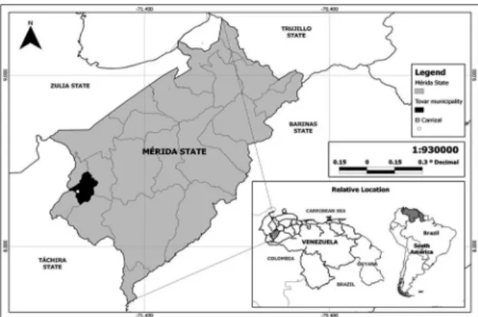
Ecological framework - The landscape of the Vene-zuelan Andean Region, which mainly includes the states of Trujillo, Merida, and Táchira, as well as the highlands of Barinas and Apure states, encompasses a cloudy high mountain forest, moorland, and a cloudy lowland tropi-cal rainforest. The climate is bimodal, with a rainy sea-son and a dry seasea-son; the main rains occurring between the months of April and May, and during the months of September to November, both with average monthly rain-fall exceeding 120 mm, whereas in other months, rain is scarce. The average annual precipitation, temperature (T), and relative humidity (RH) are, 1891 mm, 19.5ºC ,and 78%, respectively (data registered by the Venezuelan Air Force, VAF). The studied village was El Carrizal which is located at 08º17´63”N, 71º45’75”W (Fig. 1).
Human samples - From January to December 2008, 61 patients attended the Dermatology Service with le-sions compatible with ACL; 31 patients consented to sampling from the lesion. Twenty-five of the patients came from Merida state, and six from the bordering An-dean Táchira state. A member of the study team (JCR), a medical doctor and head of the Dermatology Service, performed the skin biopsies. Samples were placed into polypropylene cryotubes with 10% dimethyl sulphoxide (DMSO), and labelled to allow tracking of the patient, location, and date. The tubes were stored at -196ºC in aluminium canes submerged in liquid nitrogen in a cryo-logical tank(15) to be transported to the BIOMED labora-tory (Universidad de Carabobo, Maracay) for parasito-logical and molecular tests.
Canine survey - In 2008, El Carrizal had a popula-tion of 624 inhabitants, living in 152 dwellings. Their main occupation was agriculture (cereals and legumes, roots and tubers, vegetables, bananas and coffee). Tour-ism and commerce were other important activities.
Sixty-eight out of the 152 families (44.73%), almost proportionally distributed across four sectors of the vil-lage, were visited and informed about cutaneous leish-maniasis, the risk of having infected dogs in the house, and the objectives of our research. Informed consent to take blood samples from the animals was obtained from 43 houses, each with up to five dogs; the other 25 dwell-ings having no dogs. A total of 69 dogs were registered (without any sign of lesions, or scars compatible with ACL), but only 28 of the dogs (40.58%) were sampled, due to limiting factors, such as absence of the dogs from the house during site visits, aggressive dogs that were dif-ficult to handle, or roaming dogs that could not be located. Blood samples were transported to the laboratory in poly-styrene containers with freezing packs to allow for the molecular detection and identification of parasites.
Sand fly survey - For the purpose of this epidemio-logical study, monthly sampling collections were car-ried out from January 2008 to January 2009, we selected those specimens that were collected during January-March 2008 and November 2008-January 2009, as these months correspond to the peaks of the sand fly popu-lation, and which would include nulliparous and par-ous sand flies. Trapping was conducted in five hpar-ouses spaced across the village whose owners agreed to col-laborate. Three of these houses reported 8, 2, and 11 cases of ACL. Three CDC light traps (John W. Hock Company, Gainesville, Florida, USA) were placed over-night (from 18:00-19:00 to 6:00-7:00) in each house for three-four consecutive days per month.
One trap was located indoors in the main sleep-ing room or the room adjacent to it. A second trap was placed outdoors (0-20 metres from the house) and close to the resting places of the domestic animals, predomi-nantly dogs or chickens. The third trap was located in the woodland, approximately 100 metres from the houses. Additionally, when the weather was favourable, a
non trap was also placed in the woodland, between 19:00 to 22:00, further from the CDC trap (10 metres). All the sand flies collected were stored in vials containing abso-lute ethanol, and labelled to allow tracking of collection date, trap number, house, and habitat.
Identification of males and females sand flies was carried out in the laboratory based on morphological characters described in the guide to the identification of Lutzomyia sandflies of Young and Duncan.(16) A quick and reliable method for the large-scale identification of females and Leishmania spp. was used to separate fe-males into pools by species, date, trap, house and habi-tat.(17) Briefly, each pool was washed three times with distilled water, before placing each specimen in a small drop of diluted phenol (40%) on a microscope slide (usu-ally ten sand flies per slide) to allow for rapid clarifica-tion. The sand flies were identified under 250 X and 400 X magnifications based on morphological characters (genitalia, genital pump, genital filaments and the ae-deagus in males, and pharynx, cibarium, horizontal and vertical teeth, spermathecae, and spermathecal ducts in females). Each female was then washed again four times in distilled water, and then pooled according to the trap, house of origin, and species, with a maximum of 20 sand flies per vial in Eppendorf tubes containing lysis buffer (0.02 M NaCl, 0.5 M EDTA pH 8.0, 1M Tris-HCl, pH 7.4) and stored at 4ºC. Blood-fed females were kept apart for a separate study on the identification of blood-meal sources (in preparation).
Parasitological tests - With the aim of isolating the parasites, a portion of each of the biopsies from patients with suspected ACL was cultured in liver infusion tryp-tose (LIT) culture medium supplemented with 20% inactivated foetal bovine serum, and in Novy-McNeal-Nicolle medium. Cultures were replicated every 8-10 days, and observed for parasite growth, and they were discarded if they remained negative after four replicate cultures have been performed. In addition, another por-tion of the biopsy was macerated in sterile PBS and inoc-ulated into the footpads of hamsters (Cricetus auratus), which were checked weekly for two months to detect infection, with the aim of isolating the parasites.
Immunofluorescence antibody test - We used an im-munofluorescence antibody test (IFAT) to detect the pres-ence of antibodies against Leishmania spp. in the sera from dogs. The antigen was prepared according to the method described by Pappas et al.,(18) using whole Leish-mania promastigotes grown in vitro from isolates ob-tained from cutaneous lesions of patients with ACL, who were diagnosed and treated at the “Laboratorio de Inmu-nodiagnóstico of the Instituto de Medicina Tropical, Uni-versidad Central de Venezuela”. The reaction was consid-ered positive when more than 50% of the parasites showed complete peripheral fluorescence (in titres > 1:16).
Molecular tests - For the detection and identification of Leishmania spp., all the samples from patients, dogs, and sand flies, either fed or unfed, were analysed by the PCR-RFLP method using the ribosomal DNA internal transcribed spacer 1 (ITS1) as the target sequence.
DNA was extracted from skin biopsy samples ob-tained from human patients, as well as blood samples from dogs and sand fly pools. The sand fly pools used for PCR-RFLP were selected randomly, taking into account the different species, areas, and types of capture repre-sented, and a Proteinase K- phenol-chloroform extraction was performed as previously described.(19) DNA pellets were dried and then re-dissolved in 50 µL of sterile dis-tilled water. The samples were kept at 4ºC until analysis. DNA was also extracted from cultures of the following Leishmania reference strains: L. (V) braziliensis MHOM/ BR/1975/M2903, L. (V) guyanensis MHOM/BR/1975/ M4147, and L. (L) mexicana MHOM/BZ/1982/BEL21.
The molecular test for ITS-1 nested PCR - HaeIII RFLP was performed according to protocols described by Schönian et al.(2) and Cruz et al.,(20) with primers LITSR (5’CTGGATCATTTTCCGATG3’) and L5.8S (5’TGATACCACTTATCGCACTT3’) for the first ampli-fication, and SAC (5’CATTTTCCGATGATTACACC3’) and VAN2 (5’GCGACACGTTATGTGAGCCG3’) for the second amplification. The PCR products were digested with the enzyme HaeIIIaccording to the manufacturer’s protocol. Restriction fragments were subjected to elec-trophoresis in 2% agarose at 100V in 0.5-TBE (0.045 M Tris-borate, 1 mM EDTA) buffer and visualised under
TABLE I
Efficiency of Leishmania detection tests and species identification on skin biopsies from patients with suspected American cutaneous leishmaniasis (ACL), Mérida and Táchira states
Technique (Nº) (%) L. braziliensis L. guyanensis L. mexicana
Cult/Hams/PCR-RFLP 8 25.81 5 3 0
Culture/PCR-RFLP 7 22.58 4 3 0
Hamster/PCR-RFLP 2 6.45 1 0 1
PCR-RFLP 8 25.81 1 3 4
Negative 6 19.35 - -
-Total 31 100.00 11 9 5
ultraviolet light after staining for 15 min with ethidium bromide (0.5 µg/mL). Negative controls were the reac-tion mixture and water; positive controls were reference strains of the most common species of Leishmania spp. circulating in the area.
Statistical analysis - Data for the study were record-ed using Microsoft Excel 2010. The frequency distribu-tions and percentages were calculated for all collected variables. Tests for homogeneity (by Chi square, X2)
were calculated to compare the proportions of infection by culture, hamster inoculation (xenodiagnoses), IFAT, and PCR-RFLP, and also to compare the distribution of Leishmania species. The significance level was 0.05 with confidence limits (CL) of 95%.
Ethical considerations - The project was approved by the Committee of Bioethics of the Institute of Biomedi-cal Research of the University of Carabobo (BIOMED-UC), following the guidelines for the care of humans and animals issued by the Commission of Bioethics of the
Ministry of Science and Technology and the Operational Guidelines for Ethics Committees that Review Biomedi-cal Research (TDR/PRD/ETHICS/2000.1). Field work was carried out in close collaboration with the Service of Dermatology of the Tovar Municipality, and the samples from patients were taken and provided according to the protocols of the Control Program of Leishmaniasis. In-formed consent was requested and signed by the owners of dogs from which blood samples were taken, according to the applicable ethical regulations.
RESULTS
Human samples - Table I shows the results of the para-sitological and molecular tests from 31 biopsies of patients with suspected ACL. As a result, 8/31 (25.81%) were posi-tive by all three techniques (culture, hamster inoculation, and PCR-RFLP), 7/31 (22.58%) were positive by culture and PCR-RFLP, 2/31 were positive by hamster inocula-tion and PCR-RFLP (6.45%), 8/31 (25.81%) were positive only by PCR-RFLP, and 6/31 (19.35) were negative by all
techniques. No statistical difference (χ2 = 1.68; df = 1; p = 0.195) was observed between the proportion of posi-tive parasite cultures (48.4%), and posiposi-tive
xenodiagno-ses (32.2%) (χ2 = 1.68, df = 1; p = 0.195), but PCR-RFLP positivity (80.6%) was significantly higher than a positive
xenodiagnosis (χ2 = 14.76, df = 1; p < 0.001), as well as a
positive culture (χ2 = 7.05, df =1; p = 0.007).
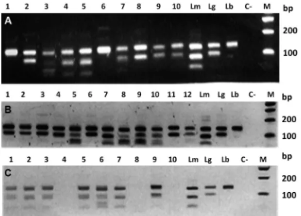
An analysis of the PCR-HaeIII-RFLP of the 25 PCR positive samples showed that three species of Leishma-nia were responsible for causing ACL in these patients: L. braziliensis in 11 cases, L. guyanensis in nine cases, and L. mexicana in five cases (Table I); Fig. 2A shows the PCR-RFLP results. A chi squared homogeneity test showed that there was no statistical difference in the dis-tribution of L. braziliensis, L. guyanensis, and L. mexi-cana in the screened samples (p = 0.2634).
Dog samples - Eighteen of the 28 dogs evaluated were positive by IFAT (64.29%), and 25 were positive by PCR (89.29%) (Table II), with PCR positivity being
sig-nificantly higher than IFAT positivity (χ2 = 4.91, df =1; p = 0.026). Seventeen (68%) of the PCR-positive dogs were infected with L. guyanensis,and eight (32%) with L. mexicana (Table II, Fig. 2B).
Fig. 2: molecular typing of Leishmania isolates from human, dogs and sand flies. The HaeIII digested polymerase chain reaction (PCR) prod-ucts were analysed by electrophoresis and ethidium bromide staining in 2% agarose gel. Lanes Lm: L. mexicana (MHOM/BZ/1982/BEL21);
Lg: L. guyanensis (MHOM/BR/1975/M4147); and Lb: L. braziliensis
(MHOM/BR/1975/M2903). Lane C-: negative control; Lane M: molec-ular weight marker (100 bp ladder, Promega). (A) Lanes 1-10: positive human clinical samples; (B) Lanes 1-12: positive canine samples; (C) Lanes 1-3, 5-7 and nine positive Lutzomyia migonei pools.
TABLE II
Results of immunofluorescence antibody test (IFAT), polymerase chain reaction (PCR) and Leishmania identification by PCR-restriction fragment length polymorphism (PCR-RFLP) on dogs blood samples from the American cutaneous
leishmaniasis (ACL) focus El Carrizal, Mérida state, in the Andean Region of Venezuela
IFAT PCR N IFAT + % PCR + % L. guyanensis L. mexicana
IFAT + (1/16) PCR + 5 17.86 17.86 3 2
IFAT + (1/32) PCR + 5 17.86 17.86 5 0
IFAT + (1/32) PCR - 2 7.14 - -
-IFAT + (1/64) PCR + 6 21.43 21.43 3 3
IFAT - PCR + 9 - 32.14 6 3
IFAT - PCR - 1 - - -
-Total (n) % (28) 100 (18) 64.29 (25) 89.29 (17) 68 (8) 32
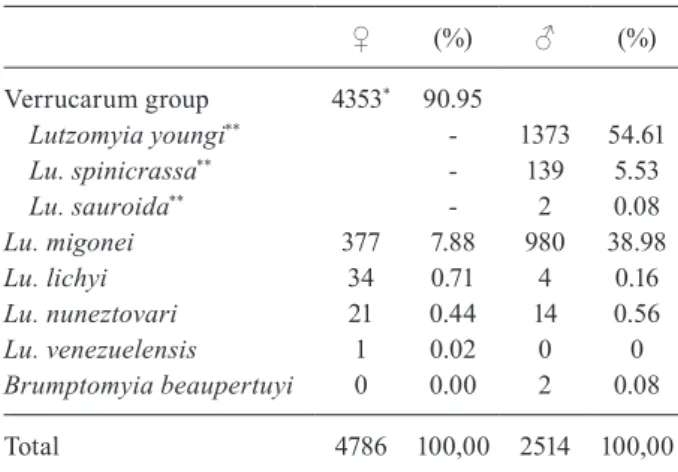
Sand flies - A total of 4,786 female and 2,514 male sand flies were collected in El Carrizal (Table III). The predominant species found were those from the Ver-rucarum group, possibly Lutzomyia youngi and Lu. spinicrassa, as revealed by the males found, since the females are indistinguishable by morphological charac-ters. Lu. migonei was also quite frequent (377 females, 980 males), whereas other species were seldom trapped.
Leishmania spp. infection in sand flies - A random sample of females was selected for the preparation of pools for Leishmania spp. detection and typing, accord-ing for differences in species, capture area, and capture method. Pools included 2,636 females from the Ver-rucarum group, 217 Lu. migonei, 21 Lu. lichyi, and 21 Lu. nuneztovari females. Results of the Leishmania in-fection in these species, as detected by PCR-RFLP per pools, and per the habitat of collection, are shown in Ta-ble IV (Fig. 2C). Females of both the Verrucarum group and Lu. migonei were caught and found infected in three habitats: indoors, outdoors, and in the woodlands. No positive PCR was obtained from the pools of Lu. lichyi and Lu. nuneztovari.
Minimum natural infection rate (MNIR) - Consider-ing that at least one specimen was infected in each of the PCR-positive pool, the minimum natural infection rate in the Verrucarum group would be 0.34% [nine pools identified as L. mexicana *(100/total females tested in the positive and negative pools) (9*(100/2,636)]. For Lu. migonei the MNIR for L. mexicana would be 4.15% [9*(100/217)], and 0.46% for L. guyanensis [1*(100/217)].
DISCUSSION
The aim of this work was to contribute to the knowl-edge of the epidemiological cycles of Leishmania spp., which is a cause of ACL in the Andean Region of Ven-ezuela. The methods commonly used to diagnose ACL according to the guidelines of the Ministry of Health, are the leishmanin skin test (LST), as a triage method, that is used in all the Regional Dermatology Services. (14) and whenever possible, the isolation of parasite
us-ing LIT-NNN culture and microscopy for confirmation. However, molecular tools have the additional advantage of increased sensitivity, and allow for definitive species identification. In previous studies in Mérida and Táchira states, both L. braziliensis and L. mexicana have been confirmed as being causative agents for ACL.(21) In this study, we confirmed those findings for L. brazilienis and L. mexicana, and further report the involvement of L. guyanensis in ACL in this region for the first time. In Andean countries, L. peruviana is the principal species that causes ACL in Peru, whereas in Ecuador the princi-pal causative species is L. mexicana.(6)
We also confirmed Leishmania infection and expo-sure in dogs from El Carrizal, Mérida state by PCR (89.3% positive) and IFAT (64.3%) respectively, pointing to the role of the dog as reservoir of ACL etiological agents.
Similar findings have been reported in other ACL re-gions. In Brazil, studies by Falqueto et al.(22) and Madeira et al.(23,24) demonstrated the presence of L. braziliensis in cutaneous lesions, and L .chagasi, isolated from different
sites, in the same animal. However, some studies indicate that humans, and not dogs, are probably the most impor-tant domestic reservoirs of L. braziliensis.(25) In a study conducted in Peru,(26) found that 81% and 31% of the dogs were positive by ELISA and PCR respectively, which sug-gests the potential role of dogs as reservoirs hosts. Cal-zada et al.(27) also observed a high positive rate by ELISA (47%) in dogs in Trinidad de las Minas, Panamá, whilst they were not able to detect any positive dogs by PCR. Nevertheless, the infection of asymptomatic dogs with sand flies in ACL endemic regions has been demonstrated by Rojas and Scorza,(28) who confirmed the transmission of Leishmania spp. to reared Lu. youngi sand flies (0.88% infection rate) by xenodiagnosis using dogs from an en-demic area in Trujillo, Venezuela.
Here, we have demonstrated the value of the PCR to reveal cryptic infections by L. guyanensis and L. mexicana in dogs from an ACL endemic area, revealing interesting changes in the classical L. guyanensis epide-miological cycle normally associated with the Amazon rain forest and or sylvatic transmission, instead of do-mestic animals hosts. The absence of L. braziliensis in the dogs that we sampled suggests that, although dogs may still be a reservoir, infections might be acquired from a sylvatic source, since all the New World cutane-ous leishmaniasis cycles are predominantly zoonotic.(13) Further studies of the DNA sequences of the Leishmania parasites present in the study samples are necessary to gain a better understanding of these findings.
Regarding the ACL vectors in the Andean Region of Venezuela, we found natural infections by L.mexicana and L. guyanensis in Lu. migonei, and by L. mexicana in females in the Verrucarum group. Naturally infected sand flies were caught in all the habitats studied (in-doors, out(in-doors, and woodland), Lu. migonei being more associated with the peri-domestic environment, as has also been found in other countries such as Brazil,(29) and Argentina.(30) As shown in a previous study, females in the Verrucarum group were collected in larger numbers
TABLE III
Species composition and abundance of phlebotomine sand flies collected at El Carrizal, Mérida state, Venezuela
♀ (%) ♂ (%)
Verrucarum group 4353* 90.95
Lutzomyia youngi** - 1373 54.61
Lu. spinicrassa** - 139 5.53
Lu. sauroida** - 2 0.08
Lu. migonei 377 7.88 980 38.98
Lu. lichyi 34 0.71 4 0.16
Lu. nuneztovari 21 0.44 14 0.56
Lu. venezuelensis 1 0.02 0 0
Brumptomyia beaupertuyi 0 0.00 2 0.08
Total 4786 100,00 2514 100,00
indoors compared to outdoors,(31) which suggests that the transmission of L. mexicana occurs frequently in the do-mestic environment.
The first record of the natural infection of sand fly species by Leishmania spp. promastigotes in Venezuela was made by Pifano and Ortiz (1952),(32) who reported Leishmania spp.infection in Lu. migonei. To the best of our knowledge, no more data have been published on this subject until our preliminary results on the role of this species in the transmission of ACL were reported at El Carrizal (Feliciangeli et al.2011).(33)
The presence of Lu. migonei has also been recorded in Colombia, Trinidad and Tobago, Argentina, Bolivia, Bra-zil, Ecuador, Paraguay, and Peru.(34) However, records of a natural infection by Leishmania spp. causing ACL are only available from Brazil, where infection by L. brazil-iensis in Lu. migonei was documented in specimens col-lected in Northeastern Brazil, in the Baturité hills,(35) and in Jacarepaguá, Rio de Janeiro.(8) Subsequently, Carvalho et al.(9) reported similar findings for Lu. migonei speci-mens collected in Praia Vermelha, Ilha Grande, and Rio de Janeiro. In Argentina, Lu. migonei, is thought to be the vector for ACL, due to the presence of L. braziliensis in several foci,(36) as well as of human and canine visceral leishmaniasis in a rural focus, less than 10 km distant from Puerto Iguazú City, Misiones, where Lu. longipalpis is apparently absent.(30) Moya et al.(10) also found L. infan-tum infected Lu. migonei in a neighbouring region.
With respect to the species in the Verrucarum group, Lu. youngi (= Lu. townsendi), has been documented in Costa Rica, Venezuela, and Colombia, whereas Lu. spin-icrassa seems to be restricted to Venezuela and
Colom-bia.(34) At El Carrizal we identified a natural infection by L. mexicana in these isomorphic species. In Venezuela, in the allopatric ACL focus of Las Calderas, Trujillo, peripyloric promastigotes were seen in Lu. youngi, and were thought to be L.braziliensis based on the criteria of Lainson and Shaw.(37) Subsequently, in an extensive work carried out in 21 localities of Mérida state at dif-ferent altitudes, Añez et al.,(38) found that among 17 par-ous sand flies that were dissected, there was a natural Leishmania spp. infection in 45% of the Lu. youngi, the dominant species at high altitudes, in 9% of Lu. spin-crassa, a species only found at median altitudes, and in 15% of Lu gomezi,a species only found at low altitudes. They concluded that, because of its abundance at > 800 metres above sea level and its high degree of endophagy, as reported by Rojas and Scorza,(28)Lu. youngi is a ma-jor vector in the Venezuelan Andes. In Colombia, Lu. spinicrassa has been proven to be a L. braziliensis vec-tor because of the massive infection by promastigotes in the pylorus and midgut in 1 out of 1,679 cryopreserved and dissected females (0.03%) collected in an allopatric population near Arboledas, north of Santander, and close to Cucuta city(15) at the border of Táchira state in Ven-ezuela. This species was also confirmed to be infected by L. braziliensis in specimens captured in the village of Catarnica, an ACL endemic focus in Táchira state.(39)
In our study we found sand flies infected in three different habitats, thus we can conclude that the trans-mission of ACL may occur in all of them. Moreover, we discovered the co-existence of two transmission cycles of ACL involving Lu. migonei as the vector and the dog as a domestic reservoir of both L. mexicana and L.
guya-TABLE IV
Leishmania identification by polymerase chain reaction-restriction fragment length polymorphism (PCR-RFLP) in phlebotomine sand flies collected at El Carrizal, Mérida state
Habitat (trap) Neg pools N° ♀ PCR + pools N° ♀ Pools RFLP + (N° ♀) Leishmania spp.
Verrucarum Indoors (CDC) 23 429 2 40 2 (40) L. mexicana
Group Outdoors (CDC) 28 501 3 60 1 (20) L. mexicana
2 (40) No identified
Woodland (CDC) 8 137 0 0 0
“ “ (Shannon) 68 1350 7 110 6 (110) L. mexicana
9 1 (9) No identified
Total ♀ = 2417 + 219 = 2636 127 2417 12 219 9 (170) L. mexicana
Lutzomyia migonei Indoors (CDC) 0 0 3 60 3 (60) L. mexicana
Outdoors (CDC) 0 0 6 84 4 (56) L. mexicana
“ “ “ 0 0 1 (8) L. guyanensis
“ “ “ 0 0 1 (20) No identified
Woodland (CDC) 2 29 1 20 1 (20) L. mexicana
“ “ (Shannon) 1 20 2 4 1(4) L. mexicana
3 49 12 168 9 (140) L. mexicana
Total ♀ = 49 +168 = 217 1 (8) L. guyanensis
nensis. Concerning the two species in the Verrucarum group, both Lu. youngi and Lu. spinicrassa might con-tribute to the transmission of L. mexicana. We have not as yet determined if any of these three sand fly species act as local vectors for L. brazilensis. Further studies with the approaches that we have employed will clarify the detailed dynamics of Leishmania spp. transmission in the ACL endemic foci of the Andean Region of Vene-zuela, and inform improved strategies for disease control.
ACKNOWLEDGEMENTS
To Rodrigo Ramirez, BSc in Biology, Centro de Estudios de Enfermedades Endémicas y Salud Ambiental (CEEESA). SA Instituto de Altos Estudios Dr Arnoldo Gabaldon (IAE-MPPS). Maracay, Venezuela, for Fig. 1 made with ArcGis, and to the inhabitants of El Carrizal for their interest and collaboration.
AUTHORS’ CONTRIBUTION
MDF, EF, MAM and IC - Formulated the study, wrote the manuscript, conceived and designed the experiments and ana-lysed the data; AT, JCR, JAB, CC and IPL - performed the experiments; HDL and OD - contributed reagents/materials/ analysis tools. All authors have read and approved the final manuscript. The authors report no conflicts of interest.
REFERENCES
1. Miles MA, Feliciangeli MD, Rojas de Arias MA. American try-panpsomiasis (Chagas disease) and the role of molecular epi-demiology in guiding control strategies. British Med J. 2003; 326(7404): 1445-8.
2. Schönian G, Nasereddin A, Dense N, Schweynoch C, Schallig HD, Presber W, et al. PCR diagnosis and characterization of Leishma-nia in local and imported clinical samples. Diagn Microbiol Infect Dis. 2003; 47(1): 349-58.
3. Fraga J, Velando N, Montalvo AM, Prat N, Boggild AK, Valencia BM, et al. Accurate and rapid species typing from cutaneous and mucocutaneous leishmaniasis lesions of the New World. Diagn Microbiol Infect Dis. 2012; 74(2): 142-50.
4. Reithinger R, Davies CR. Is the domestic dog (Canis familiaris) a res-ervoir host of American cutaneous leishmaniasis? A critical review of the current evidence. Am J Trop Med Hyg. 1999; 61(4): 530-41.
5. Trevisan DA, Lonardoni MV, Demarchi IG. Diagnostic methods to cutaneous leishmaniasis detection in domestic dogs and cats. An Bras Dermatol. 2015; 90(6): 868-72.
6. Hashiguchi Y, Gómez EA, Cáceres AG, Vélez LN, Villegas NV, Hashiguchi K, et al. Andean cutaneous leishmaniasis (Ande-an-CL, uta) in Peru and Ecuador: the vector Lutzomyia sand flies and reservoir mammals. Acta Trop. 2018; 178: 264-75.
7. de Andrade HM, Reis AB, dos Santos SL, Volpini AC, Marques MJ, Romana AJ. Use of PCR-RFLP to identify Leishmania species in naturally-infected dogs. Vet Parasitol. 2006; 140(3-4): 231-8.
8. Pita-Pereira D, Alves CR, Souza MB, Brazil RP, Bertho AL, de Figueiredo-Barbosa A, et al. Identification of naturally infected
Lutzomyia intermedia and Lutzomyia migonei with Leishmania
(Viannia) braziliensis in Rio de Janeiro (Brazil) revealed by a PCR multiplex non-isotopic hybridisation assay. Trans R Soc Trop Med Hyg.2005; 99(12): 905-13.
9. Carvalho BM, Máximo M, Costa WA, de Santana AL, da Costa SM, da Costa-Rego TA, et al. Leishmaniasis transmission in an ecotour-ism area: potential vectors in Ilha Grande, Rio de Janeiro state, Bra-zil. Parasit Vectors. 2013; 6(1): 325. doi: 10.1186/1756-3305-6-325.
10. Moya SL, Giuliani MG, Acosta MM, Salomón OD, Liotta DJ. First description of Migonemyia migonei (frança) and Nyssomyia
whitmani (Antunes and Coutinho) (psychodidae: phlebotominae)
natural infected by Leishmania infantum in Argentina. Acta Trop. 2015; 152: 181-4.
11. Killick-Kendrick R. Phlebotomine vectors of the leishmaniases: a review. Med Vet Entomol. 1990; 4(1): 1-24.
12. Shimabukuro PHF, Andrade AJ, Galati EAB. Checklist of Ameri-can sand flies (Diptera, Psychodidae, Phlebotominae): genera, species, and their distribution. ZooKeys. 2017; 660: 67-106.
13. WHO - World Health Organization. Control of the leishmaniases: report of a meeting of the WHO Expert Committee on the Control of Leishmaniases, Geneva, 22-26 March 2010. WHO Technical Report Series 949. Geneva: WHO; 2010.
14. de Lima H, Borges RH, Escobar J, Convit J. [American cutaneous leishmaniasis in Venezuela: an epidemiological clinical analysis at the national level and by federal entity, 1988-2007]. Bol Ma-lariol Salud Amb. 2010; 50(2): 283-99.
15. Young DG, Morales A, Kreutzer RD, Alexander JB, Corredor A, Tesh RB, et al. Isolations of (Kinetoplastida: Trypanosomatidae) from cryopreserved Colombian sand flies (Diptera: Psychodidae). J Med Entomol. 1987; 24(5): 587-9.
16. Young DG, Duncan MA. Guide to the identification and geo-graphic distribution of Lutzomyia sandflies in Mexico, the West Indies, Central and South America (Diptera: Psychodidae). Mem Am Entomol Inst. 1994; 54: 881 pp.
17. Feliciangeli MD, Rodríguez N, Cardona M, Bravo A. A reliable method for the identification of vectors and Leishmania spp. In large-scale epidemiological studies. J Am Mosq Control Assoc. 1999; 15: 411.
18. Papás MG, Hajkowski R, Hockmeyer WT. Dot enzyme-linked im-munosorbent assay (Dot-ELISA): a micro technique for the rapid diagnosis of visceral leishmaniasis. JImmunol Methods. 1983; 64(1-2): 205-14.
19. Sambrook J, Russel D. Molecular cloning: a laboratory manual. 3rd ed. United States of America: Cold Spring Harbor Laboratory Press; 2001.
20. Cruz I, Millet A, Carrillo E, Chenik M, Salitre P, Verla S, et al. An approach for interlaboratory comparison of conventional and real-time PCR assays for diagnosis of human leishmaniasis. Exp Parasitol. 2013; 134(3): 281-9.
21. Rodríguez N, Cardona M, Zerpa O, Barrios M, Sosa A, Fernández A. [Aplication of molecular tools in the diagnosis and character-ization of Leishmania spp. in endemic areas of Venezuela]. Bol Malariol Salud Amb. 2001; 41(2): 21-6.
22. Falqueto A, Sessa PA, Varejão JBM, Barros GC, Momen H, Grimaldi Jr G. Leishmaniasis due to Leishmania braziliensis in Espírito Santo state, Brazil: further evidence on the role of dogs as a reservoir of infection for humans. Mem Inst Oswaldo Cruz. 1991; 86(4): 499-500.
23. Madeira M, Schubach A, Schubach TM, Serra CM, Pereira SA, Figueiredo FB, et al. Is Leishmania (Viannia) braziliensis pref-erentially restricted to the cutaneous lesions of naturally infected dogs? Parasitol Res. 2005; 97(1): 73-6.
24. Madeira M, Schubach A, Schubach TM, Pereira SA, Figueiredo FB, Baptista C, et al. Post mortem parasitological evaluation of dogs seroreactive for Leishmania from Rio de Janeiro, Brazil. Vet Parasitol. 2006; 138(3-4): 366-70.
26. Reithinger R, Espínola JC, Davies CR. The transmission dynam-ics of canine American cutaneous leishmaniasis in Huánuco, Peru. Am J Trop Med Hyg. 2003; 69(5): 473-80.
27. Calzada JE, Saldaña A, González K, Rigg C, Piñeda V, Santamaría AM, et al. Cutaneous leishmaniasis in dogs: is high seroprevalence indicative of a reservoir role? Parasitology. 2015; 142(9): 1202-14.
28. Rojas E, Scorza JV. Xenodiagnóstico con Lutzomyia youngi en casos Venezolanos de leishmaniasis cutanea por Leishmania
bra-ziliensis. Mem Inst Oswaldo Cruz. 1989; 84(1): 29-34.
29. Reinhold-Castro KR, de Carvalho-Gasparotto J, Neitzke-Abreu HC, Teodoro U. Larval habitats of sand flies in rural areas of southern Brazil. J Vector Eco. 2015; 40(2): 269-76.
30. Salomón OD, Quintana MG, Bezzi G, Moran ML, Bebedor E, Valdez DV. Lutzomyia migonei as putative vector of visceral lei-shmaniasis in La Banda, Argentina. Acta Trop. 2010; 113(1): 84-7.
31. Cuccarese A, Pérez-Ybarra L, Bravo JA, Torrellas A, Flores K, Rangel C, et al. Aspects of the bionomics of three phlebotomine vector species (Diptera: Psychodidae) at El Carrizal, an endemic focus of cutaneous leishmaniasis (CL) in the Venezuelan Andean Region. Bol Malariol Salud Amb. 2016; 56(2): 145-59.
32. Pífano F, Ortiz I. [Venezuelan representatives of the genus Phle-botomus Rondani, 1940 (Diptera: Psychodidae)]. Rev Venez Sanid Asist Soc. 1952; 17: 136-51.
33. Feliciangeli MD, Villegas P, Torrellas A, Bravo A, de Lima H, Car-reño J, et al. Molecular epidemiology reveals that Lutzomyia migo-nei and dogs (Canis familiaris) maintain at least two transmission
cycles of cutaneous leishmaniasis in Venezuela. Females in the Ver-rucarum group are also involved in the CL transmission in the study area. In: 7th International Symposium on Phlebotomine Sandflies, 25-30 April, Kusadasi [Abstract book]. Kusadasi: 2011. p. 74-5.
34. Bejarano EE, Estrada LG. Family psychodidae. Zootaxa. 2016; 4122(1): 187-238.
35. Azevedo ACR, Rangel EF, Queiroz RG. Lutzomyia migonei (França 1920) naturally infected with peripylarian flagellates in Baturité, a focus of cutaneous leishmaniasis in Ceará state, Brazil. Mem Inst Oswaldo Cruz. 1990; 85(4): 479.
36. Salomón OD, Rosa JR, Stein M, Quintana MG, Fernández MS, Visintin AM, et al. Phlebotominae (Diptera: Psychodidae) fauna in the Chaco region and cutaneous leishmaniasis transmission pat-terns in Argentina. Mem Inst OswaldoCruz. 2008; 103(6): 578-84.
37. Scorza JV, Márquez M, Márquez JC. [Finding of Lutzomyia
townsendi (Ortiz, 1959) naturally infected with Leishmania
bra-ziliensis, in the suburban area of Trujillo, Venezuela]. Bol Dir Mal-ariol San Amb. 1984; 24(2): 21-8.
38. Añez N, Cazorla D, Oviedo M, Lugo AY, Valera M. Epidemiology of cutaneous leishmaniasis in Mérida, Venezuela. III. Altitudinal distribution, age structure, natural infection and feeding behav-ior of sandflies and their relation to the risk of transmission. Ann Trop Med Parasitol. 1994; 88(3): 279-87.
39. Perruolo G, Rodríguez N, Feliciangeli MD. Isolation of
Leishma-nia (Viannia) braziliensis from Lutzomyia spinicrassa (species