Angiogenesis/osteogenesis in bisphosphonate-related osteonecrosis
of the jaw:
In vitro
assessment in co-cultures of human endothelial
and osteoblastic cells
Monografia de Investigação
“Education is a progressive discovery of our own ignorance.”
“Every science begins as philosophy and ends as art.”
To my Grandparents…
To my Parents…
Thank you for all the support and encouragement to my growth as a person and as a professional.
Research work supervised by: Maria Helena Raposo Fernandes, PhD
Faculty of Dental Medicine, University of Porto
Research work co-supervised by: Pedro de Sousa Gomes, DDS, MSc
Preface
Bisphosphonate-related osteonecrosis of the jaw is a current adverse effect of long-term therapies with bisphophonates (BPs). Decreased bone turnover and the anti-angiogenic properties of the BPs might play a role in their anti-resorptive activity. It has been proposed that the adverse effects of bisphosphonates may be concentrated in the jaws due to higher blood supply and bone turnover than other bones because of the daily constant activity.
Angiogenesis and osteogenesis are intimately related. Bone formation is always preceded by vascular invasion and osteogenesis occurs in the vicinity of the new blood vessels. In this way, the anti-angiogenic properties of BPs might also have a direct effect on the osteoblastic activity, decreasing bone formation events. However, this mechanism, as a contributing factor to the osteonecrosis of the jaws associated with BPs, has received little attention.
The aim of this project is to analyze the effect of representative BPs – alendronate and zoledronate, two widely used BPs in therapeutics - in the behavior of a co-culture system of human endothelial cells and MG63 osteoblastic cells, aiming to simulate more closely the in vivo intimate relationship between angiogenesis and osteogenesis during bone formation events. This work is expected to provide information regarding the relevance of the interaction angiogenesis/osteogenesis in the pathogenesis of bisphosphonate-related osteonecrosis of the jaw.
Acknowledgements
I would like to express my gratitude to Professor Maria Helena Fernandes. I’m thankful for all the things she taught me. I really appreciate her guidance. Special thanks to Dr. Pedro de Sousa Gomes, who help me with cell cultures and other several things. Also, thanks to Dr. Raquel Palmas.
To all my friends and relatives who always support me. Without them everything was more difficult.
Publications
Publications: Review Articles
• Osteonecrose dos maxilares associada ao uso de Bifosfonatos. Parte I: etiologia e apresentação clínica. Artigo submetido à Revista da Sociedade Portuguesa de Estomatologia e Medicina Dentária (SPEMD).
• Osteonecrose dos maxilares associada ao uso de Bifosfonatos. Parte II: linhas de orientação na consulta de Medicina Dentária. Artigo submetido à Revista da Sociedade Portuguesa de Estomatologia e Medicina Dentária (SPEMD).
Poster Presentations in scientific events
• Angiogenesis/Osteogenesis in Bisphosphonate-associated jaw osteonecrosis: In vitro assessment in co-cultures of human alveolar bone and endothelial cells. IJUP 09 – 2º Meeting of Young Researchers at UP, 2009. Porto, Portugal.
Contents
Resumo ………1
Abstract ……… 2
Introduction ………. 3
Bisphosphonates ……… 3
Bisphosphonate-related osteonecrosis of the jaw ……… 4
Bone remodeling/repair ………. 6
Bisphosphonates effects on osteoblastic and endothelial cells ……… 6
Objectives ……… 7
Material and Methods ……… 8
Results ……….... 12
Discussion ………. 18
Conclusion ………. 20
References ………. 21
Resumo
Introdução. A inter-relação angiogénese/osteogénese é crucial na manutenção da integridade do tecido ósseo. Desta forma, as propriedades anti-angiogénicas dos bifosfonatos podem ter um efeito directo na actividade osteoblástica, diminuindo a formação óssea. No entanto, este mecanismo, como factor etiológico/contributivo para a osteonecrose dos maxilares associada aos bifosfonatos, tem sido pouco investigado.
Objectivos. Este estudo descreve os resultados relativos ao efeito do Alendronato no comportamento de células osteoblásticas e células endoteliais humanas.
Métodos. As células osteoblásticas MG-63 e as células endoteliais humanas (provenientes do cordão umbilical) foram cultivadas durante 7 dias, em meios de cultura específicos, na ausência (controlo) e na presença de Alendronato, 10-12 M a 10-4 M. A viabilidade/proliferação celular foi avaliada pelo ensaio do MTT. O impacto na morfologia celular e nas características fenotípicas foi avaliado pela visualização celular (microscopia confocal), utilizando uma coloração imunohistoquímica específica.
Resultados. As culturas de células MG-63 apresentaram proliferação gradual e expressaram marcadores associados ao fenótipo osteoblástico. As culturas de células endoteliais mostraram um padrão de crescimento típico e expressão da molécula de adesão PECAM-1. O Alendronato, em concentrações inferiores a 10-6 M não interferiu no comportamento das células MG-63, mas, em concentrações superiores, causou inibição do crescimento celular e alterações significativas na morfologia. O efeito do Alendronato nas culturas de células endoteliais manifestou-se por uma inibição do crescimento para níveis superiores a 10-8 M e diminuição da expressão de PECAM-1 para níveis iguais e superiores a 10-10 M.
Conclusão. A inibição da expressão da proteína de adesão endotelial PECAM-1, na presença de concentrações baixas de Alendronato, pode constituir um mecanismo contributivo para a alteração da angiogénese, com eventual repercussão no processo de formação óssea.
Abstract
Introduction. The interplay between angiogenesis and osteogenesis is of paramount importance to maintain skeletal integrity (bone development and repair). Bone formation is always preceded by vascular invasion and osteogenesis occurs in the vicinity of the new blood vessels. In this way, the anti-angiogenic properties of bisphosphonates (BPs) might also have a direct effect on the osteoblastic activity, decreasing bone formation events. However, this mechanism, as a contributing factor to the osteonecrosis of the jaws associated with BPs, has received little attention.
Objectives. This study describes the effects of Alendronate in the behaviour of human osteoblastic and endothelial cells.
Methods. MG-63 osteoblastic-like cells and human umbilical vein endothelial cells were cultured during 7 days, in specific media, in the absence (control) and in the presence of alendronate, 10-12 M to 10-4 M. Cell viability/proliferation was evaluated by the MTT assay. The impact on cell morphology and on phenotypic characteristics were assessed by cell visualization (confocal laser scanning microscopy), following a specific immunostaining.
Results. MG-63 cells proliferated gradually and expressed osteoblast-associated markers. Endothelial cell cultures presented a typical pattern of cell growth and expressed the adhesion protein PECAM-1. Alendronate, at concentrations lower that 10-6 M did not affect the behaviour of MG-63 cells, but higher levels caused inhibition of cell proliferation and alterations in the cell morphology. In endothelial cell cultures, Alendronate inhibited cell growth at concentrations higher that 10-8 M and the expression of PECAM-1 at levels similar and higher than 10-10 M.
Conclusion. The inhibition of the expression of the endothelial adhesion protein PECAM-1, in the presence of low concentrations of Alendronate, might be a contributing mechanism to an impairment of the angiogenesis, with an eventual repercussion in the bone formation process.
1. Introduction
Bisphosphonates
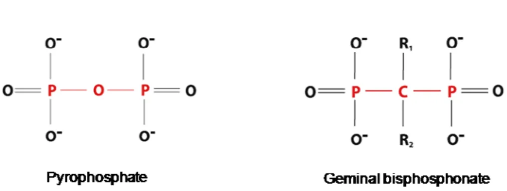
Bisphosphonates (BPs) are synthetic and stable analogues of pyrophosphate (P-C-P) that are widely used to treat skeletal diseases involving excessive bone resorption (1, 2). BPs bind selectively and avidly to bone and suppress osteoclastic bone resorption (3). The high avidity of BPs for Ca2+ ions is the basis of the bone targeting property of these compounds. Other effects have been reported, including anti-angiogenic properties (1, 2). Their chemical structure is resistant to enzymatic splitting by pyrophosphatases, to heat and in an acidic medium (1). This characteristic allows these compounds to be retained at bone during a long period of time and exert effects continuously (1, 3). Attached to the P-C-P backbone are two covalently bonded side chains (R1 and R2), (Figure 1), (1, 3). Variations in the R1 and R2 produce different BPs, which have different affinities for hydroxyapatite crystals (1). The values range from 0.6 (clodronate) to 2.9 (alendronate) and 3.5 (zoledronate) (1). The presence of an OH substitution in R1 increases their binding to bone mineral surface, and this action was independent of the structure of the R2. On the other hand, BPs lacking an R1 substitution or compounds with other substitutions such as H (etidronato) and Cl (clodronate) had significantly lower binding affinities (1).
BPs have a short plasma half-life and a low exposure of visceral tissues, and this could be the reason of a low incidence of adverse effects (2). Because their selectivity to bone and high capacity to suppress osteoclast-mediated bone resorption, BPs are the first-line therapy to treat benign and malignant skeletal diseases such as osteoporosis, Paget’s disease, hypercalcemia of malignancy and osteolytic metastases of solid tumors (1, 2). Clinically, BPs have several advantages in this kind of patients because they reduce fractures, bone pain and the need of radiation or stabilizing operations and also prevent hypercalcemic episodes, which together improve the patients’ quality of life (1).
Figure 1- Chemical structure of pyrophosphate and bisphosphonates (1).
Bisphosphonate-related osteonecrosis of the jaw
Bisphosphonates have been used over the past 30 years for the treatment of bone disorders and anomalies of calcium metabolism (1). Since 2003, BPs have been related to osteonecrosis of the jaw, and has since increased in frequency (7-9). It started when Marx identified 36 cases of painful bone exposure in the mandible, maxilla, or both, that did not respond to surgical or medical treatments (7). The common point of all these patients was that they were taking a potent BP, pamidronate or zoledronate (7). After that, BPs’ manufacturers included osteonecrosis of the jaw as an adverse effect of treatment with these compounds.
osteoblast-mediated osteoclastic resorption and has antiangiogenic properties (5, 6). The end result is a bone that shows little physiologic remodeling, because bone turnover becomes profoundly suppressed by BPs. The bone becomes brittle and unable to repair physiologic microfractures that occur in the human skeleton with daily activity (9). Masticatory forces are a constant stress applied to maxilla and mandible which implies that physiologic microdamage and microfractures occur daily in the oral cavity (9-11). It is hypothesised that in a patient taking a BP, bone are unable to repair microdamage, because of its reduced capacity to remodel and turnover and because hipovascularity, which results in osteonecrosis (9). Suppressed bone repair can lead to accumulation of microdamage (10, 11).After one-year of alendronate treatment, intracortical remodeling was significantly suppressed (68%) and microdamage accumulation increased (322%), in a dog model (10).
Bone remodeling/repair
Physiologic microdamage and microfracture of the bone tissue occur daily in the oral cavity (15). Bone remodeling removes microdamage (resorption) and replaces damaged bone with new elastic osseous tissue (formation). Different cell types are involved in bone remodeling process like osteoblasts, osteoclasts and endothelial cells. Interaction between osteoclasts and osteoblasts is essential for bone remodeling and repair (1). Likewise, proper bone remodeling requires an active process of angiogenesis which supplies the necessary stem cells and growth factors. The microvascular endothelium is an essential part of skeletal tissue, where the intercellular signaling between endothelium and bone cells plays a critical role in the homeostasis of bone integrity (17). Cross-talk between osteoblasts and endothelial cells is critical to the coordinated cell behaviour necessary for bone development and remodeling (17, 18). Vascular endothelial growth factor (VEGF), a major promoter of both physiological and pathological angiogenesis, is an important regulator of this cellular communication (17, 19, 20). Endogenous VEGF plays a critical role in bone development and repair (17) and is essential for normal angiogenesis and appropriate callus architecture and mineralization in response to bone injury (17) .
Bisphosphonates effects on osteoblastic and endothelial cells
reported regarding the effects on osteoblastic differentiation and function. The reason for this is unclear but may be related with differences in the cell types and model system, duration of BPs exposure, and type and concentrations of BP used in the different studies (3).
BPs are also known to have anti-angiogenic properties (1, 6) and this is supported by the fact that they influence negatively the number of circulating endothelial progenitor cells (14). Furthermore, zoledronate decreased the circulating levels of angiogenic factors (e.g. vascular endothelial growth factor, platelet-derived growth factor) in cancer patients with bone metastasis (1, 14). A variety of in vitro studies addressed the activity of BPs in endothelial cells, reporting effects on the adhesion, migration and expression of phenotype characteristics (36-39).
It is known that there is a reciprocal regulation and functional relationship between endothelial cells and osteoblastic cells during osteogenesis, a process tightly regulated by systemic hormones and paracrine growth factors or cytokines, and strongly affected by other bioactive molecules present in the bone environment (17). Regarding this, few studies addressed the interplay between endothelial and osteoblastic cells. These studies suggested that osteoblastic lineage cells have a different behaviour in terms of proliferation and differentiation when cultured in association with endothelial cells, in various co-culture models (co-culture with or without direct contact), compared to that observed when they are cultured alone (19, 20, 40). However, information regarding the effect of BPs in the interplay between these two cell types has not been reported.
Objectives
2. Materials and Methods
2.1. Characterization of the cell culture models
2.1.1. MG-63 human osteoblastic-like cells
MG-63 human osteoblastic-like cells (ATCC) were cultured in α-Minimum
Essential Medium (α-MEM, Gibco) supplemented with 10% fetal bovine serum (Gibco),
50 µg/mL ascorbic acid (Sigma), 100 IU/mL penicillin, 2.5 µg/mL streptomycin (Gibco) and 2.5 µg/mL amphotericin B (Gibco). Cultures were maintained in a 5% CO2
humidified atmosphere at 37 ºC until near confluence. At this stage, cells were enzymatically released (0.04% trypsin in 0.25% EDTA) and the resulting cell suspension was plated (104 cells/cm2) in standard cell culture plates for 7 days. Cultures were characterized for cell viability/proliferation, gene expression by RT-PCR and F-actin cytoskeleton organization.
2.1.2. Human Umbilical Vein Endothelial Cells (HUVECs)
Endothelial cells were isolated from human umbilical cord (41). Briefly, umbilical cords were perfused with isotonic solution to remove blood and cellular debris and the endothelial cells were released from the umbilical vein with 0.1% collagenase in medium M199 (7 min, 37ºC, 5% CO2/air) and the resultant cell suspension was
centrifuged (1500 rpm, 5 min). Cells were ressuspended and seeded in culture plates pre-coated with 1% gelatine. Cultures were established in fully supplemented EGM-2 culture medium (Lonza®) in a 5% CO2 humidified atmosphere at 37 ºC until 70 – 80%
2.1.3 Co-cultures of MG-63 cells and endothelial cells
Co-cultures of MG-63 cells and endothelial cells were co-cultured, in different proportions (1:5; 1: 10), in ECGM medium, for 48h, in a 5% CO2 humidified atmosphere
at 37 ºC. Co-cultures were evaluated by CLSM after being stained for PECAM-1 and nucleus.
2.2 Effect of Alendronate in the behaviour of osteoblastic and endothelial cells
Alendronate was purchased from Sigma-Aldrich. The solutions were prepared from a 10-2 M stock solution diluted in phosphate saline buffer (PBS).
MG-63 cells (104 cells/cm2) were seeded onto 96-well culture plates, and cultured as described above. After overnight incubation, medium was changed and alendronate was added at concentrations ranging from 10-12 to 10-4 M, and cultures were continued for 7 days. The concentration range tested was based on the information reported in the literature regarding similar studies and, also, in preliminary dose-response assays which enable to exclude the levels that caused rapid cell death. Control cultures (absence of alendronate) were performed in parallel. The culture medium, containing the bisphosphonate tested, was renewed twice during the culture time, at days 1 and 4. Cultures were characterized for cell viability/proliferation and organization of the F-actin cytoskeleton.
2.3. Characterization of the cell behaviour
2.3.1. MTT colorimetric assay
MTT assay – reduction of 3-(4,5-dimethylthiazol-2-yl)-2,5-diphenyltetrazolium bromide to a purple formazan product by viable cells – was used to estimate cell viability/proliferation. Samples were incubated with 0.5 mg/ml of MTT for the last 4 h of the culture period tested; the medium was then decanted, formazan salts were dissolved with dimethylsulphoxide and the absorbance (A) was determined at λ = 600
nm on a microplate reader.
2.3.2 Immunofluorescent staining of F-actin cytoskeleton filaments and PECAM-1
Cultures were fixed in 4% formaldehyde (methanol free), permeabilized in 0.1% Triton (5 min, RT) and incubated in 10 mg/mL bovine serum albumin (1h, RT) with 100 µg/mL RNAse. F-actin filaments were stained with Alexa-Fluor-conjugated phalloidin (1:100, 1h, RT) and nuclei were counterstained with 10 µg/mL propidium iodide (10 min, RT).
Endothelial cells were also stained for the expression of PECAM-1. Cultures, after fixation and permeabilization, were incubated overnight with primary PECAM-1 antibody (1:100, 4ºC), followed by the addition of the secondary antibody (1:1000, anti-mouse Alexa-Fluor; 1 h, RT), and then counterstained with 10 µg/ml propidium iodide (10 min, RT).
Fluorescent stained cultures were examined by confocal laser scanning microscopy (CLSM, Leica TCP SP2 AOBS confocal microscope).
2.3.3 Total RNA extraction and RT-PCR analysis
instructions. The concentration and purity of total RNA in each sample were determined by UV spectrophotometry at 260 nm and by calculating the A260nm/A280nm ratio,
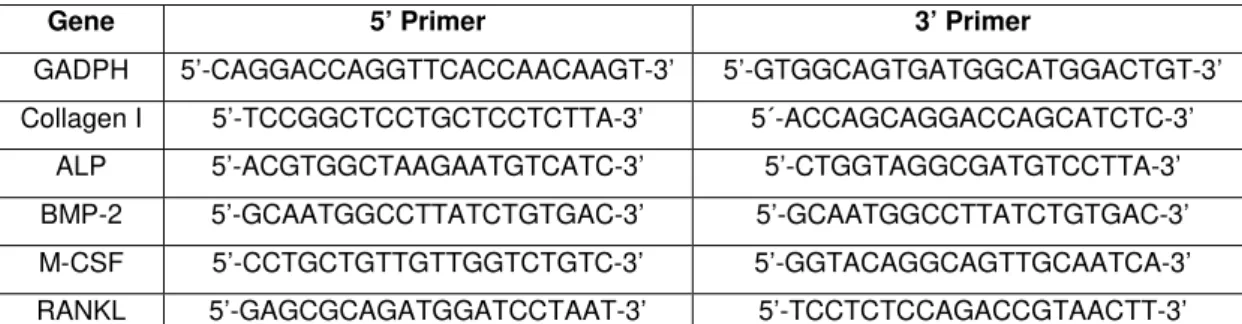
respectively. RT-PCR was done using the Titan One Tube RT-PCR System from Roche Applied Science, according to the manufacturer’s instructions. Briefly, 0.5 µg of total RNA from each sample was reverse transcribed into cDNA (30 minutes at 50 ºC), while PCR was performed with an annealing temperature of 55ºC, for 25 cycles. The primers used are listed on Table 1. Following, the PCR products were electrophoresed in a 1% agarose gel and stained with ethidium bromide.
Table 1 - Primers used on RT-PCR analysis of MG-63 cell cultures.
2.5. Statistical Analysis
Three independent experiments were performed. In each experiment, for the quantitative assays, six replicates were accomplished. Data were expressed as the mean ± standard deviation. Statistical significances were analyzed by student’s t-test. P-values ≤ 0.05 were considered significant. Qualitative assays were performed in
triplicate.
Gene 5’ Primer 3’ Primer
GADPH 5’-CAGGACCAGGTTCACCAACAAGT-3’ 5’-GTGGCAGTGATGGCATGGACTGT-3’
Collagen I 5’-TCCGGCTCCTGCTCCTCTTA-3’ 5´-ACCAGCAGGACCAGCATCTC-3’
ALP 5’-ACGTGGCTAAGAATGTCATC-3’ 5’-CTGGTAGGCGATGTCCTTA-3’
BMP-2 5’-GCAATGGCCTTATCTGTGAC-3’ 5’-GCAATGGCCTTATCTGTGAC-3’
M-CSF 5’-CCTGCTGTTGTTGGTCTGTC-3’ 5’-GGTACAGGCAGTTGCAATCA-3’
3. Results
3.1 Characterization of the cell culture models
MG-63 cell cultures
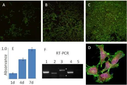
MG-63 osteoblast-like cells proliferated during the 7-day culture period, with a high growth rate, as observed in the MTT assay and CLSM. High magnification CLSM images showed well-spread cells with a prominent nucleus and a normal morphology and cytoskeleton organization, in addition to established cell-to cell contacts. MG63 cells displayed the expression of the osteoblastic-related markers Collagen type I, ALP and BMP-2 and, also, of genes involved in the communication between osteoblastic and osteoclast cells during bone modelling and remodeling, namely the osteoclastogenic modulators M-CSF and RANKL (42, 43). Results are displayed in Figure 2.
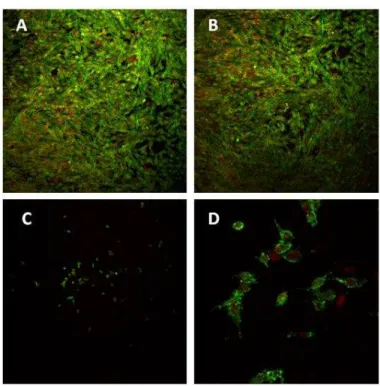
Endothelial cell cultures
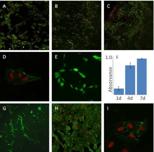
Figure 3 presents the behaviour of endothelial cell cultures regarding proliferation and function. Cells proliferated throughout the culture time, especially during the first days. Observation by phase contrast microscopy showed a tendency for a circular orientation during the proliferative phase. Cells presented a normal morphology, with a well defined nucleus, cytoplasmic extensions and cell-to-cell contact. In addition, endothelial cells expressed the adhesion protein PECAM-1.
Co-cultures of osteoblastic and endothelial cells
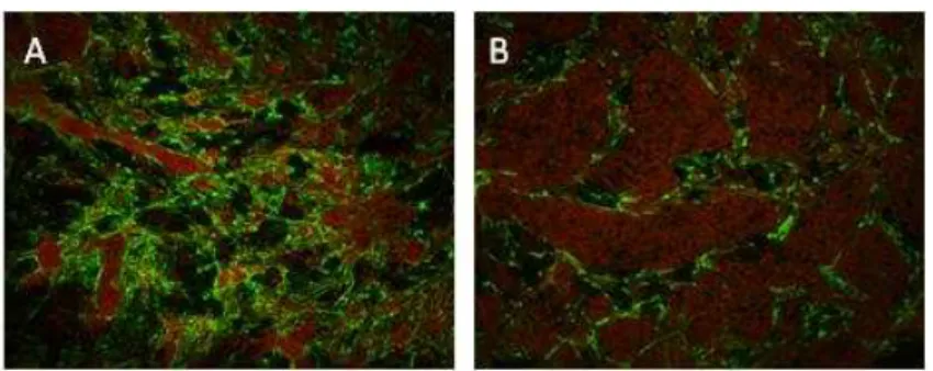
MG-63 cells and endothelial cells were co-cultured in the proportion of 1:5 and 1:10. Figure 4 shows representative CLSM images of the co-cultures, at 48 h, stained for PECAM-1 and nucleus. In both systems, endothelial cells presented a well defined organization, reflected by the formation of tubular structures surrounding clusters of osteoblastic cells.
Figure 4 - Behaviour of co-cultures of human osteoblastic and endothelial cells cultured for 48 h in the proportion of 1:5 (A) and 1:10 (B), respectively. CLSM images of cultures stained for PECAM-1 (green) and nucleus (red), 100x.
3.2 Effect of Alendronate in the behaviour of osteoblastic and endothelial cells
MG-63 and endothelial cells were exposed to Alendronate, in the concentration range 10-12 to 10-4 M, for 7 days. Cell behaviour was characterized for cell viability/proliferation, morphology, pattern of cell growth and functional activity.
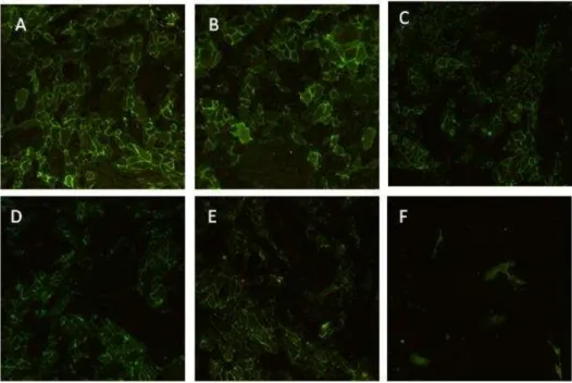
3.2.1 MG-63 osteoblast-like cells
MG-63 cells grew actively throughout the culture time. Exposure to Alendronate, at concentrations between 10-12 M and 10-6 M, induced similar behaviour to that of control, regarding proliferation and cellular morphology, as reported by the MTT assay and CLSM images. High concentrations of Alendronate, ranging from 10-5 M to 10-4 M, were harshly deleterious to this cell population, and induced significant dose- and time-dependent effects is cell morphology. Results are shown in Figures 5 and 6.
3.2.2 Human Umbilical Vein Endothelial Cells (HUVEC)
Alendronate, at 10-12 to 10-4 M, caused dose-dependent effects in the cell viability/proliferation, as assessed by the MTT assay, Figure 7. At concentrations ≤ 10-7
Figure 5 - Cell viability/proliferation (MTT assay) of MG-63 human osteoblastic-like cells cultured in the absence – control – and presence of Alendronate, in the concentration range 10-12 to 10-4 M. *p≤0.05, significantly different from control.
Figure 7 - Cell viability/proliferation (MTT assay) of human endothelial cells cultured in the absence - control conditions – and presence of Alendronate in the concentration range 10-12 to 10-4 M. *p≤0.05, significantly different from control.
4. Discussion
Different theories about the development of BRONJ are being discussed in the literature. However, the most common theory attributes the condition to reduced bone remodeling due to BP induced osteoclast inhibition and the subsequent accumulation of microfractures. Moreover, BPs have an anti-angiogenic effect, resulting in avascular necrosis (1, 2, 6, 14).
The inhibitory effects of BPs on osteoclast formation and activity, both in vivo and
in vitro are well documented on the literature, however, their effects on osteoblast function and bone formation (osteogenesis) are less understood. Bone formation is a multi-step coordinated process which involves several biological factors. In addition, angiogenesis plays a critical role in bone development and repair, and the interaction between endothelial cells and osteoblasts has a pivotal role in bone remodeling/repair process (17). There are a variety of studies showing that BPs might affect the proliferation and function of osteoblastic cells (22-34) and endothelial cells (36-40). However, the effect of BPs in the interplay between these two cell types has not been addressed yet.
The present work reports the dose-dependent effects of Alendronate in the behaviour of MG-63 osteoblast-like cells and human endothelial cells, exposed during 7 days in similar experimental conditions. The concentration range tested, 10-12 to 10-4 M, was based in previous similar in vitro studies involving BPs and these cell types (22-34, 36-40) and, also, in the levels estimated to be present in the bone environment during BPs therapies, that are reported to be around 10-9 to 10-12 M (1, 2, 3).
The first part of this work, dealing with the characterization of the cell culture models, showed that MG-63 and endothelial cell cultures presented phenotype features. MG-63 cell cultures expressed Collagen type I, ALP and BMP-2, which are osteoblastic associated markers (42), and, they also expressed M-CSF and RANKL, which are proteins essential in the coordinated activities of osteoblastic and osteoclastic cells during bone remodeling (43). Endothelial cell cultures presented a typical cell growth pattern, with the formation of circular cellular arrangements and cord-like structures, in addition to the expression of PECAM-I, an established endothelial marker (17).
with Alendronate concentrations ≤ 10-6 M exhibited a behaviour similar to control. The
effect of Alendronate in MG-63 cells was previously studied by Im et al (2004) (10-12 M to 10-4 M) (25) and Xiong et al (2009) (10-10 M to 10-6 M) (34). These studies reported stimulation of the cell growth at low concentrations and inhibition at high levels. The present work did not show stimulatory effects in MG-63 cells treated with Alendronate, which might be related to differences in the cell culture conditions, namely, culture medium, cell platting density and exposure conditions. Other previous studies addressed the activity of different BPs in various osteoblastic cell systems, namely the effect of Zoledronate in MC3T3-E1 and MG-63 cells (32), human mesenchymal stem cells (27) and the effect of Alendronate, Risedronate and Zoledronate in bone marrow stromal cells (27). These studies reported mixed dose-dependent effects in the presence of BPs regarding osteoblastic cell proliferation and function, due to the different models and condition used (3).
Results of this study also showed that Alendronate affected the behaviour of human endothelial cells. Exposure to concentrations between 10-12 M and 10-7 M resulted in a slight stimulation of cell growth, and higher levels caused inhibition in this parameter. Therefore, in the experimental conditions used, endothelial cells were more sensitive to the dose-effects of Alendronate than MG-63 cells. In addition, levels ≥ 10-10
M were associated with a dose-dependent decrease in the expression of PECAM-1. Previous studies, also showed that endothelial cells are target cells for these compounds, and dose-dependent effects on cell growth, migration and expression of phenotype characteristics have been reported in the presence of Alendronate (36) and Zoledronate (6, 37-39).
In vitro studies addressing the activity of BPs in various cell types used a large concentration range, usually within 10-12 to 10-4 M. It is worth to mention that the concentration range of BPs which osteoblasts, endothelial cells and other cell types in the bone environment are exposed to, under pharmacological conditions, are unknown (3), but estimated levels are usually ≤ 10-9 M (1, 3). However, it has been suggested
that jaw bones are more susceptible to deposition of an excessive amount of BPs due to the higher bone turnover (1, 2), and higher concentrations of BPs might be present, especially after a dental surgical procedure (12), when bone repair takes place. The present work showed that at relatively low concentrations, Alendronate, ≥ 10-10 M,
PECAM-1 is a 130-kDa transmembrane glycoprotein found in large amounts on endothelial cells. It plays a major role in several cellular interactions, namely in the adhesion cascade between endothelial cells and other cell types involved in the inflammatory process, as well as, between adjacent endothelial cells during the angiogenic process (44). These cellular junctions are crucial to maintain the endothelial layer’s integrity and play an essential role in the vessel’s sprouting and elongation processes (44). According to some studies, the cellular distribution of PECAM-1 represents a very sensitive marker of the endothelial cell function (44). In this way, this deleterious effect, observed at such low Alendronate concentrations, might be a contributing mechanism leading to decreased angiogenesis in the bone environment, with eventual repercussions in the bone formation and remodeling events.
It is worth to mention that the effects of BPs in vitro have been studied mainly on single cell type culture system, which provide limited data, and the majority of published studies were carried out in conditions that poorly represent in vivo exposure (3). The characterization of the effects of BPs in a co-culture system of endothelial/osteoblastic cells would be a more representative model, providing information regarding the relevance of the interaction angiogenesis/osteogenesis in the development of BRONJ. This study is underway.
5. Conclusion
Alendronate, in a concentration range around those reported to inhibit bone resorption in vivo (≤ 10-9 M) did not cause significant modifications regarding the
proliferation of human osteoblastic-like cells, neither induced changes in the morphology of the cells. Furthermore, in this concentration range, a slight stimulation in the proliferative behaviour of human endothelial cells was observed, nevertheless the impairment of the phenotypic function suggested by the reduced expression of PECAM-1. This may be an eventual mechanism by which Alendronate impairs the angiogenesis process with repercussions in the bone metabolism. These results support the hypothesis that impairment/inhibition of angiogenesis induced by BPs might have an important role in development and maintenance of BRONJ.
1. Bartl R FB, von Tresckow E, Bartl C. Bisphosphonates in Medical Practice: Actions, Side Effects, Indications, Strategies. 1st ed. ed. Berlin (Germany): Springer; 2007.
2. Green JR. Bisphosphonates: preclinical review. Oncologist. 2004; 4:3-13.
3. Schindeler A, Little DG. Bisphosphonate action: revelations and deceptions from in vitro studies. J Pharm Sci. 2007; 96:1872-8.
4. Rogers MJ, Gordon S, Benford HL, Coxon FP, Luckman SP, Monkkonen J, et al. Cellular and molecular mechanisms of action of bisphosphonates. Cancer. 2000; 88:2961-78.
5. Bukowski JF, Dascher CC, Das H. Alternative bisphosphonate targets and mechanisms of action. Biochem Biophys Res Commun. 2005; 328:746-50.
6. Wood J, Bonjean K, Ruetz S, Bellahcene A, Devy L, Foidart JM, et al. Novel antiangiogenic effects of the bisphosphonate compound zoledronic acid. J Pharmacol Exp Ther. 2002; 302:1055-61.
7. Marx RE. Pamidronate (Aredia) and zoledronate (Zometa) induced avascular necrosis of the jaws: a growing epidemic. J Oral Maxillofac Surg. 2003; 61:1115-7.
8. Ruggiero SL, Mehrotra B, Rosenberg TJ, Engroff SL. Osteonecrosis of the jaws associated with the use of bisphosphonates: a review of 63 cases. J Oral Maxillofac Surg. 2004; 62:527-34.
9. Sarin J, DeRossi SS, Akintoye SO. Updates on bisphosphonates and potential pathobiology of bisphosphonate-induced jaw osteonecrosis. Oral Dis. 2008; 14:277-85.
10. Mashiba T, Hirano T, Turner CH, Forwood MR, Johnston CC, Burr DB. Suppressed bone turnover by bisphosphonates increases microdamage accumulation and reduces some biomechanical properties in dog rib. J Bone Miner Res. 2000; 15:613-20.
11. Komatsubara S, Mori S, Mashiba T, Li J, Nonaka K, Kaji Y, et al. Suppressed bone turnover by long-term bisphosphonate treatment accumulates microdamage but maintains intrinsic material properties in cortical bone of dog rib. J Bone Miner Res. 2004; 19:999-1005.
12. Sarasquete ME, Gonzalez M, San Miguel JF, Garcia-Sanz R. Bisphosphonate-related osteonecrosis: genetic and acquired risk factors. Oral Dis. 2009; 15:382-7.
13. Lehrer S, Montazem A, Ramanathan L, Pessin-Minsley M, Pfail J, Stock RG, et al. Normal serum bone markers in bisphosphonate-induced osteonecrosis of the jaws. Oral Surg Oral Med Oral Pathol Oral Radiol Endod. 2008; 106:389-91.
14. Allegra A, Oteri G, Nastro E, Alonci A, Bellomo G, Del Fabro V, et al. Patients with bisphosphonates-associated osteonecrosis of the jaw have reduced circulating endothelial cells. Hematol Oncol. 2007; 25:164-9.
15. Migliorati CA, Casiglia J, Epstein J, Jacobsen PL, Siegel MA, Woo SB. Managing the care of patients with bisphosphonate-associated osteonecrosis: an American Academy of Oral Medicine position paper. J Am Dent Assoc. 2005; 136:1658-68.
16. Ruggiero SL, Dodson TB, Assael LA, Landesberg R, Marx RE, Mehrotra B. American Association of Oral and Maxillofacial Surgeons position paper on bisphosphonate-related osteonecrosis of the jaws--2009 update. J Oral Maxillofac Surg. jaws--2009; 67:2-12.
17. Carano RA, Filvaroff EH. Angiogenesis and bone repair. Drug Discov Today. 2003; 8:980-9.
18. Kanczler JM, Oreffo RO. Osteogenesis and angiogenesis: the potential for engineering bone. Eur Cell Mater. 2008; 15:100-14.
19. Clarkin CE, Emery RJ, Pitsillides AA, Wheeler-Jones CP. Evaluation of VEGF-mediated signaling in primary human cells reveals a paracrine action for VEGF in osteoblast-mediated crosstalk to endothelial cells. J Cell Physiol. 2008; 214:537-44.
20. Grellier M, Ferreira-Tojais N, Bourget C, Bareille R, Guillemot F, Amedee J. Role of vascular endothelial growth factor in the communication between human osteoprogenitors and endothelial cells. J Cell Biochem. 2009; 106:390-8.
21. Cao Y, Mori S, Mashiba T, Westmore MS, Ma L, Sato M, et al. Raloxifene, estrogen, and alendronate affect the processes of fracture repair differently in ovariectomized rats. J Bone Miner Res. 2002; 17:2237-46.
22. Garcia-Moreno C, Serrano S, Nacher M, Farre M, Diez A, Marinoso ML, et al. Effect of alendronate on cultured normal human osteoblasts. Bone. 1998; 22:233-9.
23. Reinholz GG, Getz B, Pederson L, Sanders ES, Subramaniam M, Ingle JN, et al. Bisphosphonates directly regulate cell proliferation, differentiation, and gene expression in human osteoblasts. Cancer Res. 2000; 60:6001-7.
24. Evans CE. Bisphosphonates modulate the effect of macrophage-like cells on osteoblast. Int J Biochem Cell Biol. 2002; 34:554-63.
25.Im GI, Qureshi SA, Kenney J, Rubash HE, Shanbhag AS. Osteoblast proliferation and maturation by bisphosphonates. Biomaterials. 2004; 25:4105-15.
27. von Knoch F, Jaquiery C, Kowalsky M, Schaeren S, Alabre C, Martin I, et al. Effects of bisphosphonates on proliferation and osteoblast differentiation of human bone marrow stromal cells. Biomaterials. 2005; 26:6941-9.
28. Duque G, Rivas D. Alendronate has an anabolic effect on bone through the differentiation of
mesenchymal stem cells. J Bone Miner Res. 2007; 22:1603-11.
29. Naidu A, Dechow PC, Spears R, Wright JM, Kessler HP, Opperman LA. The effects of
bisphosphonates on osteoblasts in vitro. Oral Surg Oral Med Oral Pathol Oral Radiol Endod. 2008; 106:5-13.
30. Stefanik D, Sarin J, Lam T, Levin L, Leboy PS, Akintoye SO. Disparate osteogenic response of mandible and iliac crest bone marrow stromal cells to pamidronate. Oral Dis. 2008; 14:465-71.
31. Idris AI, Rojas J, Greig IR, Van't Hof RJ, Ralston SH. Aminobisphosphonates cause osteoblast apoptosis and inhibit bone nodule formation in vitro. Calcif Tissue Int. 2008; 82:191-201.
32. Orriss IR, Key ML, Colston KW, Arnett TR. Inhibition of osteoblast function in vitro by aminobisphosphonates. J Cell Biochem. 2009; 106:109-18.
33. Simon MJ, Niehoff P, Kimmig B, Wiltfang J, Acil Y. Expression profile and synthesis of different collagen types I, II, III, and V of human gingival fibroblasts, osteoblasts, and SaOS-2 cells after bisphosphonate treatment. Clin Oral Investig. 2009; DOI: 10.1007/s00784-009-0312-2.
34. Xiong Y, Yang HJ, Feng J, Shi ZL, Wu LD. Effects of alendronate on the proliferation and osteogenic differentiation of MG-63 cells. J Int Med Res. 2009; 37:407-16.
35. Schindeler A, Little DG. Osteoclasts but not osteoblasts are affected by a calcified surface treated with zoledronic acid in vitro. Biochem Biophys Res Commun. 2005; 338:710-6.
36. Hashimoto K, Morishige K, Sawada K, Tahara M, Shimizu S, Ogata S, et al. Alendronate suppresses tumor angiogenesis by inhibiting Rho activation of endothelial cells. Biochem Biophys Res Commun. 2007; 354:478-84.
37. Ziebart T, Pabst A, Klein MO, Kammerer P, Gauss L, Brullmann D, et al. Bisphosphonates: restrictions for vasculogenesis and angiogenesis: inhibition of cell function of endothelial progenitor cells and mature endothelial cells in vitro. Clin Oral Investig. 2009; DOI: 10.1007/s00784-009-0365-2.
38. Yamada J, Tsuno NH, Kitayama J, Tsuchiya T, Yoneyama S, Asakage M, et al. Anti-angiogenic property of zoledronic acid by inhibition of endothelial progenitor cell differentiation. J Surg Res. 2009; 151:115-20.
39. Walter C, Klein MO, Pabst A, Al-Nawas B, Duschner H, Ziebart T. Influence of bisphosphonates on endothelial cells, fibroblasts, and osteogenic cells. Clin Oral Investig. 2009; in press.
40. Villars F, Guillotin B, Amedee T, Dutoya S, Bordenave L, Bareille R, et al. Effect of HUVEC on human osteoprogenitor cell differentiation needs heterotypic gap junction communication. Am J Physiol Cell Physiol. 2002; 282:C775-85.
41. Marin V, Kaplanski G, Gres S, Farnarier C, Bongrand P. Endothelial cell culture: protocol to obtain and cultivate human umbilical endothelial cells. J Immunol Methods. 2001; 254:183-90.
42. Chau JF, Leong WF, Li B. Signaling pathways governing osteoblast proliferation, differentiation and function. Histol Histopathol. 2009; 24:1593-606.
SCIENTIFIC PUBLICATIONS
Osteonecrose dos maxilares associada ao uso de Bifosfonatos. Parte I: etiologia e apresentação clínica♣
Ana Isabel Coelho, Pedro de Sousa Gomes, Maria Helena Fernandes
Resumo
Os bifosfonatos (BFs) são análogos estruturais do pirofosfato, que apresentam elevada afinidade para o tecido ósseo. Estes fármacos diminuem a actividade osteoclástica e são utilizados no tratamento de doenças metabólicas do tecido ósseo que envolvem uma reabsorção óssea elevada. A utilização prolongada de BFs está associada ao desenvolvimento de osteonecrose dos maxilares. A osteonecrose dos maxilares associada à terapêutica com bifosfonatos (OMAB) é uma entidade clínica caracterizada por uma região de osso exposto necrótico, na área maxilofacial, que persiste mais de oito semanas sem cicatrizar (após a identificação por um profissional de saúde), em pacientes que tomaram ou estão a tomar BFs, e que não têm história clínica de radioterapia cervico-facial. A maioria dos casos de osteonecrose dos maxilares tem sido associada à utilização de formulações intravenosas, que apresentam potência e biodisponibilidade mais elevadas que as formulações orais. Esta revisão bibliográfica descreve os diversos factores etiológicos relacionados com o desenvolvimento de OMAB, bem como a sua apresentação clínica, com referência aos vários estádios e respectivas abordagens terapêuticas. Este trabalho sistemático teve por base a literatura cientifico-médica disponível em jornais indexados na PUBMED®, de 2000 a 2009. Não foram adoptados critérios de inclusão ou de exclusão devido ao número limitado de artigos científicos disponíveis sobre este tema recente.
Palavras-chave: Bifosfonatos; osteonecrose dos maxilares associada aos bifosfonatos; factores de risco; apresentação clínica.
Osteonecrose dos maxilares associada ao uso de Bifosfonatos. Parte II: linhas de orientação na consulta de Medicina Dentária
Ana Isabel Coelho, Pedro de Sousa Gomes, Maria Helena Fernandes
Resumo
Desde 2003, a terapêutica com bifosfonatos constitui uma fonte de preocupação na área da medicina dentária, com o relato dos primeiros casos conhecidos de osteonecrose dos maxilares associada à terapêutica com bifosfonatos de administração intravenosa (pamidronato e zolendronato). A maioria das situações relatadas está associada a um procedimento cirúrgico dentoalveolar, geralmente uma exodontia, que precipita o surgimento da osteonecrose.
Os pacientes em tratamento com bifosfonatos podem surgir na consulta de Medicina Dentária numa fase de prevenção, de diagnóstico ou de necessidade de tratamento para a osteonecrose. Este artigo de revisão descreve as linhas de orientação a observar na consulta de medicina dentária, na abordagem de pacientes em tratamento com bifosfonatos, focando as medidas de prevenção e as estratégias de tratamento de acordo com o estadio clínico que o paciente apresenta.
Palavras-chave: Osteonecrose dos maxilares associada aos bifosfonatos, osteonecrose dos maxilares; linhas de orientação na consulta de medicina dentária.