Anita da Silva Lourenço
Licenciada em Bioquímica
Improved polymeric medical devices for
active substances delivery
Dissertação para obtenção do Grau de Mestre em
Biotecnologia
Orientador: Dr. Teresa Casimiro, FCT-UNL
Co-orientadores: Dr. António Soares, BeyonDevices, Lda
Prof. Dr. Ana Aguiar-Ricardo, FCT-UNL
Júri:
Presidente:
Arguente:
Vogais:
Prof. Dr. Pedro Simões
Dr. Ana Matias
Improved polymeric medical devices for active substances delivery
Copyright © Anita da Silva Lourenço, Faculdade de Ciências e Tecnologia, Universidade
Nova de Lisboa.
A Faculdade de Ciências e Tecnologia e a Universidade Nova de Lisboa têm o direito,
perpétuo e sem limites geográficos, de arquivar e publicar esta dissertação através de
exemplares impressos reproduzidos em papel ou de forma digital, ou por qualquer outro
meio conhecido ou que venha a ser inventado, e de a divulgar através de repositórios
científicos e de admitir a sua cópia e distribuição com objetivos educacionais ou de
Acknowledgments
I would like to express my sincere gratitude to many people that spent this last year with me and helped me in this final phase of my master’s graduation.
My first acknowledgement is for Dr. Teresa Casimiro, for all the commitment, guidance,
support, constructive advices and incentives. There is no words to express my gratitude
for all the support during the time that I spent in this laboratory.
I also wish to thank to Prof. Ana Aguiar-Ricardo, my co-supervisor, for always
challenging me to go further, for the interesting suggestions and for the precious
contribution in the development of this thesis.
Thank you both for kindly hosted me in your lab, it was a great pleasure to work with you!
I want to thank also to Dr. António Soares, for accepting my proposal to work with us, for
the availability to co-supervise my thesis and for all the support as intermediary between
the university and the company.
To BeyonDevices, for giving me the opportunity to develop my thesis in a collaboration
university-company and for kindly provide me the material that I needed for the
development of this project.
Funding from Fundação para a Ciência e Tecnologia (FC&T-Lisbon) through contracts
PEst-C/EQB/LA0006/2013 and PTDC/EQU/116097/2009 is acknowledged.
To Prof. Ilda Sanches and Dr. Rosario Mato, my very special thank for receiving me at your lab at DCV, for providing me a ‘microbial part’ in this work and for giving me all the support I needed during its development.
To Prof. Alexandra Fernandes and Pedro Martins, for analyzing my samples and for all
the extra-h spent with my work.
To Prof. Pedro Simões and Pedro Lisboa, that gently gave me the lavender oil.
To Prof. Ana Rego, for the XPS assays.
To Mrs. Maria José Carapinha, Mrs. Conceição and Mrs. Idalina for all the help.
Further, I would like to thank to 510 lab team, Dr. Vasco Bonifácio, Rita Restani, Sofia
Silva, Telma Barroso, Renato Cabral, Tiago Reis, Rita Pires, José Pinto and Raquel
Viveiros. A special thanks to Vanessa Correia and Patrícia Morgado, for all the help,
To my dear master colleague and friend Márcia Tavares, for the support, friendship and
all the good moments.
And finally, I would like to thank to my parents, the most important people of my life
without whom this was not possible. Thank you for always believing in me. You are the
best!
Abstract
The work presented in this thesis was developed in collaboration with a Portuguese
company, BeyonDevices, devoted to pharmaceutical packaging, medical technology and
device industry. Specifically, the composition impact and surface modification of two
polymeric medical devices from the company were studied: inhalers and vaginal
applicators.
The polyethylene-based vaginal applicator was modified using supercritical fluid
technology to acquire self-cleaning properties and prevent the transport of bacteria and
yeasts to vaginal flora. For that, in-situ polymerization of 2-substituted oxazolines was
performed within the polyethylene matrix using supercritical carbon dioxide. The cationic
ring-opening polymerization process was followed by end-capping with N,N
-dimethyldodecylamine. Furthermore, for the same propose, the polyethylene matrix was
impregnated with lavender oil in supercritical medium.
The obtained materials were characterized physical and morphologically and the
antimicrobial activity against bacteria and yeasts was accessed. Materials modified using
2-substituted oxazolines showed an effective killing ability for all the tested
microorganisms, while the materials modified with lavender oil did not show antimicrobial
activity. Only materials modified with oligo(2-ethyl-2-oxazoline) maintain the activity
during the long term stability. Furthermore, the cytotoxicity of the materials was tested,
confirming their biocompatibilty.
Regarding the inhaler, its surface was modified in order to improve powder flowability
and consequently, to reduce powder retention in the inhaler´s nozzle. New dry powder inhalers (DPIs), with different needle’s diameters, were evaluated in terms of internal resistance and uniformity of the emitted dose. It was observed that they present a mean
resistance of 0.06 cmH2O0.5/(L/min) and the maximum emitted dose obtained was 68.9%
for the inhaler with higher needle´s diameter (2 mm). Thus, this inhaler was used as a
test and modified by the coating with a commonly-used force control agent, magnesium
stearate, dried with supercritical carbon dioxide (scCO2) and the uniformity of delivered
dose tests were repeated. The modified inhaler showed an increase in emitted dose from
68.9% to 71.3% for lactose and from 30.0% to 33.7% for Foradil.
Key-words: Polyethylene, oligo(2-oxazoline)s, supercritical CO2, lavender oil,
antimicrobial activity, self-disinfecting, dry powder inhaler, internal resistance, emitted
dose, surface modification, powder retention, force control agent.
Resumo
O trabalho apresentado nesta tese foi desenvolvido em colaboração com uma empresa
de Portuguesa, BeyonDevices, dedicada à produção de embalagens farmacêuticas,
tecnologia médica e indústria de dispositivos. Especificamente, estudou-se o efeito da
modificação de superfície e composição de dois dispositivos médicos poliméricos da
empresa: aplicadores vaginais e inaladores.
O aplicador vaginal de polietileno de alta densidade foi modificado utilizando tecnologia
de fluídos supercríticos para adquirir propriedades de auto-limpeza e evitar o transporte
de bactérias e fungos para a flora vaginal. Para isso, foi realizada uma polimerização
in-situ de 2-oxazolinas na matriz do polietileno, utilizando dióxido de carbono supercrítico.
O processo de polimerização catiónica de abertura de anel foi seguido por uma
terminação usando N,N-dimetildodecilamina. Além disso, para o mesmo propósito, a
matriz de polietileno foi impregnada com óleo de alfazema em meio supercrítico.
Os materiais obtidos foram caracterizados física e morfologicamente, e a atividade
antimicrobiana contra bactérias e fungos foi avaliada. Materiais modificados usando
oxazolinas-2-subtituída mostraram-se efetivos na morte de todos os microrganismos
testados, enquanto os materiais modificados com óleo de alfazema não apresentaram
atividade antimicrobiana. Apenas os materiais modificados com oligo(2-etil-2-oxazolina)
mantêm a atividade durante os testes de estabilidade a longo prazo. Além disso,
estudou-se a citotoxicidade dos materiais, confirmando que todos são biocompatíveis.
Em relação ao inalador, a sua superfície foi modificada com o objetivo de melhorar a
fluidez do pó e, consequentemente, reduzir a retenção de pó no bocal dos inaladores.
Avaliaram-se os novos inaladores de pó seco (DPIs), com diferentes diâmetros de
agulhas, em termos de resistência interna e uniformidade da dose emitida. Concluiu-se
que eles apresentam uma resistência média de 0,06 cmH2O0.5/(L/min) e que a máxima
dose emitida foi de 68,9% para o inalador com maior diâmetro de agulha (2 mm). Assim,
este inalador foi usado como teste e a superfície modificada com um agente de controlo
da força vulgarmente utilizado, estearato de magnésio. Secou-se com dióxido de
carbono supercrítico e repetiram-se estudos de dose emitida. O inalador modificado
mostrou um aumento na dose emitida de 68,9% para 71,3% para a lactose e de 30,0%
para 33,7% para Foradil.
Palavras-chave: Polietileno, oligo(2-oxazolina)s, CO2 supercrítico, óleo de alfazema,
atividade antimicrobiana, auto-desinfecção, inaladores de pó seco, resistência interna,
Abbreviation list
BF3.Et2O – Boron trifluoride diethyl etherate
C. albicans – Candida albicans
CFU – Colony forming cells
C. glabrata – Candida glabrata
CROP – Cationic ring-opening polymerization
DPI – Dry poder inhaler
DSC – Differential scanning calorimetry
DUSA – Dose uniformity sampling apparatus
E. coli – Escherichia coli
EtOx – Oligo(2-ethyl-oxazoline)
MeOx – Oligo(2-methyl-oxazoline)
MgSt – Magnesium stearate
MHA – Muller-Hinton agar
MHB – Muller-Hinton broth
PE – Polyethylene
POx – Polyoxazolines
S. aureus – Staphylococcus aureus
SEM – Scanning electron microscopy
scCO2– Supercritical carbon dioxide
SCFs – Supercritical fluids
XPS – X-ray photoelectron spectroscopy
YMB – Yeast mannitol broth
Contents
Acknowledgments ... i
Abstract……. ... iii
Resumo…… ... v
Abbreviation list ... vii
Contents……. ... ix
Index of Figures ... xiii
Index of Tables ... xvii
Chapter 1: Antimicrobial self-clean of a Vaginal Applicator ... 1
1.1. Introduction ... 1
1.1.1. Medical devices ... 1
1.1.2. Vaginal applicators ... 1
1.1.3. Self-disinfecting surfaces ... 3
1.1.4. Antimicrobial polymers ... 4
1.1.5. Poly(2-oxazoline)s ... 4
1.1.6. Mechanism of action of end-capped poly(2-oxazoline)s ... 5
1.1.7. Lavender oil ... 6
1.1.8. Supercritical fluids (SCFs) ... 7
1.1.9. scCO2-assisted in-situ polymerization in polyethylene ... 8
1.1.10. ScCO2-assisted impregnation ... 9
1.2. Experimental ... 11
1.2.1. Materials ... 11
1.2.2. Instrumentation ... 11
1.2.3. In-situ polymerization of 2-substituted oxazolines ... 12
1.2.3.1. Experimental apparatus ... 12
1.2.3.2. Pre-treatment of the PE samples ... 13
1.2.3.3. Synthesis of living oligomers ... 13
1.2.3.4. End-capping with N,N-dimethyldodecylamine ... 14
1.2.3.5. Impregnation of Lavender oil ... 14
1.2.4. Evaluation of the antimicrobial activity ... 15
1.2.4.1. Microorganisms growth conditions ... 15
1.2.4.2. Disc diffusion ... 15
1.2.4.3. Microdilution ... 15
1.2.5. Evaluation of the anti-biofouling activity ... 17
1.2.6. Biocompatibility tests ... 17
1.2.6.2. Polyethylene impregnated with lavender oil ... 18
1.2.6.2.1. Proliferation of human fibroblast cells in the presence of samples ……….18
1.2.6.2.2. Characterization of the cytotoxic profile of the samples ... 18
1.3. Results and discussion ... 21
1.3.1. Synthesized materials ... 21
1.3.2. Materials characterization ... 21
1.3.2.1. Gravimetric determination of loading ... 21
1.3.2.2. Morphological characterization ... 23
1.3.2.3. Determination of materials composition ... 24
1.3.2.4. Thermal properties ... 25
1.3.3. Evaluation of the antimicrobial activity ... 26
1.3.3.1. Disc diffusion ... 26
1.3.3.2. Microdilution ... 27
1.3.3.3. Evaluation of the anti-biofouling activity ... 29
1.3.3.4. Bactericidal or bacteriostatic ... 32
1.3.4. Biocompatibility tests ... 33
1.3.4.2. Polyethylene impregnated with 2-substituted oxazolines ... 34
1.4. Conclusion ... 39
Chapter 2: Surface modification to reduce powder retention in a Dry Powder Inhaler 41 2.1. Introduction ... 41
2.1.1. Inhalers ... 41
2.1.2. Dry powder inhalers ... 42
2.1.3. Reduction of powder retention ... 43
2.1.4. ScCO2-assisted drying ... 44
2.2. Experimental ... 45
2.2.1. Materials ... 45
2.2.2. Characterization of the different Dry Powder Inhalers ... 45
2.2.2.1. Experimental apparatus ... 45
2.2.2.2. Flux tests ... 46
2.2.2.3. Uniformity of delivered dose ... 46
2.2.3. Modification of Dry Power Inhaler ... 46
2.2.3.1. Coating with magnesium stearate ... 46
2.2.3.2. Drying of DPI with scCO2 ... 47
2.3. Results and discussion ... 49
2.3.1. Different Dry Powder Inhalers ... 49
2.3.1.1. Flux tests ... 49
2.3.2. Comparison of optimized device performance ... 52
2.3.2.1. Powder retention ... 52
2.4. Conclusion ... 53
Final conclusions ... 55
References ……….57
Index of Figures
Chapter 1
Figure 1.1BeyonDevices’ Vaginal Applicator.6 ... 2
Figure 1.2 Reaction scheme to general strategy to synthesize oxazolines (e.g. 2-methyloxazoline) by cationic ring-opening polymerization (CROP), yielding a living polymer.29 ... 5
Figure 1.3 One proposed interaction between an antimicrobial polymer and a Gram-positive bacterium.22 ... 6
Figure 1.4 Schematic pressure-temperature phase diagram for a pure component showing the supercritical Fluid (SCF) region.39 ... 7
Figure 1.5 Carbon dioxide density-temperature phase diagram.40 ... 7
Figure 1.6 General representation for the in-situ polymerization in scCO2. The monomer, initiator and impregnating-material are placed in a high-pressure cell and the CO2 is added. A supercritical phase occurs, the monomer and initiator solubilize and the polymerization starts. After the end of polymerization, the non-impregnated polymer precipitates and CO2 is released. ... 9
Figure 1.7 General representation for a scCO2-assisted impregnation. The compound and impregnating-material are placed in a high-pressure cell and the CO2 is added. A supercritical phase occurs, the compound solubilize and the impregnation starts. After depressurization, the non-impregnated polymer precipitates. ... 10
Figure 1.8 Schematic representation of the experimental apparatus. 1-CO2 cylinder; 2-high pressure pump; 3-line filter; 4-check valve; 5-2-high pressure transducer; 6-rupture disk; 7-thermostatted bath; 8-syringe; 9-HPLC high pressure valve; 10-high-pressure cell, 11-stirrer; 12-shlenk; 13-vent; V1 to V10-high pressure valves (adapted from reference29) ... 12
Figure 1.9 Real experimental apparatus, during the in-situ polymerization. ... 12
Figure 1.10 Real apparatus of a high-pressure cell. ... 13
Figure 1.11 Real experimental apparatus, inside the high-pressure cell. ... 13
Figure 1.12 Synthesis of oligo(2-methyl-2-oxazoline) end-capped with N,N-dimethyldodecylamine using CROP polymerization in scCO2 medium. ... 14
Figure 1.13 Schematic representation of the methodology used to evaluate the antimicrobial activity of samples. ... 16
Figure 1.15 SEM micrographs of surface and cross section of the unmodified PE and
PE modified with antimicrobial compounds: PE_MeOx, PE_EtOx and PE_OA. ... 23
Figure 1.16 DSC thermograms of unmodified PE, of oligomers synthesized outside the
PE matrix and of modified PE samples. ... 25
Figure 1.17 Inhibition of E. coli, S. aureus, C. albicans and C. glabrata replication (left to
right, top to bottom) by direct exposure to different materials with antimicrobial activity.
The results are presented as the reduction in number of viable cells in a logarithmic
scale. ... 28
Figure 1.18 Evaluation of the long term stability of samples, by assessment of the
antimicrobial activity using the same method as before, after 52 days at 60 ºC. Killing of
E. coli, S. aureus, C. albicans and C. glabrata (left to right, top to bottom) by direct
exposure to different materials with antimicrobial activity. ... 29
Figure 1.19 SEM micrographs of surface of the PE, PE_MeOx, PE_EtOx and PE_OA
after contact with E. coli and S. aureus. ... 30
Figure 1.20 SEM micrographs of surface of the PE, PE_MeOx, PE_EtOx and PE_OA
after contact with C. albicans and C. glabrata. ... 31
Figure 1.21 Photomicrographs of fibroblast cells after being in contact with PE_OA
material (a) and untreated PE (b). Negative control (c), without addition of any
component, and positive control (d), with addition of ethanol, is also presented. ... 34
Figure 1.22 Cellular activity measured by the resazurin assay. Fibroblast cells in the
presence of PE_OA material and untreated PE, negative control without addition of any
component and positive control with addition of ethanol. ... 34
Figure 1.23 Visual observation of the microplates 45 min after addition of MTS for
Fibroblast (above) and HTC-116 (bellow) after 1 h and 24 h of incubation, in the presence
of PE_MeOx and PE_EtOx materials, untreated PE and MeOx and EtOx solutions and
the negative control, respectively. ... 35
Figure 1.24 Photomicrographs of fibroblast cells after being seeded in contact with
PE_MeOx (a), PE_EtOx (b), untreated PE (c), MeOx (d) and EtOx (e) during 24 h.
Negative control (f), without addition of any component, is also presented. ... 35
Figure 1.25 Cellular activity measured by the MTS assay. Fibroblast (left) and HTC-116
(right) after 1 h of incubation in the presence of PE_MeOx and PE_EtOx materials,
untreated PE and MeOx and EtOx solutions. ... 36
Figure 1.26 Cellular activity measured by the MTS assay. Fibroblast (left) and HTC-116
(right) after 24 h of incubation in the presence of PE_MeOx and PE_EtOx materials,
Chapter 2
Figure 2.1 Examples of pressurised metered dose inhaler (pMDI), nebuliser and dry
powder inhaler (DPIs), respectively.71–73 ... 42
Figure 2.2 BeyonDevices’ Inhaler86and schematic representation of different needle’s
diameters (a) 1.5 pencil, (b) 1.25, (c) 1.5 and (d) 2 mm. ... 45
Figure 2.3 Schematic representation of the DUSA apparatus, adapted from Copley
Scientific.87 ... 45
Index of Tables
Chapter 1
Table 1.1 Reference and composition of materials. ... 21
Table 1.2 Percentage of different elements in the surface of each sample. Data obtained
by XPS... 24
Table 1.3 Percentage of different elements in each sample, evaluated by elemental
analysis... 24
Table 1.4 Antimicrobial activity evaluated by disk diffusion for different synthesized
oligomers and lavender oil, against different gram positive and negative bacteria and
yeasts. ... 26
Table 1.5 Evaluation of the presence of growing using samples of medium obtained in
microdilution test in new medium. ... 32
Chapter 2
Table 2.1 Emitted dose for the four different inhalers and two different lactoses, with the
needles in opposite position. ... 50
Table 2.2 Emitted dose for the four different inhalers and lactose MM50, with the needles
in same position. ... 51
Table 2.3Emitted dose for the inhaler with needle diameter’s of 2 mm and Foradil, with
the needles in same position. ... 51
Table 2.4 Emitted dose for the inhaler with needle diameter’s of 2 mm, with the inside surface of the inhaler nozzle’s coated with MgSt, using lactose MM50 and the needles in same position. ... 52
Chapter 1: Antimicrobial self-clean of a Vaginal Applicator
1.1. Introduction
This chapter describes the adaptation strategy of this thesis for modification of the
surface of a vaginal applicator in order to induce self-disinfecting ability, reduce the
contamination and consequently the risk of bacterial and fungal infection during the
handling of the vaginal applicator.
1.1.1. Medical devices
The use of medical devices is one of the biggest growing areas of medicine and an
increasing source of healthcare associated infections (HAIs).1
Medical device infections are typically related with microbe colonization in devices and
this contamination mostly happens by inoculation with only a few microorganisms from
the consumer’s skin or mucous during use.2
The ability to adhere to materials and promote formation of a biofilm is an important
feature of the pathogenicity of microorganism involved in contamination. Biofilm
formation occurs in two phases: a rapid attachment of the microorganism to the surface
and a prolonged accumulation stage that involves cell proliferation and intracellular
adhesion. Factors involved in microbial adherence include physicochemical forces as
polarity, London-van der Waal’s forces and hydrophobic interactions.3 Once adhered to
the surface, microorganisms multiply and accumulate in multilayered cell clusters, which
requires intercellular adhesion, chemical interactions or quorum sensing mechanisms.4
Therefore, improving the bacterial resistance of polymer surfaces is a major goal in the
development of the medical devices. In order to reduce the frequency and level of
contamination of surfaces, the use of self-disinfecting surfaces is a promising method.5
1.1.2. Vaginal applicators
Intravaginal drug delivery has been conventionally restricted to the delivery of
antinfectives to the local vaginal cavity. The vaginal route is a potential way for the
delivery of therapeutically biological molecules, vaccines and hormones, mostly in the
Applicators are planned to be introduced in the vagina, effectively deliver the therapeutic
product, and then be removed (see Figure 1.1).6
Figure 1.1 BeyonDevices’ Vaginal Applicator.6
The healthy human vagina is dominated by lactobacilli, which plays an important role in
protecting the host against urogenital infections. Vulvovaginal candidiasis (VVC) is an
infection of the female genital tract caused by the abnormal growth of yeast-like yeasts.
VVC is caused mainly by the genus Candida, where 80–90% of cases are due to
Candida albicans and 10–20% due to the other species called not-C. albicans (Candida
tropicalis, Candida glabrata, Candida krusei, Candida parapsilosis, Candida
pseudotropicalis, and Candida lusitaniae). C. glabrata is the second specie most
frequent in the VVC. The manifestations of VVC are often painful and uncomfortable and
by affecting millions of women annually, being considered an important global public
health problem.7
Urinary tract infections (UTIs) are one of the most frequent clinical bacterial
contaminations in women, accounting for nearly 25% of all infections. Around 50–60%
of women will develop UTIs in their lifetimes and Escherichia coli is the organism that
causes UTIs in most patients. Despite the fact that most E. coli is eradicated by the host
defense mechanisms within days, only small clusters of intracellular E. coli are observed
to persist for several months in an antibiotic-resistant state.8
The self-disinfecting surface of a vaginal applicator can promote the reduction of the risk
of infections caused by spread of microorganisms from external environment in vaginal
flora.
1.1.3. Self-disinfecting surfaces
Innovative technologies have identified new approaches to develop self-disinfecting
surfaces to minimize HAIs. These include changing the structure or surface to minimize
the attachment of microbes or to delay the development of biofilms. The surface
modification with long-term or even permanent antimicrobial activity is one key strategy
for the provision of improved hygienic living conditions.
One of the strategies used for surface modification is to increase the surface
hydrophilicity by grafting polar and hydrophilic groups such as alcohols or ethers and this
can be performed by plasma treatment9 and/or by grafting hydrophilic polymers.10
Another strategy consists in the impregnation or coating the surface with heavy metals
(eg, silver or copper), however the poor biocompatibility of the agents (for example silver
ions) is a challenge in the design and development of bactericide surfaces.11,12
Immobilization or impregnation of low molecular weight antibiotics, cationic antimicrobial
peptides, lysozyme, chitosan and charged polymers on different surfaces has also been
tested in the development of bactericidal surfaces. The use of antibiotics such as
penicillin, ampicillin or gentamycin has provided long-term antimicrobial activity and low
risk of side effects, but inducing bacterial resistance is the downside of this strategy.13
Antimicrobial peptides (AMPs) are short natural or synthetic peptides which consist of
less than 50 amino acids, mostly with overall positive charge, and the mechanism to kill
bacteria is mainly through a cell membrane disruption. The study and use of AMPs is
increasing due to their broad spectrum antimicrobial activity, speed of action and low
propensity for developing resistant mutants owing to their mechanism of bacteria killing.14
Antimicrobial properties are achieved by the immobilization of AMPs to polymer, however
the conjugation of AMPs may compromise their anti-biofouling properties, once a
decrease in peptide concentration resulted in an increased non-fouling character with a
decrease of bactericidal activity, may lead to their poor long-term performance.15 The
development of bactericidal surfaces with lysozyme and chitosan is valuable and
emerging, as natural agents are environmentally benign, but the bland bactericidal
activity of chitosan and its influence on pH may limit its application.16 Charged polymers,
particularly quaternary ammonium polymers, can make potent antimicrobial coatings,
however their toxicity and biocompatibility may limit their applications in materials for
biomedical use.17
Generally, the death of the bacteria is a result of either bacterial membrane disruption or
of a specific interaction of the immobilized antibacterial agent with a target biomolecule
1.1.4. Antimicrobial polymers
Antimicrobial polymers are promising substitutes of low-molecular-weight antibiotics,
environmentally problematic biocides and disinfectants. The majority of antimicrobial
polymers are amphiphilic polycations, containing quaternary ammonium, phosphonium,
or thertiary sulfonium groups. These macromolecules can be classified as
biocide-releasing polymers, polymeric biocides and biocidal polymers.18 Macromolecules
belonging to first two classes are considered biodegradable and mostly release biocides
that will destroy the vaginal flora. In opposition, biocidal polymers act as one molecule
that is able to destabilize and eventually destroy the cell membrane of microorganisms
resulting in cell lysis and cell death.19 Even antibiotic-resistant bacteria such as
methicillin-resistance Staphylococcus aureus (MRSA) can be killed effectively. Some
examples of potentially biodegradable biocidal polymers have been described in the past
few years.20 An alternative to the traditional polymers are biocides coupled as end groups
to inactive polymers.21 If the polymer contains a second, nonantimicrobially active end
group mentioned to as a satellite group, the antimicrobial activity of these polymers can
be controlled. It was shown recently that polyoxazolines with an antimicrobial group are
antimicrobially active.22
1.1.5. Poly(2-oxazoline)s
Poly(2-oxazoline)s (POx) constitute a polymer class with exceptional properties and
great versatility with use in a wide range of biomedical applications and have been
proposed as a versatile platform for the development of new medicines.23 POx is a
neutral hydrophilic polymer with non-fouling properties studied for inhibiting bacterial
adhesion.24
Polyethylene glycol (PEG) is the most extensive used polymer in biomedicine, being
used mainly to increase half-life and immunogenicity of proteins.25 Although PEG
remains the gold-standard in polymer based biomedical applications, based on its low
polydispersity, biocompatibility and stealth behavior, it has some disadvantages and
limitations. The polyether backbone of PEG is susceptible to oxidative degradation
representing strong drawbacks particularly for long term applications as antifouling
surfaces for implants, as well as possible induction of PEG-mediated complement
activation has been recently reported.26
Thus, POx constitutes a probable candidate to overcome the drawbacks of PEG,
biocompatibility, stealth behavior, low dispersity, responsiveness, high functionalization
possibilities, and high versatility attainable by copolymerization.23
Poly(2-methyl-2-oxazoline) (PMeOx) and poly(2-ethyl-2-Poly(2-methyl-2-oxazoline) (PEtOx) have analogous stealth
behavior as PEG while offering useful properties such as thermo-responsiveness, low
viscosity and high stability.27
POx are readily obtained via the cationic ring-opening polymerization (CROP) of
2-oxazolines (see Figure 1.2). One huge advantage of the cationic ring-opening
polymerization of 2-oxazoline monomers arises from the fact that they can be
synthesized in a way that side-reactions such as the termination of chain growth and/or
chain-coupling are suppressed (because of the control of this type of polymerization, it
has been described as living polymerization).28
Figure 1.2 Reaction scheme to general strategy to synthesize 2-oxazolines (e.g. 2-methyloxazoline) by cationic ring-opening polymerization (CROP), yielding a living polymer.29
The living polymers of POx could be end-functionalized with various quaternary
ammonium groups and the obtained oligomers exhibit antimicrobial activity due to this
satellite groups. This strategy allow to obtain polymers with antimicrobial activity, low
cytotoxicity, fast killing rate and broad-spectrum biocidal activity, representing a good
approach to avoid and overcome bacterial resistance.30
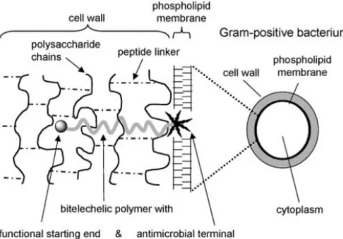
1.1.6. Mechanism of action of end-capped poly(2-oxazoline)s
The biocidal mechanism, of such polymers is still not entirely clear. Such compounds
have an antimicrobial action if they contain an alkyl chain of adequate length to interact
with the bacterial membrane by binding to it. Then the membrane’s equilibrium is
disturbed due to the positive charge of the bioactive function eventually leading to cell
death (see Figure 1.3).31,32 POx derivatives only show bactericide activity if equipped
Figure 1.3 One proposed interaction between an antimicrobial polymer and a Gram-positive bacterium.22
1.1.7. Lavender oil
In addition to Pox, essential oils are well recognized in traditional medicine as
antimicrobial agents and they are characterized by a wide spectrum of activity against
bacteria and yeasts.
Lavender oil is well known for its application in aromatherapy, cosmetics, soaps and
perfumes. Lavender essential oil is usually produced by steam distillation, from both the
flower heads and foliage, but the chemical composition differs greatly, with the sweeter
and most aromatic oil being derived from the flowers.33 Today, the pure oil is most often
used in aromatherapy or incorporated into soaps and other products as a pleasant
fragrance or as an antimicrobial agent.
Lavender oil (primarily Lavandula angustifolia) has been found to be active against many
species of bacteria and yeasts.34 It has also been suggested that essential oils, including
lavender, may be useful in treating bacterial infections that are resistant to antibiotics.
For example, L. angustifolia oil has demonstrated to have in vitro activity against both
MRSA (methicillin-resistant Staphylococcus aureus) and VRE (vancomycin-resistant
enterococci).35
Some cases of activity against bacteria and yeasts have been reported in literature,
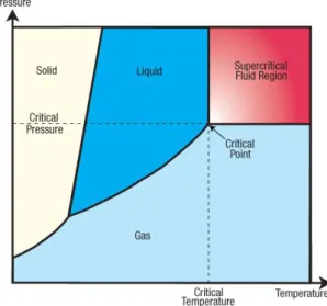
1.1.8. Supercritical fluids (SCFs)
Supercritical fluids have gained interest as reaction solvent or processing agent.
Recently, they have been applied in polymerization, swelling, impregnation,
fractionation, purification and formation of powdered polymers. By definition, SCFs are
substances at a temperature and pressure higher than their critical values, and which
have a density close to or higher than its critical density (Figure 1.4and Figure 1.5).37
SCFs may be relatively dense and dissolve certain solids while being miscible with
permanent gases and exhibiting high diffusivity and low viscosity. In addition, SCFs are
highly compressible and the density dependent properties (and therefore solvent
properties) can be “tuned” over a wide range by varying pressure.38
Figure 1.4 Schematic pressure-temperature phase diagram for a pure component showing the supercritical Fluid (SCF) region.39
In particular supercritical carbon dioxide (scCO2) has been studied extensively for the
synthesis and processing of polymers. CO2 is naturally occurring, abundant and existing
in natural reservoirs. In comparison to other substances, CO2 has an easily accessible
critical point with a critical temperature (Tc) of 31.1 °C and a critical pressure (pc) of 7.38
MPa. It is an ambient gas, and can be recycled after use. Finally, it is inexpensive and
non-flammable.41
CO2 can be separated from a reaction mixture by reducing the pressure, yielding a dry
polymer product and in one-step process. It is possible the production of materials with
high purity, since the final depressurization and extraction enables the removal of
unreacted monomers and initiator from the polymer matrix. Also, it is possible to obtain
polymers by a relatively simple and inexpensive process.
SCFs have also been recognized as excellent extraction solvent, due its availability and
safety being considered as a GRAS (Generally Recognized as Safe) solvent. Such
advantages make scCO2 a solvent of choice in food and fragrances industry proven by
coffee decaffeination, hop and essential oil extraction processes.42
1.1.9. scCO2-assisted in-situ polymerization in polyethylene
Polyethylene (PE) is considered the most important and extensively used thermoplastic
because of its low cost, good processability and several range of applications. However,
PE has some disadvantages such as low surface energy, lack of chemical functionality,
difficulty in dyeing and poor compatibility with synthetic polar polymers. The
characteristics modification of PE has been widely investigated for electrical, coating,
bonding, and biomedical applications.
The density of SCFs, and therefore their solvent strength, is tunable from gas to liquid
by changing pressure and temperature. This offers the ability to control the degree of polymers’ swelling, as well as the partitioning of small molecules penetrating between swollen polymer and fluid phases.43 The low viscosity and almost inexistent surface
tension of SCFs allow for rapid mass transfer into a swollen polymer.
Since the capacity of a solvent to dissolve solutes mainly depends on its density,
changes in temperature or pressure may significantly change the dissolution properties
of a supercritical solvent without variation of its composition. Additionally, the viscosity of
scCO2 is much lower than that of liquid solvents, and also varies strongly with deviations
in pressure and temperature as stated before. Because of these features, diffusion
small change in temperature or pressure, in particular near the critical point, has a large
effect on the diffusivities.44
ScCO2 has been used to impregnate polymers matrices with different additives. Using
scCO2 as a swelling agent, is possible to develop a synthetic method to produce new
polymer composites.45 Both the monomer and initiator are dissolved in scCO 2,
impregnated into the polymer substrate, and subsequently polymerized (see Figure 1.6).
Using this method, it is possible to obtain polymer composites, even when using
polymers that are immiscible and cannot be obtained through conventional methods.
Figure 1.6 General representation for the in-situ polymerization in scCO2. The monomer, initiator and
impregnating-material are placed in a high-pressure cell and the CO2 is added. A supercritical phase occurs,
the monomer and initiator solubilize and the polymerization starts. After the end of polymerization, the non-impregnated polymer precipitates and CO2 is released.
In this work, an in-situ polymerization was performed in order to synthesize
oligo(2-oxazoline)s terminated with a quaternary ammonium inside the PE matrix, in scCO2.
During scCO2 assisted process, PE swells and POx polymerizes inside the PE matrix.
At the end of the procedure, the reactor is vented, PE returns to its initial form and trapps
the antimicrobial POx inside its structure.46
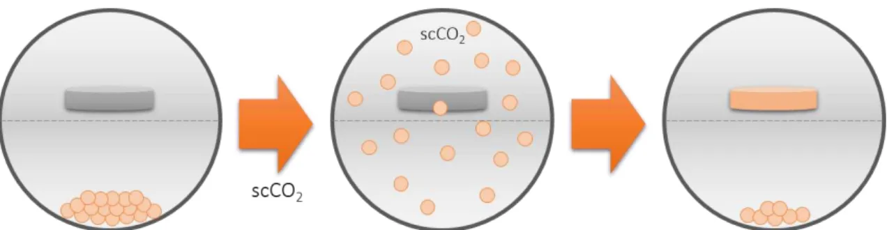
1.1.10. ScCO2-assisted impregnation
The polymer sorption and swelling processes in supercritical media and, in particular in
scCO2, became recently an area of increasing attention. Swelling and plasticization by
scCO2 sorption lead to the polymer transition from a glassy state into a rubbery state. In
a rubbery state, the polymer chains can move more freely, which makes impurity
extraction and polymer impregnation with additives more efficiently.
The impregnation process is feasible when the active substance (the solute) is soluble
in the supercritical fluid, the polymer is swollen by the supercritical solution and the
partition coefficient is favorable enough to allow the matrix to be charged with enough
Figure 1.7 General representation for a scCO2-assisted impregnation. The compound and
impregnating-material are placed in a high-pressure cell and the CO2 is added. A supercritical phase occurs, the compound
solubilize and the impregnation starts. After depressurization, the non-impregnated polymer precipitates.
In this work, the lavender oil was impregnated in the PE matrix, using scCO2, leading to
1.2. Experimental
1.2.1. Materials
The high density polyethylene was supplied by BeyonDevices and used without further
purification. The monomers 2-methyl-oxazoline and 2-ethyl-oxazoline, the initiator boron
trifluoride diethyl etherate (BF3.Et2O) and N,N-dimethyldodecylamine were purchased
from Sigma-Aldrich. All commercial reagents were used as received. Carbon dioxide was
supplied by Air Liquide with a purity lighter than 99.998%. Dulbecco's modified Eagle's
medium (DMEM-F12) and resazurin based in vitro toxicology assay kit were purchased
from Sigma-Aldrich. Human fibroblast cells (Normal human dermal fibroblasts adult,
cryopreserved cells) were purchased from PromoCell. Fetal bovine serum (FBS) was
purchased from Biochrom AG.
1.2.2. Instrumentation
The thermal behavior of the samples was measured using Differential Scanning
Calorimetry (DSC) in a Perkin Elmer DSC 7. The samples (~12mg) were analyzed under
a flow of nitrogen gas and taken between 20oC and 200oC at a scan rate of 10 oC/min.
The morphology of the samples was recorded using Scanning Electron Microscopy
(SEM) in a Hitachi S-2400 instrument, with an accelerating voltage set to 15 kV. All
samples were gold coated before analysis.
X-ray photoelectron spectroscopy (XPS) analysis were performed on a XSAM800 X-ray
spectrometer, operated in the fixed analyser transmission (FAT) mode, with a pass
energy of 10 eV, a power of 120 W and using a non-monochromatic radiation (energy
of 1486.6 eV). Spectra were collected with a step of 0.1 eV, using a Sun SPARC Station
4 with Vision software (Kratos). The curve fitting for component peaks was carried out
with a non-linear least-squares algorithm using a commercial program – the
XPSPEAK41. For the bond energy correction (EL) for accumulated charge, was
considered the bond energy of bonded carbon only for other carbons and for hydrogen
of 285 eV. Sensitivity factors used were: C 1s - 0.25, O 1s 0.66, N 1s - 0.42 and Si 2p –
0.27.
Elementary analysis was achieved using a FlashEA 1112 Series CHNS Analyzer. The
tests were performed with the combustion reactor temperature of 900 oC, the GC column
temperature of 65 oC, the Helium flow rate of 130 ml/min, the oxygen flow rate of 250
ml/min. The oxygen injection time was 7 s, the sample injection time was 12 s and the
standard was BBOT [2,5-Bis (5-tert-butyl-benzoxaxol-2-yl) thiophene] 6.51% N, 72.53%
C, 6.09% H e 7.44% S, with K factor as calibration method.
1.2.3. In-situ polymerization of 2-substituted oxazolines
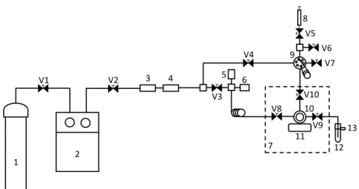
1.2.3.1. Experimental apparatus
The in-situ polymerization was undertaken in a high-pressure apparatus schematically
presented in Figure 1.8. The real apparatus is shown on Figure 1.9, Figure 1.10 and
Figure 1.11.
Figure 1.8 Schematic representation of the experimental apparatus. 1-CO2 cylinder; 2-high pressure pump;
3-line filter; 4-check valve; 5-high pressure transducer; 6-rupture disk; 7-thermostatted bath; 8-syringe; 9-HPLC high pressure valve; 10-high-pressure cell, 11-stirrer; 12-shlenk; 13-vent; V1 to V10-high pressure valves (adapted from reference29)
Figure 1.9 Real experimental apparatus, during the in-situ polymerization.
1
2
3 4 5 6
7 9
8
11 10
12 13
V1 V2
V3 V4
V5
V6
V7
V8
Figure 1.10 Real apparatus of a high-pressure cell.
Figure 1.11Real experimental apparatus, inside the high-pressure cell.
1.2.3.2. Pre-treatment of the PE samples
The polyethylene-based vaginal applicator was cut into similar pieces (~25 mg) and the
samples were washed with ethanol under stirring overnight.
1.2.3.3. Synthesis of living oligomers
In situ-polymerization reactions were carried out in a 33 ml high-pressure cell. Two
different 2-substituted oxazoline oligomers were synthesized (MeOx and EtOx) and
used were [M]/[I]=15 (2-methyl-2-oxazoline) and [M]/[I]=12 (2-ethyl-2-oxazoline),
according with previous studies.29
The PE samples (~25 mg), the 2-substituted oxazoline and the initiator were placed in a
high-pressure cell with a magnetic stirring bar, with a physical division between the PE
samples (see Figure 1.11) and the reagents, and then immersed in a thermostatized oil
bath. The polymerizations were performed at 115 ºC. The desired pressure of 18 MPa
was achieved using carbon dioxide pressurized in the high-pressure cell. After 20 h of
reaction, the living oligomer was obtained and able to be terminated with different
molecules.
1.2.3.4. End-capping with N,N-dimethyldodecylamine
The termination of living oligomer was performed with addition of a tenfold excess of
amine in relation to the added amount of initiator,48 using a HPLC high-pressure valve.
The reaction was maintained at the temperature of polymerization under stirring during
24 h. After this, the temperature was lowered to 45oC and the oligomer was washed
using fresh CO2 during 2 h, in order to remove unreacted reagents. The obtained
samples were placed in ethanol overnight under stirring, in order to remove the oligomer
adsorbed to the surface (see Figure 1.12).
Figure 1.12 Synthesis of oligo(2-methyl-2-oxazoline) end-capped with N,N-dimethyldodecylamine using CROP polymerization in scCO2 medium.
1.2.3.5. Impregnation of Lavender oil
PE samples (~25 mg) were impregnated with lavender oil in scCO2 using a high-pressure
cell. The impregnation was performed at 115 ºC and 18 MPa using an excess of oil. After
20 h of continuous stirring the high pressure vessel was rapidly depressurized.
The lavender oil used was extracted using scCO2 in the aim of a doctoral thesis1 at the
host laboratory and was used in the development of these materials. Composition of
lavender oil extracted can be found in Appendix 1.
1.2.4. Evaluation of the antimicrobial activity
1.2.4.1. Microorganisms growth conditions
Escherichia coli DH5α (gram-negative bacteria), Staphylococcus aureus ATCC25923
and Staphylococcus aureus COL MRSA (both gram-positive bacteria) strains were
grown at 37oC in Mueller-Hinton broth medium (MHB, DIFCO, USA). Candida albicans
PYCC 3436 and Candida glabrata PYCC2814 (both yeasts) strains were grown at 37oC
in Yeast Mannitol broth medium (YMB, composition in g/L: Potassium hydrogen
phosphate 0.5; magnesium sulfate heptahydrate 0.2; sodium chloride 0.2; calcium
chloride hexahydrate 0.2; mannitol 10; yeast extract 0.4). Cultures grown overnight at
37oC were diluted in the same media to 105 CFU/mL to carry out the tests referred to
below.
1.2.4.2. Disc diffusion
This test was performed using the non-impregnated oligomers recovered from the
bottom of the reactor after the in-situ polymerizations. Cells were cultivated using a swab
impregnated with a solution of each microorganism (105 cells) in standard growth
medium, Mueller-Hinton Agar (MHA, DIFCO, USA).
Oligomers were dissolved in sterile water in a concentration of 100 mg/ml. Lavender oil
was used without dissolution.
Paper discs (no. 231039, Becton and Dickinson, USA) were then impregnated with 10
µl of oligomer solution or lavender oil and placed on the surface of the agar plates
containing growth medium. A negative control was also performed using a disc
impregnated with sterile water. The plates were incubated for 24 h at 37oC for bacterial
strains and 48 h at 37oC for yeast. Experiments were executed in duplicate and after
incubation the inhibition zone was evaluated.
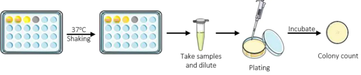
1.2.4.3. Microdilution
The tests were performed using four different microorganisms referred to above: E. coli,
S. aureus COL, C. albicans and C. glabrata. One impregnated polyethylene sample (~25
mg) was placed in each well of a 24-well tissue culture plate, containing 1 ml of medium
and 5 µl of the microorganism suspension (105 cells), and incubated at 37 °C with
shaking for 24 h for bacterial strains and 48 h for yeast. The samples were diluted (10-1
and Yeast Mannitol Agar (YMA, composition in g/L: Potassium hydrogen phosphate 0.5;
magnesium sulfate heptahydrate 0.2; sodium chloride 0.2; calcium chloride hexahydrate
0.2; mannitol 10; yeast extract 0.4; agar 10) for yeasts, and incubated at 37°C for 24 h.
After this, the number of colonies was determined (colony forming units – CFU/ml) and
the results were normalized for reduction of the viable cells in log stages (see Figure
1.13). Experiments were performed in duplicated in two independent assays.
Figure 1.13 Schematic representation of the methodology used to evaluate the antimicrobial activity of samples.
The long term stability of the samples was evaluated using an Accelerated Aging
procedure (ASTM F1980). Data obtained from the study is based in the conditions that
simulate the effects of aging on the materials.
Accelerated Aging calculation is based on Arrhenius’ equation which simply states that
a 10 °C increase in temperature doubles the rate of chemical reaction. Four variables
were used in calculating the accelerated aging test duration: test temperature (Te),
ambient temperature (Ta), Reaction rate factor (Q10=2 for medical devices) and real-time
equivalent (DRT).
𝐴𝐴𝑅(𝐴𝑐𝑐𝑒𝑙𝑒𝑟𝑎𝑡𝑒𝑑 𝐴𝑔𝑔𝑖𝑛𝑔 𝑅𝑎𝑡𝑒) = 𝑄10((𝑇𝑒−𝑇𝑎)/10) eq. 1
𝐴𝐴𝑇𝐷(𝐴𝑐𝑐𝑒𝑙𝑒𝑟𝑎𝑡𝑒𝑑 𝐴𝑔𝑔𝑖𝑛𝑔 𝑇𝑖𝑚𝑒 𝐷𝑢𝑟𝑎𝑡𝑖𝑜𝑛) =𝐷𝑒𝑠𝑖𝑟𝑒𝑑 𝑅𝑒𝑎𝑙 𝑇𝑖𝑚𝑒𝐴𝐴𝑅 eq. 2
Using equations 1 and 2, it was possible to determine the conditions for the accelerated
aging test. In this case, the samples were placed at 60 °C during 52 days. After this time,
1.2.4.4. Bactericidal vs bacteriostatic
Using the mediums from microdiluition test where bacterial growth was not verified, 10
µl were diluted in 1 ml of new medium and incubated at 37 oC during 24 h. After this time,
the presence of growing was evaluated.
1.2.5. Evaluation of the anti-biofouling activity
The samples used in the microdilution tests were washed with PBS and fixed with
glutaraldehyde (2.5%) during 10 minutes. After that, the samples were washed with
water, dehydrated with ethanol 70%, 80% and 90% sequentially during 10 minutes and
stored in ethanol 100%. SEM was used to assess the bacterial adhesion and proliferation
during the 24 h of the microdilution assay for bacteria or 48h for yeasts.
1.2.6. Biocompatibility tests
1.2.6.1. Polyethylene impregnated with 2-substituted oxazolines
In vitro cytotoxicity assays were performed with cell line HCT-116 (colorectal carcinoma
cell line) and human fibroblasts through the use the CellTiter 96® AQueous
Non-Radioactive Cell Proliferation Assay (Promega, Madison, USA), a colorimetric method
for determining the number of viable cells.
To the maintenance and splitting of the cell line, and at 80 % confluence, cells were
harvested and centrifuged during 5 minutes. The supernatant was discarded, and the
cell pellet resuspended in 2 ml of medium. For growth inhibition assays, 0.75x10⁵
cells/mL were plated into flat bottomed 96-well plates (Costar, Corning, NY) and
incubated at 37 oC, 99% humidity and 5% CO
2 (v/v). Cell density was evaluated as the
total number of viable cells within the grids on the hemacytometer (Hirschmann,
Eberstadt, Germany) using trypan blue exclusion method. For this procedure 350 µl of
medium was pipetted to a 2 mL eppendorf together with 50 µl of the 2ml cell suspension
followed by 100 µl of 0.4 % (v/v) trypan blue solution (Sigma). The hemacytometer was
loaded and examined immediately under the microscope at low resolution, and cell
viability was determined through the following equation:
𝑁º 𝑜𝑓 𝑐𝑒𝑙𝑙𝑠
𝑚𝑙 =
∑𝑐𝑒𝑙𝑙𝑠 𝑝𝑒𝑟 𝑞𝑢𝑎𝑑𝑟𝑎𝑛𝑡
4 ∗ 10
After 24 h, MeOx and EtOx solutions (0.45 mg/mL), and PE, PE_MeOx and PE_EtOx
material samples were added (after removal of depleted medium), and the cells were
incubated for 1 h and 24 h [37 oC, 99% humidity and 5% CO
2 (v/v)]. Subsequently a
reaction mix of medium, MTS
(3-(4,5-dimethylthiazol-2-yl)-5-(3-carboxymethoxyphenyl)-2-(4-sulfophenyl)-2H-tetrazolium, inner salt), and PMS (phenazine methosulfate) (Kit
Promega) in a ratio of 100:19:1 was added to each well and further incubated for 45 min.
During this period, MTS is bioreduced into formazan, by dehydrogenase enzymes
present in metabolically active cells, which in turn is susceptible of being measured at
490 nm absorbance by Tecan Infinite F200 Microplate Reader (Tecan, Männedorf,
Switzerland), directly from the 96-well assay plate, so that the quantity of formazan
product measured is directly proportional to the number of living cells in culture. The cell
viability results for each concentration were normalized relatively to the control samples
and obtained accordingly to the following formula:
𝑆𝑎𝑚𝑝𝑙𝑒 𝐴𝑏𝑠𝑜𝑟𝑣𝑎𝑛𝑐𝑒 (490 𝑛𝑚)
𝐶𝑜𝑛𝑡𝑟𝑜𝑙 𝐴𝑏𝑠𝑜𝑟𝑣𝑎𝑛𝑐𝑒 (490 𝑛𝑚)∗ 100 = 𝐶𝑒𝑙𝑙 𝑉𝑖𝑎𝑏𝑖𝑙𝑖𝑡𝑦 (%) eq. 4
1.2.6.2. Polyethylene impregnated with lavender oil
1.2.6.2.1. Proliferation of human fibroblast cells in the presence of samples
To evaluate cell behavior in the presence of the samples, human fibroblasts cells (4x10⁴
cells/well) were seeded with materials in a 96-well plate and cultured with DMEM-F12
supplemented with fetal bovine serum (FBS), for 48 h, at 37°C under a 5% CO2
humidified atmosphere. Wells containing cells in the culture medium without materials
were used as negative control. EtOH 96% was also added to some wells and they were
used as a positive control. Previously to cell seeding, materials were sterilized using UV
irradiation during 30 minutes. Cell growth was monitored using an Olympus CX41
inverted light microscope (Tokyo, Japan) equipped with an Olympus SP-500 UZ digital
camera.49,50,51
1.2.6.2.2. Characterization of the cytotoxic profile of the samples
Human fibroblast cells were seeded, in contact with sterilized materials, at a density of
4x10⁴ cells/well and cultured with DMEM-F12. Subsequently, the mitochondrial redox
activity of viable cells was assessed through a resazurin assay (n=5). At 24 and 48 h,
CO2 humidified incubator, for 24 h, at 37 ºC. Fluorescence of metabolized resazurin was
measured using a Gemini EM spectrophotometer at an excitation/emission wavelength
of λ=545/590nm, respectively.52 Wells containing cells in the culture medium without
materials were used as negative control (K-). Ethanol (96%) was added to wells that
1.3. Results and discussion
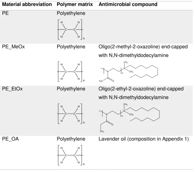
1.3.1. Synthesized materials
Figure 1.1 summarizes all synthesized and used materials and their respective
compositions.
Table 1.1 Reference and composition of materials.
Material abbreviation Polymer matrix Antimicrobial compound
PE Polyethylene
PE_MeOx Polyethylene Oligo(2-methyl-2-oxazoline) end-capped
with N,N-dimethyldodecylamine
PE_EtOx Polyethylene Oligo(2-ethyl-2-oxazoline) end-capped
with N,N-dimethyldodecylamine
PE_OA Polyethylene Lavender oil (composition in Appendix 1)
1.3.2. Materials characterization
1.3.2.1. Gravimetric determination of loading
All polymerizations were carried out at 115 ºC and 18 MPa during 20 h and more 24 h
for oligomer end-capping.30,54 High density polyethylene is extremely difficult to process
The gravimetric data were obtained by the following equation:
𝑀𝑎𝑠𝑠 𝑔𝑎𝑖𝑛 (%) =𝑊𝑡−𝑊0
𝑊0 × 100 eq. 4
where W0 is the initial weight of the PE sample and Wt is the weight of the modified PE
sample after washing and drying. The reported mass gain was the mean value of six
different samples.
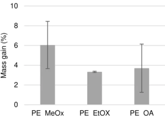
Figure 1.14 Variation of the mass gain (%) in modified PE samples for different antimicrobial compounds used.
Observing the results for the modified PE samples with oligo(2-oxazoline)s (Figure 1.14),
there is a decrease in the mass gain of PE samples with the increase in the size of the
side chain of the oligo(2-oxazoline). The amount of monomer used was calculated in
order to achieve the saturation of medium with monomer, ensuring the maximum loading
into the PE matrix. The PE modified with oligo(2-methyl-2-oxazoline), comprising a
methyl side chain, presents a higher mass gain comparing with PE modified with
oligo(2-ethyl-2-oxazoline), which contains an ethyl side chain. This suggests that the rate of
diffusion through the PE matrix plays a role in the loading of the monomer, once all the
reaction conditions were the same, the free volume of PE is identical and hence the
larger monomers diffuse through the matrix slower than smaller ones, leading to a small
amount of oligomer polymerized inside the PE matrix.
In the case of PE impregnated with lavender oil, there is no previous work describing the
supercritical-assisted impregnation in literature, so it is not possible to compare this result
with previous data. Although, PE impregnated with lavender oil presents similar values
of mass gain to the modified PE samples with oligo(2-oxazoline)s showing that probably
that is the maximum amount possible to impregnate in this polyethylene matrix. 0 2 4 6 8 10
PE_MeOx PE_EtOX PE_OA
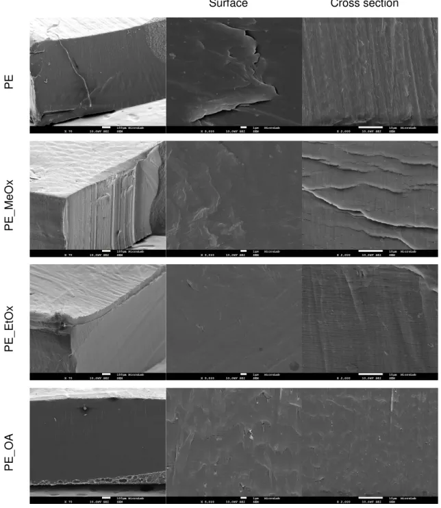
1.3.2.2. Morphological characterization
SEM micrographs were obtained for unmodified PE, PE modified with two
2-oxazoline-based oligomers and PE impregnated with lavender oil (see Figure 1.15).
Surface Cross section
PE
P
E
_MeO
x
P
E
_E
tO
x
P
E
_OA
Figure 1.15 SEM micrographs of surface and cross section of the unmodified PE and PE modified with antimicrobial compounds: PE_MeOx, PE_EtOx and PE_OA.
The SEM images show that all materials present the same morphology, without changes
material in high pressure and temperature conditions do not cause morphological
modifications in the structure. Moreover, the presence of oligomers or compounds are
not visible at the PE matrix surface, suggesting that the antimicrobial compounds are
inside the PE matrix, which was swelled during the scCO2 process and back to original
form after depressurization, confining the new compound inside and avoiding its
leaching.46
1.3.2.3. Determination of materials composition
In order to confirm the success of oligomers polymerization, NMR analysis was made
using the non-impregnated oligomers recovered from the bottom of the reactor after the
in-situ polymerizations (see Appendix 2 and Appendix 3).30
The effectiveness of the supercritical in-situ polymerization and impregnation procedures
was evaluated by XPS and elementary analysis.
Table 1.2 Percentage of different elements in the surface of each sample. Data obtained by XPS.
Sample Nitrogen (%) Carbon (%) Oxygen (%) Silicon (%)
PE 2.1 80.0 16.5 1.4
PE_MeOx 3.7 81.9 11.7 2.7
PE_EtOx 0.3 83.8 14.1 1.7
PE_OA 0.9 84.4 13.0 1.6
XPS is a technique able to identify and quantify the elemental composition at the surface
region with an analysis depth of the order of 3-10 nm. Observing the results (see Table
1.2), no significant changes were observed comparing the untreated PE with modified
PE materials. This technique only analyses the surface, and most of this differences
could be due to the presence of some impurities at the samples surface, which are not
significant when considering the overall sample.
Table 1.3 Percentage of different elements in each sample, evaluated by elemental analysis.
Sample Nitrogen (%) Carbon (%) Hydrogen (%) Sulphur (%)
PE 0.03 83.12 11.86 0
PE_MeOx 0.12 82.92 13.58 0
PE_EtOx 0.07 83.09 12.80 0
Complementing the XPS analysis, elemental analysis based on sample combustion
dynamics was performed. Analyzing the Table 1.3, we could notice an increase in the
amount of nitrogen and hydrogen, confirming the introduction of a new compound
containing these elements in its composition. Also, it is possible to see a decrease in the
percentage of carbon (the element present in more quantity in PE), which is an expected
result, since the addition of new compounds to the PE matrix reduces the proportion of
this element in the overall matrix.
1.3.2.4. Thermal properties
DSC thermograms of unmodified PE, of 2-oxazoline-based oligomers synthesized
outside the PE matrix and of modified PE samples are shown in Figure 1.16.
20 40 60 80 100 120 140 160 180
EtOx
Temperature (oC)
MeOx PE PE_EtOx
PE_MeOx
Figure 1.16 DSC thermograms of unmodified PE, of oligomers synthesized outside the PE matrix and of modified PE samples.
The DSC of unmodified PE displays the expected single melting peak at 139 ºC. By
analysing the thermograms, we can conclude that the presence of oligo(2-oxazoline)s
oligomers in the PE matrix shift melting temperature (Tm) to values more close of
oligomer Tm and also this last one disappear. It can be seen that in each thermogram
an interaction between the oligomer and PE matrix, and consequently a successfully
impregnation. These results also suggest that the treatment with scCO2 do not change
the degree of crystallinity compared with original PE.
1.3.3. Evaluation of the antimicrobial activity
1.3.3.1. Disc diffusion
In order to evaluate the susceptibility of bacteria and yeasts to the developed
antimicrobial compounds, solutions of 100 mg/mL in water of each oligomer were placed
in paper discs and tested for different microorganisms. Also, lavender oil extracted using
scCO2 was tested against the same microorganisms, without any dilution. This method
allows a quick determination of the antimicrobial activity of compounds.
Table 1.4 Antimicrobial activity evaluated by disk diffusion for different synthesized oligomers and lavender oil, against different gram positive and negative bacteria and yeasts.
Diameter of growth inhibition zone (mm)
Microorganism MeOx EtOx Lavender oil
E. coli DH5α 15 17 12
S. aureus COL 20 23 23
S. aureus ATCC 25923 29 31 22
C. albicans PYCC 3436 29 36 10
C. glabrata PYCC 2814 15 23 0
From the Table 1.4 it is possible to say that the synthesized oligomers,
oligo(2-methyl-2-oxazoline) and oligo(2-ehtyl-oligo(2-methyl-2-oxazoline), have a significant antimicrobial activity
against all the 5 microorganisms tested. Oligo(2-ethyl-2-oxazoline presents for all the
microorganisms the highest growth inhibition zone. This results are in agreement with
previous reports using similar oligomers.30 This kind of test only gives a qualitative result,
since the limitations of the technique should be considered, such as the diffusion of the
solution from the disk due to the viscosity or possible interactions between the sample
and the disk.
In the case of lavender oil, antimicrobial activity was observed against all
microorganisms, with exception of C. glabrata. The chemical composition of lavender oil
components. Linalool has been reported as the component responsible for the
yeastscidal activity and the activity of lavender oil against microorganisms could be
depend on an additive effect of its major components; however, the contribution of minor
constituents to the antimicrobial activity cannot be disconsidered.55 A previous study
already reported the antimicrobial and antifungal activity of lavender oil.56 The result
related with C. glabrata is unexpected, once the activity against yeasts is already
reported. However there is no previous study reporting the antimicrobial activity of the
lavender oil against this microorganism. This occurrence could be due to the previously
mentioned limitations of the technique and a negative result can not mean the absence
of activity and this result should be confirmed with tests performed in liquid medium.
It is important to note that the tested compounds present antimicrobial activity against a
multi-resistant S. aureus, which is a very promising result, since there are not many
compounds effective against this microorganism.57
Unfortunately, there is no zone diameter interpretative standards for the tested
compounds, so it is not possible to evaluate if the microorganisms are susceptible,
intermediate or resistant.
1.3.3.2. Microdilution
To evaluate the ability of the materials to kill bacteria and yeasts upon contact, cells
viability after direct exposure to PE modified with antimicrobial compounds was assessed
using E. coli, S. aureus, C. albicans and C. glabrata.
The materials were incubated in medium containing cells and, after incubation, samples
medium were diluted, plated and the number of colony-forming units (CFU) was counted.
Incubation of bacteria and yeasts cells, with stirring, in the presence of the unmodified
PE did not influence the normal growth of the E. coli, S. aureus and C. glabrata cells.
These results are the expected, since PE in its native form does not have antimicrobial
activity (Figure 1.17).58 However, unmodified PE killed >99.9% of the C. albicans cells (3
log stages reduction=a reduction of 99.9%). This effect was not predictable, so other
tests are required to explain this result, such as the use of another strain of C. albicans
to clarify if this antimicrobial activity is only for this strain in particular or species-specific.
The addition of 2-oxazoline-based antimicrobial oligomers to the PE matrix improved
drastically the material antimicrobial performance. After 18 h for bacteria and 24 h for
yeasts, the materials reduce the cells viability over 99.9%. These results are in