Objective: To propose and analyze the test-retest reliability of an instrument to verify the presence and intensity of pain in the cervical, thoracic and lumbar spine in Brazilian young people.
Methods: This reliability study enrolled a sample of 458 participants (13 to 20 years). Two groups were formed for each sex according to the range of days for the test-retest (10±3 and 28±2 days). For analysis of spinal pain, a drawing of the human body with cervical, thoracic and lumbar spine areas delimited was presented. The following question was presented: during a normal day, do you feel pain in any of these regions of your spine? If so, what is the intensity from 0 to 10 (mark on the line)? The starting point, with the number 0, corresponded to no pain, and the number 10 to severe pain. The agreement of frequency and of intensity of pain was verified by Kappa test and Bland-Altman plot, respectively.
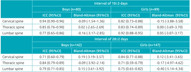
Results: Intraclass correlation coefficients ranged from 0.71 (confidence interval of 95% — 95%CI — 0.59–0.79) to 0.94 (95%CI 0.90–0.96). The results concerning the agreement of pain scores showed the mean differences to be close to 0, and the largest mean difference was -0.40 (95%CI -5.14–4.34). The agreement in reported pain ranged from 72.2 (Kappa 0.43; 95%CI 0.28–0.58) to 90.1% (Kappa 0.76; 95%CI 0.60–0.92).
Conclusions: This instrument was shown to be a reliable manner to verify the pain in different regions of the spine in Brazilian young people.
Keywords: Adolescent; Child; Neck pain; Low back pain; Pain measurement.
Objetivo: Propor e analisar a reprodutibilidade de um instrumento para verificar a presença e a intensidade da dor na coluna cervical, torácica e lombar em jovens brasileiros.
Métodos: Estudo de reprodutibilidade com uma amostra de 458 participantes (13 a 20 anos). Dois grupos foram formados para cada sexo de acordo com o intervalo de dias entre teste e reteste (10±3 e 28±2 dias). Para a análise da dor na coluna, foi apresentada a figura de um corpo humano com as áreas da coluna cervical, torácica e lombar delimitadas. A seguinte pergunta foi realizada: durante um dia comum, você sente dor em alguma dessas regiões da coluna? Se sim, qual é a intensidade de 0 a 10 (marque um traço)? A extremidade com o número 0 correspondia à ausência de dor e o número 10, à dor muito intensa. A concordância na frequência e intensidade da dor foi verificada por meio do teste Kappa e da plotagem de Bland-Altman, respectivamente.
Resultados: Os coeficientes de correlação intraclasse variaram de 0,71 (intervalo de confiança de 95% — IC95% — 0,59–0,79) a 0,94 (IC95% 0,90–0,96). Os resultados relativos à concordância no escore de dor mostraram que as diferenças médias foram próximas de 0 e a maior diferença média foi de -0,40 (IC95% -5,14–4,34). A concordância no relato de dor variou de 72,2 (Kappa 0,43; IC95% 0,28–0,58) a 90,1% (Kappa 0,76; IC95% 0,60–0,92).
Conclusões: O instrumento demonstrou ser uma forma reprodutível de verificar a dor em diferentes regiões da coluna vertebral em jovens brasileiros.
Palavras-chave: Adolescente; Criança; Cervicalgia; Dor lombar; Medição da dor.
ABSTRACT
RESUMO
*Corresponding author. E-mail: arrudaga@yahoo.com.br (G.A. Arruda). aUniversidade Estadual de Londrina, Londrina, PR, Brazil.
bInstituto Federal de Educação, Ciência e Tecnologia de São Paulo, Boituva, SP, Brazil. cBrunel University London, Uxbridge, Greater London, England.
Received on February 07, 2018; approved on June 24, 2018; available online on December 20, 2018.
ProPosal and test-retest reliability of a
scale for cervical, thoracic, and lumbar
sPine Pain in brazilian young PeoPle
Proposição e reprodutibilidade de uma escala de dor na
coluna cervical, torácica e lombar em jovens brasileiros
Gustavo Aires de Arruda
a*
, Diogo Henrique Constantino Coledam
b,
Arli Ramos de Oliveira
a, Fernanda dos Santos Neri
a,
INTRODUCTION
Among the regions in the human body affected by musculo-skeletal pain, the lumbar spine has been widely investigated. A systematic review observed prevalence of low back pain vary-ing from 9 to 65% in young people from different regions of
the world.1 Recent studies described that the prevalence of low
back pain in Brazilian children and adolescents ranges from
15.5 to 18%.2-4 The high prevalence of back pain can be
con-sidered a public health problem.5 In addition to low back pain,
there is high prevalence (>20%) of young people that report
pain on cervical and thoracic spine regions.3 Multiple pain sites
are associated to disabilities in adolescents,6 and concomitant
neck and low back pain increases the risk of mental health
problems when compared to single pain.7 Early prevention is
recommended, as low back pain in adolescence can predict low
back pain in adulthood.8
Questionnaires are extensively used to assess back pain in children and adolescents. In Brazil, studies that aimed to assess back pain in children and adolescents failed to report
the process of translation and cross-culturally adaptation2,9
and did not report its reproducibility data in Brazilian young
people.2-4,9-11 Another usual limitation of questionnaires used
in Brazilian studies is that pain is analyzed as a dichotomous
way, i.e., presence or absence of pain. Therefore, the intensity
of pain cannot be estimated.3,4,11
The visual analogue scale is an instrument that can overcome the issue of dichotomous pain reports. This scale is frequently used to evaluate the intensity of the pain and has been largely used as a reference procedure in the validation of instruments
for pain verification.12-14 Noll et al. proposed a Brazilian scale
to assess back pain that can point out information regarding intensity, but it does not discriminate cervical, thoracic or low
back pain.15
Low back pain assessment is necessary, as it has a complex
etiology and can emerge from many causes,5 and the estimated
total health cost of people living with chronic back pain seems to be doubled, when compared to those who do not mention
any pain.16 However, high prevalence of pain in other regions
of spine may affect children and adolescents. A Danish study described that neck pain was the most common spinal pain
region, followed by mid back and low back pain.17 Still,
con-sequences of multiple pain sites are not clear, as this issue has received sparse attention.
An instrument to analyze the frequency and intensity of pain in different regions of the spine would be relevant for professionals and researchers that need to identify the prevalence and to check the efficacy of intervention pro-grams that aim to prevent or reduce cervical, thoracic and low back pain in children and adolescents. Therefore, the
purpose of this study was to analyze the test-retest reliabil-ity of an instrument to verify the presence and intensreliabil-ity of pain in the cervical, thoracic and lumbar spine in Brazilian young people.
METHOD
This is a reliability study that was part of a larger project that involved children and adolescents from Londrina, Paraná, Brazil. The larger project included information about physi-cal activity, eating habits and consumption of alcoholic bever-ages, smoking, spinal pain, socioeconomic and demographic information by questionnaires, after that anthropometric measures, blood pressure and heart rate were collected, and it was performed the Fitnessgram motor tests. Study protocols were approved by the Ethics in Research Committee from the university where the study took place (Protocol no. 234/10). Parents or guardians of students who agreed to participate in the study signed a consent form wherein all the procedures, researcher contact details, and possible risks and benefits of the study were described.
Londrina city had 48,688 students enrolled in state schools (publicly administered institutions) at the beginning of the
study, from 5th grade of elementary school to the 3rd grade
of high school. The total of 30,777 students were attending
the 5th to 8th grades. Regarding the 1st, 2nd and 3rd high school
years, 17,911 students were enrolled in state schools (data from the City Department of Education of Londrina, referring to the year 2009). In the present study, the schools with 400 to 800 enrolled students were considered medium-sized schools, and the schools with more than 800 enrolled students were considered large. The number of enrolments was proportion-ally distributed among small, medium and large schools in the city. Two state schools in the city of Londrina were randomly selected for the composition of the sample in the present study: a medium-sized (central region) and a large one (northern region). Classrooms were randomly selected in each school (conglomerate). The sample involved approximately 50% of the participants of each school. Participants in this study were composed of 458 people (236 girls and 222 boys), in the age range from 13 to 20 years old.
Two groups were formed for each sex, according to the days for the test-retest. The first group consisted of 80 boys with
the mean interval of test-retest of the pain scale 10±3 days,
and the second group was composed of 142 boys and the
mean interval between test-retest was 28±2 days. The same
considering as an alternative hypothesis the intraclass correla-tion coefficients (ICC) value of 0.70 and for the null hypoth-esis the value of 0.40, using the F-test with the significance level of 0.01. Under the same conditions, sample sizes of 89, 142 and 147 subjects had 96.5, 99.8 and 99.9% of power, respectively. All data was calculated using Power Analysis and Sample Size Software 15.
The following procedures were conducted to develop the instrument to evaluate back pain. First version of the instru-ment was developed, and its content was analyzed by four experts. Suggestions were examined and incorporated in a second version of the instrument, and experts then carried out a new content analysis. This version was used in young people to verify their understanding regarding the instru-ment and reproducibility.
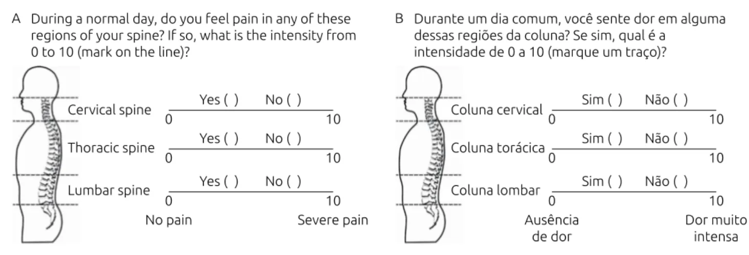
A drawing of the human body (lateral position), which made it possible to visualize the spine, was presented to the students to explore the presence of spinal pain (Figures 1A and 1B). The areas of the cervical, thoracic and lumbar spine were delimited by a dashed line and the name of the region
indicated. The following question with the options yes or no
was presented to students: during a normal day, do you feel pain in any of these regions of your spine? If so, what is the intensity from 0 to 10 (mark on the line)? The visual analog scale measured 10 cm. The starting point, with the number 0, corresponded to no pain; and the number 10, to severe pain. The instrument was applied in the classroom during phys-ical education classes. Only the students participating in the research remained in the room. Prior to responding the ques-tionnaire, an explanation was given regarding the purpose of the instrument. While participants answered the question-naire, possible doubts were explained. One of the research-ers (GAA) was present during the whole procedures of data
collection, and s/he received assistance from other researchers previously trained to perform the procedures in a standard-ized way. Participants were advised to disregard pains in other regions of the body other than the spine. Also, they were advised to ignore sporadic pain caused by recent trauma such as falls, knocks, etc., reporting only usual pain.
Additionally, in this study the translation of the instru-ment to English was carried out according to previous
rec-ommendations.18 Firstly, two professional translators
trans-lated the original Portuguese version to English (translations). During the translation process, equality of meaning was prioritized instead of equality of word. Subsequently, two Brazilian researchers in the field of health translated this version from English to Portuguese (back-translations). Finally, the research team reached a consensus regarding the final version of the instrument based on its first and second translation. This procedure was performed to facilitate the use of the instrument in other countries, thus making it pos-sible to compare information about prevalence and intensity of spine pain in young people.
Normal distribution of the data was analyzed by the Kolmogorov-Smirnov’s test. Descriptive analyses used mean and standard deviation (SD). The Student’s unpaired t-test was performed to compare the characteristics between groups for boys, and equality of variances was averiguated by Levene’s test. The same tests were used to compare characteristics between girls. The test-retest reliability of pain scores was verified by the ICC one-way random effect and their respective 95% confi-dence intervals (95%CI). The interpretation was performed according to the values: <0.40 = poor; 0.40 to <0.75 = good;
≥0.75 = excellent.19 The agreement between the scores for
test-retest was verified with the Bland-Altman plot method. The bias between the mean values of pain and interval of days
Figure 1 Scale for verifying the presence of spinal pain: (A) English version; (B) Portuguese version.
Durante um dia comum, você sente dor em alguma dessas regiões da coluna? Se sim, qual é a
intensidade de 0 a 10 (marque um traço)? B
Coluna cervical
Coluna torácica
Coluna lombar
0 10
Sim ( ) Não ( )
0 10
Sim ( ) Não ( )
0 10
Sim ( ) Não ( )
Ausência de dor
Dor muito intensa During a normal day, do you feel pain in any of these
regions of your spine? If so, what is the intensity from 0 to 10 (mark on the line)?
A
Cervical spine
Thoracic spine
Lumbar spine
0 10
Yes ( ) No ( )
0 10
Yes ( ) No ( )
0 10
Yes ( ) No ( )
(test-retest) was verified by regression models (linear, quadratic and cubic) and R-squared. The same procedure was used to check the bias between the mean values of pain and differences (test-retest). The agreement of reports for the presence of
spi-nal pain according to the region was verified using the Kappa
index, and the interpretation performed according to values:
≤0.20 = poor; 0.21 to 0.40 = regular; 0.41 to 0.60 =
moder-ate; 0.61 to 0.80 = good; >0.80 = very good.20 The relative
fre-quency and 95%CI of spinal pain according to the region were calculated. The comparisons of frequencies between test-re-test for each group were performed using the McNemar’s test-re-test.
Results were considered statistically significant when p≤0.05.
All data were analyzed using Statistical Package for the Social
Sciences (SPSS) 20.0.
RESULTS
Table 1 presents the sample characteristics according to the gender and mean interval days (10 or 28 days) of spinal pain scale application. No differences were found between inter-val day groups in boys or girls (p>0.05). The mean age of all groups was 15 years old.
Table 2 exhibits the ICC for the pain scale according to the spinal region. Table 3 contains the relative frequency of pain in the test and retest moments for each spinal region, while Table 4 shows the agreement in the indication of pain presence between test and retest instrument administration moments.
With the interval of 10 days between test and retest appli-cation of the pain scale, all values of ICC were considered
excellent (ICC ≥0.77) for boys and girls. When the interval
between applications was higher, the reliability for the boys was good and excellent for the cervical, thoracic and lumbar spine regions. Among girls, the test-retest reliability was excel-lent for the cervical spine and good for the thoracic and lum-bar spine. The agreement of pain scores showed that the mean differences were close to 0. The largest mean difference in the 10-day period was observed among boys for the lumbar spine with -0.16 (95%CI -3.17–2.85). For 28 days, the highest average
Table 1 Characteristics of the sample according to gender
and interval of days between pain scale application.
BMI: body mass index; SD: standard deviation; p>0.05 for comparisons between groups of days for boys and girls.
10±3 days
Boys (n=80) Mean±SD
Girls (n=89) Mean±SD
Age (years) 15.1±1.7 14.9±1.7
Body mass (kg) 63.5±16.0 52.9±11.8 Height (cm) 169.2±10.3 160.0±6.4
BMI (kg/m2) 22.1±4.7 20.6±3.9
28±2 days
Boys (n=142) Mean±SD
Girls (n=147) Mean±SD
Age (years) 15.5±1.5 15.4±1.4
Body mass (kg) 61.9±12.3 54.7±11.1
Height (cm) 170.3±8.9 161.4±6.6
BMI (kg/m2) 21.2±3.1 20.9±3.9
Table 2 Intraclass correlation coefficient and Bland-Altman plot for the pain scale, according to gender and interval
of days between test-retest.
p<0.01 for all intraclass correlation coefficient values; 95%CI: 95% confidence interval. Interval of 10±3 days
Boys (n=80) Girls (n=89)
ICC (95%CI) Bland-Altman (95%CI) ICC (95%CI) Bland-Altman (95%CI) Cervical spine 0.94 (0.90–0.96) -0.09 (-1.54–1.36) 0.82 (0.73–0.88) -0.15 (-3.88–3.58)
Thoracic spine 0.85 (0.76–0.90) 0.02 (-2.65–2.69) 0.83 (0.74–0.89) 0.00 (-3.69–3.70)
Lumbar spine 0.77 (0.65–0.86) -0.16 (-3.17–2.85) 0.92 (0.88–0.95) 0.05 (-3.07–3.17)
Interval of 28±2 days
Boys (n=142) Girls (n=147)
ICC (95%CI) Bland-Altman (95%CI) ICC (95%CI) Bland-Altman (95%CI)
Cervical spine 0.71 (0.60–0.79) 0.19 (-3.19–3.57) 0.84 (0.77–0.88) 0.12 (-3.41–3.65)
Thoracic spine 0.84 (0.79–0.89) -0.09 (-3.92–2.14) 0.71 (0.59–0.79) -0.17 (-4.97–4.62)
value of the difference was observed for the girls in the lumbar spine, with -0.40 (95%CI -5.14–4.34) (Table 2).
The number of days between test-retest had a slight influence on the magnitude of the differences. In all cases, the models with the best fits were cubic; the highest varia-tion explained only 2.2% of the differences. These findings suggest that the differences are independent on the num-ber of days between test-retest in this study (Figures 2A, 2B and 2C). The bias for the differences between test-retest and mean values of pain was analyzed considering only the strat-ification by sex. In general, the models with the best fits were cubic, except for the cervical spine in boys. For this, the best fit was the quadratic model, explaining less than 7% of the variance of the results, and being the highest value obtained (Figures 2D, 2E and 2F).
Table 3 shows that there were no significant differences in frequency of individuals who reported pain in the cervical, thoracic and lumbar spine between test-retest. This fact was evidenced by the McNemar’s test and can also be seen by the overlap of 95%CI in frequencies. The major difference in the frequency of reporting pain in the test-retest interval of 10 days for the boys was found in the lumbar spine with 7.5 percentage points, and for girls in the cervical spine with 4.5 percentage points. In the period from 28 days, the same regions had the greatest variation with a difference of 8.4 percentage points for boys and 5.4 percentage points for girls.
With the exception of the cervical spine for boys (26.1 vs.
24.6%) in the interval of 28 days, the frequency of reported pain was slightly higher in the retest moment (Table 3). Generally, higher frequencies of pain were reported among
girls in both the test and retest of the instrument for all regions independently on the group of days range. Considering the same interval of days for application, the only region that showed no overlap of 95%CI between gender was the tho-racic spine, with the application interval of 28 days between
Table 3 Relative frequencies of spine pain according to gender and interval of days between test-retest for the
pain scale.
p>0.05 for all comparisons of relative frequencies between test-retest by McNemar’s test; %: relative frequencies of spine pain; 95%CI: 95% confidence interval.
Interval of 10±3 days
Boys (n=80) Girls (n=89)
Test % (95%CI) Retest % (95%CI) Test % (95%CI) Retest % (95%CI)
Cervical spine 26.3 (16.6–35.9) 31.3 (21.1–41.4) 39.3 (29.2–49.5) 43.8 (33.5–54.1)
Thoracic spine 36.3 (25.7–46.8) 37.5 (26.9–48.1) 42.7 (32.4–53.0) 43.8 (33.5–54.1)
Lumbar spine 30.0 (20.0–40.0) 37.5 (26.9–48.1) 42.7 (32.4–53.0) 46.1 (35.7–56.4)
Interval of 28±2 days
Boys (n=142) Girls (n=147)
Test % (95%CI) Retest % (95%CI) Test % (95%CI) Retest % (95%CI)
Cervical spine 26.1 (18.8–33.3) 24.6 (17.6–31.7) 38.8 (30.9–46.7) 44.2 (36.19–52.3)
Thoracic spine 23.2 (16.3–30.2) 26.8 (19.5–34.0) 38.1 (30.2–46.0) 38.8 (30.90–46.7)
Lumbar spine 28.9 (21.4–36.3) 37.3 (29.4–45.3) 41.5 (33.5–49.5) 44.2 (36.19–52.3)
Interval of 10±3 days
Boys (n=80) Girls (n=89)
Kappa(95%CI) % Kappa(95%CI) % Cervical
spine 0.76 (0.60–0.92) 90.1 0.77 (0.64–0.90) 88.8
Thoracic
spine 0.71 (0.54–0.87) 86.3 0.75 (0.61–0.89) 87.7
Lumbar
spine 0.61 (0.43–0.79) 82.5 0.70 (0.56–0.85) 85.4
Interval of 28±2 days
Boys (n=142) Girls (n=147)
Kappa(95%CI) % Kappa(95%CI) % Cervical
spine 0.44 (0.27–0.61) 78.9 0.64 (0.51–0.76) 82.4
Thoracic
spine 0.61 (0.46–0.76) 85.2 0.43 (0.28–0.58) 72.2
Lumbar
spine 0.50 (0.35–0.64) 77.4 0.53 (0.39–0.67) 76.9
Table 4 Agreement with the Kappa index and relative frequencies in reported pain according to gender and interval of days between test-retest.
Figure 2 Analysis of the influence of the number of days between test-retest (A, B and C) and average values of
pain (D, E and F) on the differences obtained between test-retest.
Boys Girls
Boys Girls Gender 10
5
5 10 15 20 25 30
0
–10 –5
P
ain intensity
difference (tes
t, retest)
Days between test and retest for cervical spine
Boys: R2 cubic = 0.022
Girls: R2 cubic = 0.013
A
10
5
5 10 15 20 25 30
0
–10 –5
P
ain intensity
difference (tes
t, retest)
Days between test and retest for thoracic spine
Boys: R2 cubic = 0.012
Girls: R2 cubic = 0.005
B
10
5
5 10 15 20 25 30
0
–10 –5
P
ain intensity
difference (tes
t, retest)
Days between test and retest for lumbar spine
Boys: R2 cubic = 0.012
Girls: R2 cubic = 0.010
C
10
5
0 2 4 6 8 10
0
–10 –5
P
ain intensity
difference (tes
t, retest)
Mean pain in cervical spine (test + retest / 2)
Boys: R2 quadratic = 0.069
Girls: R2 cubic = 0.003
D
10
5
5 2 4 6 8 10
0
–10 –5
P
ain intensity
difference (tes
t, retest)
Mean pain in thoracic spine (test + retest / 2)
Boys: R2 cubic = 0.024
Girls: R2 cubic = 0.004
E
10
5
0 2 4 6 8 10
0
–10 –5
P
ain intensity
difference (tes
t, retest)
Mean pain in cervical spine (test + retest / 2)
Boys: R2 cubic = 0.028
Girls: R2 cubic = 0.018
test moments — boys: 23.2% (95%CI 16.29–30.19) versus girls: 38.1% (95%CI 30.24–45.95).
The agreement in reported pain (Table 4) for 10 days was
considered good for boys and girls. Kappa statistics ranged from
0.61 (82.5%) to 0.76 (90.1%) among boys, and 0.70 (85.4%) to 0.77 (88.8%) among girls, according to the examined region. For boys, in the 28 days period between the test-retest, the agree-ment for the pain scale in both the lumbar spine and cervical
spine were moderate [Kappa 0.44 (78.9%) and 0.50 (77.4%),
respectively], and the thoracic spine was good [Kappa 0.61
(85.2%)]. For the girls, in this same 28-day interval the
agree-ment was good for the cervical spine [Kappa 0.64 (82.4%)]
and moderate for the thoracic and lumbar spine [Kappa 0.43
(72.2%) and 0.53 (76.9%), respectively].
DISCUSSION
The main findings of this study were acceptable values of the instrument on test-retest reliability and agreement of pain frequency and pain intensity independently on day intervals. It should be considered that it was not expected to find per-fect reproducibility. Aspects such as memory, seasonality of the investigated phenomenon or the clinical condition of the par-ticipant on the assessment of day might influence the obtained information. In the present study, it was found that the differ-ences between the test-retest were not affected by the number of days. Such information may be of great interest when the instrument is used for multiple measurements.
To identify the reproducibility of an instrument proposed to verify the pain, it is a presumption to start using such method. However, some methodological considerations should be done regarding studies that verified back pain in Brazilian young
people.2-4,10,11,21 The Nordic musculoskeletal questionnaire has
been widely used to analyze back pain among Brazilians,3,4,10,11
Despite the fact that it has a Portuguese version,22 the
repro-ducibility of this Nordic questionnaire was not tested in
Brazilian youths.3,4,10,11 Dorneles et al.2 used a questionnaire
that has no Portuguese version, and the reliability data was not described neither for original instrument nor for the study
conducted.23 Also, in studies that reproducibility was analyzed,
the interval of test-retest assessment is usually seven days.15,21
Therefore, it is not possible to know whether the instruments are reproducible in larger time intervals. The scale proposed in the present study showed reproducibility during a period of more than 10 days, providing support for the use of the spine pain instrument.
Although the instruments described before provides valuable information for observational studies, such as prevalence of
musculoskeletal pain, only categorical outcome (e.g., presence
vs. absence and frequency of pain) limits the utilization of
these scales in intervention studies. In experimental studies that investigated interventions for back pain treatment, it is necessary to check how procedures can reduce pain
inten-sity.24,25 Noll et al.partially reduced this limitation and
pro-posed an instrument that, in addition to closed questions, had visual analogue scale (0 to 10) to estimate pain
inten-sity.15 However, pain intensity is assessed considering general
back pain and do not specifies region. In the present study, the instrument was developed to assess presence and inten-sity of pain using a visual analogue scale on three regions: cervical, thoracic and lumbar regions. A human body draw has been previously used in studies and it is suggested as an
ideal method to identify body regions.26-28 These
characteris-tics, such as simplicity and possibility of identifying the pain region, contribute to instrument applicability in epidemio-logical and experimental studies.
The main limitation of the present study is the fact that the validity of the scale was not described. In young people, the criterion validity process of pain scales is conducted matching the results to their clinical diagnose records or to secondary
outcomes (i.e., disability).29,30 Unfortunately, no information
about clinical records of the sample was analyzed. Other lim-itation of this study involved the fact that it was not a popu-lation-based survey, with a representative sample. Despite the limitation, the scale is recommended, as it is self-administered, easy to use and understand, as well as it has low cost, being suitable to use in epidemiological studies.
The results of this study support the possibility of using this instrument to screen Brazilian adolescents with spinal pain, and to supply an indicator of the intensity of the pain. It also enables the diagnosis of possible factors associated with the presence of spinal pain or analysis of the effects of intervention to reduce spinal pain. Future studies are sug-gested to verify the accuracy of the scale in diagnosing cer-vical, thoracic and lumbar spinal pain in adolescents when compared with a clinical examination and the relationship between spinal pain and postural deviations (examined by imaging methods such as X-rays), spinal injuries and bad posture habits.
In conclusion, the proposed instrument is a reliable tool to verify both presence and intensity of pain in different regions of the spine in Brazilian young people.
funding
The study did not receive funding.
conflicts of interests
REFERENCES
1. Calvo-Muñoz I, Gómez-Conesa A, Sánchez-Meca J. Prevalence of low back pain in children and adolescents: a meta-analysis. BMC Pediatr. 2013;13:14.
2. Dorneles RC, Oliveira HL, Bergmann ML, Bergmann GG. Flexibility and muscle strength/resistance indicators and screening of low back pain in adolescents. Rev Bras Cineantropom Desempenho Hum. 2016;18:93-102. 3. Saes MD, Soares MC. Factors associated with back pain in
adolescents from public schools in one city from South Brazil. Rev Salud Publica. 2017;19:171-80.
4. Scarabottolo CC, Pinto RZ, Oliveira CB, Zanuto EF, Cardoso JR, Christofaro DG. Back and neck pain prevalence and their association with physical inactivity domains in adolescents. Eur Spine J. 2017;26:2274-80.
5. Kordi R, Rostami M. Low back pain in children and adolescents: an algorithmic clinical approach. Iran J Pediatr. 2011;21:259-70.
6. Hoftun GB, Romundstad PR, Rygg M. Factors associated with adolescent chronic non-specific pain, chronic multisite pain, and chronic pain with high disability: the Young-HUNT Study 2008. J Pain. 2012;13:874-83.
7. Rees CS, Smith AJ, O’Sullivan PB, Kendall GE, Straker LM. Back and neck pain are related to mental health problems in adolescence. BMC Public Health. 2011;11:382. 8. Hestbaek L, Leboeuf-Y de C, Kyvik KO, Manniche C. The
course of low back pain from adolescence to adulthood: eight-year follow-up of 9600 twins. Spine (Phila Pa 1976). 2006;31:468-72.
9. Zapata AL, Moraes AJ, Leone C, Doria-Filho U, Silva CA. Pain and musculoskeletal pain syndromes in adolescents. J Adolesc Health. 2006;38:769-71.
10. Fassa AG, Facchini LA, Dall’Agnol MM, Christiani DC. Child labor and musculoskeletal disorders: the Pelotas (Brazil) epidemiological survey. Public Health Rep. 2005;120:665-73. 11. Vitta A, Martinez MG, Piza NT, Simeão SF, Ferreira NP.
Prevalence of lower back pain and associated factors in students. Cad Saude Publica. 2011;27:1520-8.
12. Sugiura S, Aoki Y, Toyooka T, Shiga T, Otsuki K, Aikawa E, et al. Characteristics of low back pain in adolescent patients with early-stage spondylolysis evaluated using a detailed visual analogue scale. Spine (Phila Pa 1976). 2015;40:E29-34. 13. Yao W, Mai X, Luo C, Ai F, Chen Q. A cross-sectional survey
of nonspecific low back pain among 2083 schoolchildren in China. Spine (Phila Pa 1976). 2011;36:1885-90.
14. Hakala PT, Saarni LA, Punamäki RL, Wallenius MA, Nygård CH, Rimpelä AH. Musculoskeletal symptoms and computer use among Finnish adolescents - pain intensity and inconvenience to everyday life: a cross-sectional study. BMC Musculoskelet Disord. 2012;13:41.
15. Noll M, Candotti CT, Vieira A, Loss JF. Back pain and body posture evaluation instrument (BackPEI): development,
content validation and reproducibility. Int J Public Health. 2013;58:565-72.
16. Hong J, Reed C, Novick D, Happich M. Costs associated with treatment of chronic low back pain: an analysis of the UK General Practice Research Database. Spine (Phila Pa 1976). 2013;38:75-82.
17. Aartun E, Hartvigsen J, Wedderkopp N, Hestbaek L. Spinal pain in adolescents: Prevalence, incidence, and course: a school-based two-year prospective cohort study in 1,300 Danes aged 11-13. BMC Musculoskelet Disord. 2014;15:187. 18. Guillemin F, Bombardier C, Beaton D. Cross-cultural adaptation of health-related quality of life measures: literature review and proposed guidelines. J Clin Epidemiol. 1993;46:1417-32.
19. Fleiss JL. The design and analysis of clinical experiments. New York: John Wiley; 1986.
20. Svanholm H, Starklint H, Gundersen HJ, Fabricius J, Barlebo H, Olsen S. Reproducibility of histomorphologic diagnoses with special reference to the Kappa statistic. APMIS. 1989;97:689-98.
21. Lemos AT, Santos FR, Moreira RB, Machado DT, Braga FC, Gaya AC. Low back pain and associated factors in children and adolescents in a private school in Southern Brazil. Cad Saude Publica. 2013;29:2177-85.
22. de Barros EN, Alexandre NM. Cross-cultural adaptation of the Nordic musculoskeletal questionnaire. Int Nurs Rev. 2003;50:101-8.
23. Sjolie AN. Low-back pain in adolescents is associated with poor hip mobility and high body mass index. Scand J Med Sci Sports. 2004;14:168-75.
24. Perich D, Burnett A, O’Sullivan P, Perkin C. Low back pain in adolescent female rowers: a multi-dimensional intervention study. Knee Surg Sports Traumatol Arthrosc. 2011;19:20-9. 25. Dias MH, Amaral E, Pai HJ, Tsai DT, Lotito AP, Leone C, et al.
Acupuncture in adolescents with juvenile fibromyalgia. Rev Paul de Pediatr. 2012;30:6-12.
26. Young IA, Haig AJ, Yamakawa SK. The association between backpack weight and low back pain in children. J Back Musculoskelet Rehabil. 2006;19:25-33.
27. Mohseni-Bandpei MA, Bagheri-Nesami M, Shayesteh-Azar M. Nonspecific low back pain in 5000 Iranian school-age children. J Pediatr Orthop. 2007;27:126-9.
28. Kistner F, Fiebert I, Roach K, Moore J. Postural compensations and subjective complaints due to backpack loads and wear time in schoolchildren. Pediatr Phys Ther. 2013;25:15-24.
29. Huguet A, Stinson JN, McGrath PJ. Measurement of self-reported pain intensity in children and adolescents. J Psychosom Res. 2010;68:329-36.
30. Legault EP, Cantin V, Descarreaux M. Assessment of musculoskeletal symptoms and their impacts in the adolescent population: adaptation and validation of a questionnaire. BMC Pediatr. 2014;14:173.