Full paper published online: February 28, 2009 ISSN 1678-9199.
ACUTE HEPATOTOXICITY OF Crotalus durissus terrificus (SOUTH AMERICAN
RATTLESNAKE) VENOM IN RATS
França RF (1), Vieira RP (2), Ferrari EF (1), Souza RA (1), Osorio RAL (1),
Prianti-Jr. ACG (1), Hyslop S (3), Zamuner SR (1), Cogo JC (1), Ribeiro W (1)
(1) Laboratory of Physiology and Pharmacodynamic, Institute of Research and Development, Vale do Paraíba University, UNIVAP, São José dos Campos, São Paulo State, Brazil; (2) Department of Pathology and Physical Therapy, School of Medicine, University of São Paulo, USP, São Paulo, São Paulo State, Brazil; (3) Department of Pharmacology, School of Medical Sciences, State University of Campinas, UNICAMP, Campinas, São Paulo State, Brazil.
ABSTRACT: Venom of the South American rattlesnake, Crotalus durissus terrificus (Cdt), presents myotoxic and neurotoxic outcomes, but reports on its effects on the liver are scarce. This study examined the hepatotoxicity resulting from Cdt venom administration (100, 200 and 300 μg/kg) in male Wistar rats. Animals were studies at 3, 6, 9 and 12 hours after venom injection. The hepatotoxicity was assessed through serum levels of aspartate aminotransferase (AST), alanine aminotransferase (ALT), alkaline phosphatase (AP), gamma glutamyl transferase (GGT), bilirrubin and also by histopathological evaluation. All the different concentrations of Cdt venom resulted in increased levels of hepatic enzymes, when compared with the control group, except for the 100 μg/kg dose, which presented normal levels at 9 and 12 hours after venom administration. Bilirrubin levels remained unchanged by Cdt venom. Histological analysis revealed endothelial damage, inflammatory cell infiltration, as well as sinusoidal and portal congestion. Based on these observations, we may conclude that Cdt venom causes dose- and time-dependent hepatic damage in rats, characterized by elevated hepatic enzyme levels and histological alterations.
KEY WORDS: Crotalus durissus terrificus, hepatotoxicity, histology, AST, ALT, rattlesnake, liver enzymes, venom.
CONFLICTS OF INTEREST: There is no conflict.
FINANCIAL SOURCE: FAPESP and FVE/UNIVAP.
CORRESPONDENCE TO:
INTRODUCTION
The South American rattlesnake, Crotalus durissus terrificus, presents a wide
distribution throughout southeastern Brazil (1). Envenomations by this species
account for 6 to 8% of all registered bites by venomous snakes in the country, with
an approximate mortality rate of 1.8%, the highest among venomous species (2). C.
d. terrificus venom contains a variety of toxins, including convulxin (3), crotamine (4),
crotoxin – its main neurotoxin, which accounts for nearly 50% of the venom dry
weight (5) – and gyroxin (6-8), as well as several enzymes common to other snake
venoms (9, 10).
Experimental and clinical envenomations by C. d. terrificus venom result mainly in
myotoxicity and neurotoxicity, while the most effective treatment is administration of
anticrotalic antivenom (11). Whereas some consequences of C. d. terrificus venom
have been extensively studied – such as myonecrosis (12-17), coagulation
disturbances (7, 8, 18-20), neuromuscular effects (21, 22), renal involvements (14,
23) and homeostatic problems (20) – considerably less attention has been paid to its
effects on the liver.
In humans, envenomation by C. d. terrificus causes hepatic lesions involving
necrosis, effects attributed to release of pro-inflammatory cytokines (24-27). Also,
hepatic steatosis and increased serum aspartate aminotransferase (AST) levels were
reported by Azevedo Marques et al. (14) in patients bitten by C. d. terrificus and
extensive hepatic necrosis by Barraviera et al. (24). In addition, Barraviera et al. (25)
showed increased serum levels of AST (86%) and ALT (66%), by hepatocyte
electron microscopy that showed mitochondrial damage, suggesting that liver injury
may occur partly in the cytoplasm and partly in the mitochondria. Bancher, Rosa and
Furlanetto (28) proved that, in mice, the venom could be detected in hepatic tissue.
The liver is a multifunctional organ, responsible for vital functions and for
maintenance of energy balance in the organism. The release of intracellular enzymes
into circulation is frequently used as an indicator of hepatocyte damage (29),
although the release of some enzymes like AST and ALT – also present in tissues
other than liver – needs to be evaluated by other injury markers to confirm any
hepatic damage. Therefore, in the present study, we examined the liver damage
induced by C. d. terrificus venom in rats through enzymatic markers of liver function
MATERIALS AND METHODS
Venoms and Reagents
The kits for the quantification of serum enzymes and bilirubin were from Labtest
Diagnóstico (Brazil). The reagents for histology were purchased from Synth (Brazil).
C. d. terrificus venom was obtained by manual milking of snakes (both sexes)
collected in the region of São José dos Campos and maintained in the snake house
of the Nature Study Center, Vale do Paraíba University. The venom was lyophilized
and stored at 4°C. Stock solutions of venom were prepared in sterile 0.9% NaCl
immediately before use.
Animals
Male Wistar rats (180-220 g) obtained from Bem-te-vi Farm (Indaiatuba, SP, Brazil)
were maintained in a 12-hour-light/dark cycle at 23 ±2°C with free access to food and
water. Animal procedures described herein were approved by the institutional Animal
Research Ethics Committee of UNIVAP (protocol number L035/2004/CEP) and
performed in accord with the guidelines of the Brazilian College for Animal
Experimentation (COBEA).
Experimental Protocols
To study the hepatotoxicity of C. d. terrificus venom, rats (283 per group) received an
intramuscular injection of sterile saline solution only or venom (100, 200 or 300
µg/kg) with saline solution in a final volume of 100 µL. The rats were subsequently
killed with an overdose of ether at 3, 6, 9 or 12 hours after saline or venom injection
(7 rats/time interval in each group) and blood was collected from the inferior vena
cava. Samples of hepatic tissue were also obtained for histological analysis.
Serum Enzyme Activities and Bilirubin Levels
Serum alanine aminotransferase (ALT), alkaline phosphatase (AP), aspartate
aminotransferase (AST) and γ-glutamyl transferase (GGT) activities were determined
spectrophotometrically, as described by Reitman and Frankel (30), Szasz (31) and
Roy (32). Bilirubin tests were made by a chosen diazo-coupling reagent and by a
Histomorphometrical Analysis
Liver fragments from rats killed at different times after venom administration were
fixed in 10% formaldehyde overnight and then dehydrated in an increasing alcohol
series, clarified with xylol and embedded in paraffin. Sections of 5-µm-thick were cut
by a microtome (American Optical Co., USA) and stained with hematoxylin and
eosin, for subsequent examination and documentation by an Olympus CX-41®
microscope (Olympus Optical, USA) fitted to an Olympus PM10SP Automatic
Photomicrographic® system (Olympus America Medical, USA). Twenty fields were
analyzed for each slide. Using a 100-point grid with a known area (10,000 μm2 at
1,000x magnification) attached to the microscope lens the number of points
contacting the Kupffer cells and leukocytes were counted within a total area (10,000
μm2). The density of congested blood vessels, endothelial area and degenerated hepatocytes were counted and the values adjusted to the total tissue area (10,000
μm2). The density of Kupfer and mononuclear cells, as well as the density of congested blood vessels, endothelial area and degenerated hepatocytes were
expressed as number of cells/mm2 of tissue. For a qualitative histological evaluation,
the following criteria were used to determine the histologic score (scale 0-10): 0 =
negative; 0.5 to 2.0 = mild; 2.5 to 5.5 = moderate; 6.0 to 8.0 = severe; 8.5 to 10.0 =
very severe (34).
Statistical Analysis
The results were expressed as mean ± SEM and statistical comparisons were
performed by Student’s t-test or one-way of variance (Anova) followed by the
Tukey-Kramer test. The p value was considered significant (p < 0.05); very significant (p <
0.01) and extremely significant (p < 0.001).
RESULTS
Serum Liver Enzyme Activities and Bilirubin Levels
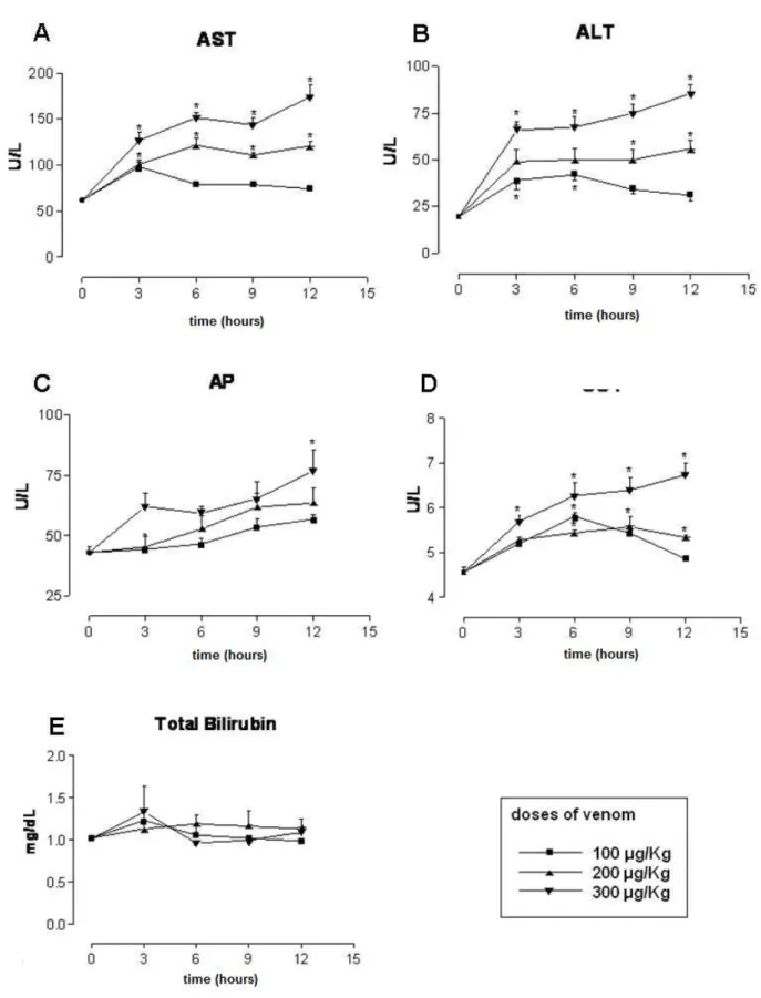
Figure 1A shows the changes in AST levels at various time intervals following the
injection of different doses of C. d. terrificus venom. The administration of 100 μg/kg
of the venom resulted in increased AST activity at 3 hours when compared to the
control group (p < 0.05), but no differences after 6, 9 and 12 hours (p > 0.05).
Regarding the 200- and 300-μg/kg venom doses, we observed an increased AST
Figure 1B reveals the changes in ALT levels at diverse intervals after administration
of different concentrations of C. d. terrificus venom. Concerning the 100-μg/kg dose,
no changes in ALT activity was found, when compared with the control group at 3, 6,
9 and 12 hours (p > 0.05). The 200-μg/kg concentration of C. d. terrificus venom
augmented ALT activity only at 9 and 12 hours relative to the control group (p <
0.05). At the dose of 300 μg/kg of C. d. terrificus venom, the ALT activity was greater
than in the control group at 3, 6, 9 and 12 hours (p < 0.05).
AP activity increased progressively after the administration of all venom doses,
although a significant change was seen only with the highest dose (300 μg/kg) at 12
hours post-venom (Figure 1C). GGT levels showed a peak increase 6 hours after
administration of the lowest dose, with a return to the pre-venom levels at 12 hours
post-venom. A moderate and persistent rise in GGT activity was registered with the
intermediate dose (200 μg/kg), while the highest one produced a progressive
increase during the 12 hours after venom administration (Figure 1D). Figure 1E
shows that total bilirubin levels were not changed by any venom dose or time
Histological Analysis
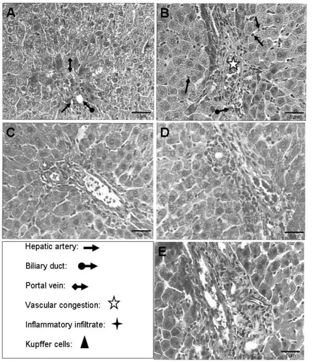
Sections of hepatic tissue from saline-treated (control) rats showed normal
hepatocytes with strongly stained nuclei and protein-rich cytoplasm. Transversal
sections showed intact sinusoids, portal spaces and a lobular central vein (Figure
2A). In contrast, rats injected with 100 µg/kg showed damage caused by an
inflammatory infiltrate 3 and 6 hours after venom injection (Figure 2B). At 9 and 12
hours post-venom, the appearance of the tissue had returned to normal (Figure 2C).
However, the morphometric analysis did not show statistical differences between the
control group and 100 µg/kg group. At the venom dose of 200 µg/kg, copious cellular
infiltrate and the extent of damage augmented with time (Figure 2D). The
morphometric analysis showed that at 200 µg/kg at 12 hours, the densities of Kupfer
cells, endothelium and degenerated hepatocytes were increased when compared
with the control group. The density of congested blood vessels at 9 hours was higher
than in the control group. At the highest venom dose (300 µg/kg), the hepatocytes
showed marked disorganization, with swelling, necrosis and a progressive increase
in damage up to 12 hours after venom administration (Figure 2E). Moreover, the
histomorphometric findings demonstrate in Table 1 that at 9 hours, the density of
Kupfer cells, congested blood vessels, endothelium densities and degenerated
hepatocyte were greater than in those of control group. At 12 hours, all parameters
Figure 2. Histological analysis. (A) Photomicrograph of hepatic tissue from the control group (HE, 400x). See hepatic artery ( ), biliary duct ( ), portal vein ( ). (B) Photomicrograph of hepatic tissue 6 hours after venom injection
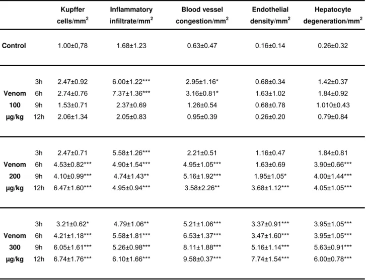
Table 1. Morphometric analysis of liver histological aspects
Kupffer
cells/mm2
Inflammatory
infiltrate/mm2
Blood vessel
congestion/mm2
Endothelial
density/mm2
Hepatocyte
degeneration/mm2
Control 1.00±0,78 1.68±1.23 0.63±0.47 0.16±0.14 0.26±0.32
3h 2.47±0.92 6.00±1.22*** 2.95±1.16* 0.68±0.34 1.42±0.37
6h 2.74±0.76 7.37±1.36*** 3.16±0.81* 1.63±1.02 1.84±0.92
9h 1.53±0.71 2.37±0.69 1.26±0.54 0.68±0.78 1.010±0.43
Venom
100
µg/kg 12h 2.06±1.34 2.05±0.83 0.95±0.39 0.26±0.20 0.79±0.84
3h 2.47±0.71 5.58±1.26*** 2.21±0.51 1.16±0.47 1.84±0.81
6h 4.53±0.82*** 4.90±1.54*** 4.95±1.05*** 1.63±0.69 3.90±0.66*** 9h 4.10±0.99*** 4.74±1.43** 5.16±1.92*** 1.95±1.05* 4.00±1.44*** Venom
200
µg/kg 12h 6.47±1.60*** 4.95±0.94*** 3.58±2.26** 3.68±1.12*** 4.05±1.05***
3h 3.21±0.62* 4.79±1.06** 5.21±1.06*** 3.37±0.91*** 3.95±1.05*** 6h 4.21±1.18*** 5.58±1.81*** 6.53±1.37*** 3.47±1.60*** 3.95±1.05*** 9h 6.05±1.61*** 5.26±0.98*** 8.11±1.88*** 5.16±1.14*** 5.63±0.91*** Venom
300
µg/kg 12h 6.74±1.76*** 6.10±1.66*** 9.58±0.37*** 7.74±1.54*** 6.00±0.78***
Morphometric analysis of liver histological aspects found in the control group and Cdt venom group
(100 µg/kg; 200 µg/kg and 300 µg/kg) 3, 6, 9 and 12 hours after administration (7 rats per group). Data
expressed as intensity score scale from 0 to 10: 0 = negative; 0.5 to 2.0 = mild; 2.5 to 5.5 = moderate;
6.0 to 8.0 = severe; 8.5 to 10.0 = very severe. *p < 0.05; **p < 0.01 and ***p < 0.001 vs. shocked rats
DISCUSSION
The present study showed that the Crotalus durissus terrificus venom augments
activity of hepatic enzymes and also produces histological alterations, suggesting
hepatotoxicity.
The liver maintains the organism’s energy supply for many functions, such as
producing substances that break down fats, converting glucose to glycogen,
producing urea, synthesizing certain amino acids, filtering harmful substances and
maintaining a proper blood glucose level. There are some intracellular enzymes that
are released into the circulation and are frequently used as an indicator of damage to
hepatocytes. The principal marker enzymes include alanine (ALT) and aspartic (AST)
aminotransferases, which catalyze the transfer of α-amino groups from alanine and
aspartate to the α-keto group of ketoglutaric acid to produce pyruvic acid and
oxaloacetic acid, respectively (29). ALT is a more specific marker of hepatocellular
injury, and occurs in the liver, kidneys, heart, skeletal muscle and pancreas, whereas
AST has a wider distribution, occurring in organs such as heart, liver, skeletal
muscles, kidneys and pancreas. In the liver, AST occurs predominantly (80%) in
mitochondria and 20% in the cytoplasm, whereas ALT is confined to the cytoplasm
(29, 35). Other enzymes such as alkaline phosphatase (AP) and γ-glutamyl
transferase (GGT) may also be used as markers of hepatic dysfunction, although
they are found in the liver, bone, the kidneys and its enhancement is related to
production elevation or excretion deficit, such as obstructive jaundice and
cholestasis. GGT is present in the liver, kidney and pancreas. Bilirubin is a pigment
derived from the catabolism of the heme radical in hemoproteins such as
hemoglobin, while increased serum levels of this pigment can indicate cholestatic
processes and hepatocellular damage.
The measurement of serum enzyme activities in clinical studies, after C. d. terrificus
venom administration, showed increased levels of creatine kinase (14, 16, 18, 36),
creatine phosphokinase (14), lactate dehydrogenase (14, 16, 36), aspartate
aminotransferase (36) and glutamic-oxaloacetic transaminase (14), primarily as
consequences of venom myotoxic action (11), as well as injury markers of cardiac
muscle disturbance similar to those of skeletal muscles (7, 36). Serum levels of
alkaline phosphatase and alanine aminotransferase were also elevated in dogs
patients bitten by C. d. terrificus show coagulation disorders primarily mediated by
thrombin-like enzymes present in this venom (8, 18).
A study in dogs revealed that C. d. terrificus venom causes marked alterations in
hematological, hemostatic and biochemical parameters that may also be seen,
through histological examination, as intravascular coagulation in cardiac and hepatic
tissues (20). These authors also showed a progressive increase of serum levels of
myoglobin, creatine kinase and aspartate aminotransferase demonstrated that
animals developed rhabdomyolysis. On the other hand, AP did not increase
expressly. Probably, this is related to cholestatic diseases, which is plausible
because, like AP, bilirubin concentration did not increase over time and, as outlined
in the preceding sections, is also related to cholestatic process (35-37).
As proven by the current study, the administration of C. d. terrificus venom to rats
resulted in increased circulating levels of enzymes (ALT, AST and GGT) that are
frequently employed as indicators of hepatic damage and cholestasis. The increased
AST levels observed in the present work are similar to elevated serum activity of the
same enzyme after human envenomations (24, 11). Changes in serum levels of
other enzymes, including creatine kinase (CK) and lactate dehydrogenase (LDH),
have also been observed in human envenomations (11). However, there are
differences in kinetic profiles of these alterations. For example, CK and LDH levels
present their peaks respectively before and after other enzymes. The hepatic
damage caused by the smallest venom dose (100 µg/kg), assessed by enzyme
release and histological analysis (Table 1), appeared to be minimal and reversible.
Higher venom doses (200 and 300 µg/kg) resulted in greater damage that was not
reversible within the time scale of the present experiments. In part, this hepatotoxicity
is probably mediated by PLA2 component of crotoxin, which presents the ability to
interfere in hepatic mitochondrial respiration (38). The presence of inflammatory
infiltrates associated with severe damage indicates that C. d. terrificus venom and its
components are capable of affecting macrophage function and stimulating cytokine
production (39, 40). These findings agree with the demonstrations of acute phase
inflammatory reactions in humans bitten by C. d. terrificus (26).
The venom components responsible for the hepatotoxicity are still undetermined, but
they probably involve PLA2 action on cell membrane lipids and mitochondrial
respiration (38), as well as various cell types such as lymphocytes (41, 42),
this conclusion, crotoxin – which presents all PLA2 activity of C. d. terrificus venom –
has been implicated in numerous effects, including neurotoxicity (22), myotoxicity
(12, 50, 51), renal toxicity (52), edema (45, 51), inhibition of macrophage activity (40,
44), immunosuppression (42), stimulation of the hypothalamo-pituitary-adrenal axis
(including increased plasma glucose, ACTH, corticosterone and TNF-α levels) and of
corticotropin-releasing hormone (CRH) and of arginine vasopressin pathways (53),
and cytostatic (54) and cytotoxic (55) effects in cancer cell lines.
Coagulation disturbances (19, 20) leading to fibrin deposition and ischemia, mainly
through the action of the thrombin-like enzyme (gyroxin) of this venom (8, 9, 19)
could also contribute to the hepatic damage registered in the present study. In
addition, considering the inter- and intrasubspecific variation in the composition and
biological activities of this venom (51, 56-61), there may be differences in hepatic
effects produced by venoms obtained from C. d. terrificus specimens from other
regions, and among other subspecies like C. d. cascavella, C. d. collilineatus, C. d.
durissus and C. d. ruruima. Finally, diverse susceptibilities of animal strains and
species (18) may also contribute to variations in hepatic effects of this venom.
The present study provides evidence that Crotalus durissus terrificus venom induces
hepatotoxicity in rats, by increasing blood liver enzymes, probably derived from liver,
since histological analysis revealed augmented endothelium density and hepatic
damage.
ACKNOWLEDGEMENTS
This work was supported by the State of São Paulo Research Foundation (FAPESP,
grant n. 2001/12486-8) and Valeparaibana Teaching Foundation (FVE/UNIVAP). R.
F. França was supported by a FAPESP studentship (grant n. 02/11642-9). R. P.
Vieira was supported by a FAPESP post-doc studentship (grant n. 2007/01026-2). S.
Hyslop is supported by a research fellowship from the National Council for Scientific
REFERENCES
1. Campbell JA, Lamar WW. The venomous reptiles of Latin America. Ithaca: Cornell
University Press; 1989. 425 p.
2. Araújo FAA, Santalúcia M, CabraL RF. Epidemiologia dos acidentes por animais
peçonhentos. In: Cardoso JLC, França FOS, Wen FH, Málaque CMS, Haddad Junior
V, editors. Animais Peçonhentos no Brasil: biologia, clínica e terapêutica dos
acidentes. São Paulo: Sarvier; 2003. p. 6-12.
3. Prado-Franceschi J, Brazil OV. Convulxin, a new toxin from the venom of the
South American rattlesnake Crotalus durissus terrificus. Toxicon. 1981;19(6):875-87.
4. Goncalves JM. Purification and properties of crotamine. In: Bucley EE, Porgee N,
editors. Venoms. Washington: American Association for the Advancement of
Science; 1956. p. 261-274.
5. Slotta KH, Fraenkel-Conrat HL. Schlangengifte, III. Mitteil.: Reinigung und
krystallisation des klapperschlangen-giftes. Ber Dtsch Chem Ges.
1938;71(5):1076-81.
6. Seki C, Vidal JC, Barrio A. Purification of gyroxin from a South American
rattlesnake (Crotalus durissus terrificus) venom. Toxicon. 1980;18(3):235-47.
7. Siqueira JE, Higuchi ML, Nabut N, Lose A, Souza JK, Nakashima M. Lesão
miocárdica em acidente ofídico pela espécie Crotalus durissus terrificus (cascavel),
relato de caso. Arq Bras Cardiol. 1990;54(5):323-5.
8. Alexander G, Grothusen J, Zepeda H, Schwartzman RJ. Gyroxin, a toxin from the
venom of Crotalus durissus terrificus, is a thrombin-like enzyme. Toxicon.
1988;26(10):953-60.
9. Raw I, Rocha MC, Esteves MI, Kamiguti AS. Isolation and characterization of a
thrombin-like enzyme from the venom of Crotalus durissus terrificus. Braz J Med Biol
Res. 1986;19(3):333-8.
10. Bercovici D, Chudziniski AM, Dias VO, Esteves MI, Hiraichi E, Oishi NY, Picarelli
ZP, Rocha MC, Ueda CMPM, Yamanouye N, Raw I. A systemic fractionation of
Crotalus durissus terrificus venom. Mem Inst Butantan. 1987;49(3):69-78.
11. Azevedo-Marques MM, Hering SE, Cupo P. Acidente crotálico. In: Cardoso JLC,
França FOS, Wen FH, Málaque CMS, Haddad Jr V, editors. Animais Peçonhentos
12. Gopalakrishnakone P, Dempster DW, Hawgood BJ, Elder HY. Cellular and
mitochondrial changes induced in the structure of murine skeletal muscle by crotoxin,
a neurotoxic phospholipase A2 complex. Toxicon. 1984;22(1):85-98.
13. Lima MR, dos Santos MC, Tambourgi DV, Marques T, da Silva WD, Kipnis T.
Susceptibility of different strains of mice to South American rattlesnake (Crotalus
durissus terrificus) venom: correlation between lethal effect and creatine kinase
release. Toxicon. 1991;29(6):783-6.
14. Azevedo-Marques MM, Cupo P, Coimbra TM, Hering SE, Rossi MA, Laure CJ.
Myonecrosis, myoglobinuria and acute renal failure induced by South American
rattlesnake (Crotalus durisus terrificus) envenomation in Brazil. Toxicon.
1985;23(4):631-6.
15. Azevedo-Marques MM, Hering SE, Cupo P. Evidence that Crotalus durissus
terrificus (South American rattlesnake) envenomation in humans causes myolysis
rather than hemolysis. Toxicon. 1987;25(11):1163-8.
16. Cupo P, Azevedo-Marques MM, Hering SE. Clinical and laboratory features of
South American rattlesnake (Crotalus durissus terrificus) envenomation in children.
Trans R Soc Trop Med Hyg. 1988;82(6):924-9.
17. Rossi MA, Peres LC, de Paola F, Cupo P, Hering SE, Azevedo-Marques MM.
Electron-microscopic study of systemic myonecrosis due to poisoning by tropical
rattlesnake (Crotalus durissus terrificus) in humans. Arch Pathol Lab Med. 1989;
113(2):169-73.
18. Jorge MT, Ribeiro LA. Epidemiologia e quadro clínico do acidente por cascavel
sul-americana (Crotalus durissus). Rev Inst Med Trop São Paulo. 1992;34:347-54.
19. Marunak SL, Acosta OC, Leiva LC, Ruiz RM, Aguirre MV, Teibler P. Mice plasma
fibrinogen consumption by thrombin-like enzyme present in rattlesnake venom from
the North-east region of Argentina. Medicina. 2004;64(6):509-17.
20. Sousa e Silva MC, Tomy SC, Tavares FL, Navajas L, Larsson MH, Lucas SR,
Kogika MM, Sano-Martins IS. Hematological, hemostatic and clinical chemistry
disturbances induced by Crotalus durissus terrificus snake venom in dogs. Hum Exp
Toxicol. 2003;22(9):491-500.
21. Barrio A, Brazil OV. Neuromuscular action of the Crotalus terrificus terrificus
(Laur.) poisons. Acta Physiol Lat Am. 1951;1(4):291-308.
22. Gopalakrishnakone P, Hawgood BJ. Morphological changes induced by crotoxin
23. Monteiro HS, da Silva IM, Martins AM, Fonteles MC. Actions of Crotalus durissus
terrificus venom and crotoxin on the isolated rat kidney. Braz J Med Biol Res.
2001;34(10):1347-52.
24. Barraviera B, Bonjorno-Júnior JC, Arakaki D, Domingues MA, Pereira PC,
Mendes RP, Machado JM, Meira DA. A retrospective study of 40 victims of Crotalus
snake bites. Analysis of the hepatic necrosis observed in one patient. Rev Soc Bras
Med Trop. 1989;22(1):5-12.
25. Barraviera B, Coelho KY, Curi PR, Meira DA. Liver dysfunction in patients bitten
by Crotalus durissus terrificus (Laurenti, 1768) snakes in Botucatu (state of São
Paulo, Brazil). Rev Inst Med Trop São Paulo. 1995;37(1):63-9.
26. Barraviera B, Lomonte B, TarkowskI A, Hanson LA, Meira DA. Acute-phase
reactions, including cytokines, in patients bitten by Bothrops and Crotalus snakes in
Brazil. J Venom Anim Toxins. 1995;1(1):11-22.
27. Wajchenberg BL, Sesso J, Inague T. Feições clínico-laboratoriais do
envenenamento crotálico humano. Rev Ass Med Bras. 1954;1:179-93.
28. Bancher W, Rosa RR, Furlanetto RS. Estudos sobre a fixação eletiva e
quantitativa do veneno de Crotalus durissus terrificus nos tecidos nervoso, renal,
hepático e muscular de Mus musculus Linneus, 1758. Mem Inst Butantan.
1973;37:139-48.
29. Rodrigues LEA. Enzimologia Clínica. Rio de Janeiro: Revinter; 2001. 157 p.
30. Reitman S, FrankeL S. A colorimetric method for the determination of serum
glutamic oxalacetic and glutamic pyruvic transaminases. Am J Clin Pathol.
1957;28(1):56-63.
31. Szasz G. A kinetic photometric method for serum gamma-glutamyl
transpeptidase. Clin Chem. 1969;15(2):124-36.
32. Roy AV. Rapid method for determining alkaline phosphatase activity in serum
with thymolphtalein monophosphate. Clin Chem. 1970;16:431-6.
33. Winsten S, Cehelyk B. A rapid micro diazo technique for measuring total bilirubin.
Clin Chim Acta. 1969;25(3):441-6.
34. Heijne WH, Lamers RJ, van Bladeren PJ, Groten JP, van Nesselrooij JH, van
Ommen B. Profiles of metabolites and gene expression in rats with chemically
induced hepatic necrosis. Toxicol Pathol. 2005;33(4):425-33.
35. Gayotto LCC, Alves VAF. Doenças do fígado e vias biliares. São Paulo: Atheneu;
36. Cupo P, Azevedo-Marques MM, Hering SE. Acute myocardial infarction-like
enzyme profile in human victims of Crotalus durissus terrificus envenoming. Trans R
Soc Trop Med Hyg. 1990;84(3):447-51.
37. Mattos AA, Dantas W. Compêndio de hepatologia. 2nd ed. São Paulo: Fundo
Editorial Byk; 2001. 919 p.
38. Valente RH, Novello JC, Marangoni S, Oliveira B, Pereira-da-Silva L, Macedo
DV. Mitochondrial swelling and oxygen consumption during respiratory state 4
induced by phospholipase A2 isoforms isolated from the South American rattlesnake
(Crotalus durissus terrificus) venom. Toxicon. 1998;36(6):901-13.
39. Sampaio SC, Sousa e Silva MCC, Borelli P, Curi R, Cury Y. Crotalus durissus
terrificus snake venom regulates macrophage metabolism and function. J Leukoc
Biol. 2001;70(4):551-8.
40. Sampaio SC, Brigatte P, Sousa e Silva MC, dos Santos EC, Rangel-Santos AC,
Curi R, Cury R. Contribution of crotoxin for the inhibitory effect of Crotalus durissus
terrificus snake venom on macrophage function. Toxicon. 2003;41(7):899-907.
41. Garcia F, Toyama MH, Castro FR, Proença PL, Marangoni S, Santos LMB.
Crotapotin induced modification of T lymphocyte proliferative response through
interference with PGE2 synthesis. Toxicon. 2003;42(4):433-7.
42. Rangel-Santos A, Lima C, Lopes-Ferreira M, Cardoso DF. Immunosuppressive
role of principal toxin (crotoxin) of Crotalus durissus terrificus venom. Toxicon.
2004;44(6):609-16.
43. Martins AMC, Lima AAM, Toyama MH, Marangoni S, Fonteles MC, Monteiro
HSA. Renal effects of supernatant from macrophages activated by Crotalus durissus
cascavella venom: the role of phospholipase A2 and cyclooxygenase. Pharmacol
Toxicol. 2003;92(1):14-20.
44. Sampaio SC, Rangel-Santos AC, Peres CM, Curi R, Cury Y. Inhibitory effect of
phospholipase A2 isolated from Crotalus durissus terrificus venom on macrophage
function. Toxicon. 2005;45(5):671-6.
45. Camara PRS, Esquisatto LCM, Camargo E, Ribela M, Toyama MH, Marangoni S,
Nucci G, Antunes E. Inflammatory edema induced by phospholipases A2 isolated
from Crotalus durissus sp. in the rat dorsal skin: a role for mast cells and sensory
46. Francischetti IM, Saliou B, Leduc M, Carlini CR, Hatmi M, Randon J, Faili A, Bon
C. Convulxin, a potent platelet-aggregating protein from Crotalus durissus terrificus
venom, specifically binds to platelets. Toxicon. 1997;35(8):1217-28.
47. Francischetti IM, Ghazaleh FA, Reis RAM, Carlini CR, Guimarães JA. Convulxin
induces platelet activation by a tyrosine kinase-dependent pathway and stimulates
tyrosine phosphorylation of PLC-Gamma2, independently of integrin αIIbβ3. Arch
Biochem Biophys. 1998;353:239-50.
48. Jandrot-Perrus M, Lagrue AH, Okuma M, Bon C. Adhesion and activation of
human platelets induced by convulxin involve glycoprotein VI and integrin α2β1. J Biol
Chem. 1997;272(43):27035-41.
49. Polgár J, Clemetson JM, KehreL BE, Wiedemann M, Magnenat EM, Wells TN,
Clemetson KJ. Platelet activation and signal transduction by convulxin, a C-type
lectin from Crotalus durissus terrificus (tropical rattlesnake) venom via the p62/GPVI
collagen receptor. J Biol Chem. 1997;272(21):13576-83.
50. Miyabara EH, Tostes RC, Selistre-de-Araújo HS, Aoki MS, Moriscot AS. Role of
nitric oxide in myotoxic activity induced by crotoxin in vivo. Toxicon,
2004;43(4):425-32.
51. Rangel-Santos A, dos Santos EC, Lopes-Ferreira M, Lima C, Cardoso DF, Mota
I. A comparative study of biological activities of crotoxin and CB fraction of venoms
from Crotalus durissus terrificus, Crotalus durissus cascavella and Crotalus durissus
collilineatus. Toxicon. 2004;43(7):801-10.
52. Martins AM, Toyama MH, Havt A, Novello JC, Marangoni S, Fonteles MC,
Monteiro HS. Determination of Crotalus durissus cascavella venom components that
induce renal toxicity in isolated rat kidneys. Toxicon. 2002;40(8):1165-71.
53. Chisari A, Spinedi E, Voirol MJ, Giovambattista A, Gaillard RC. A phospholipase
A2-related snake venom (from Crotalus durissus terrificus) stimulates
neuroendocrine and immune functions: determination of different sites of action.
Endocrinology. 1998;139(2):617-25.
54. Donato NJ, Martin CA, Perez M, Newman RA, Vidal JC, Etcheverry M.
Regulation of epidermal growth factor receptor activity by crotoxin, a snake venom
phospholipase A2 toxin. A novel growth inhibitory mechanism. Biochem. Pharmacol.
1996;51(11):1535-43.
55. Corin RE, Viskatis LJ, Vidal JC, Etcheverry MA. Cytotoxicity of crotoxin on murine
56. Faure G, Bon C. Crotoxin, a phospholipase A2 neurotoxin from the South
American rattlesnake Crotalus durissus terrificus: purification of several isoforms and
comparison of their molecular structure and of their biological activities. Biochemistry.
1988;27(2):730-8.
57. Faure G, Bon C. Several isoforms of crotoxin are present in individual venoms
from the South American rattlesnake Crotalus durissus terrificus. Toxicon.
1987;25(2):229-34.
58. Faure G, Choumet V, Bouchier C, Camoin L, Guillaume JL, Monegier B,
Vuilhorgne M, Bon C. The origin of the diversity of crotoxin isoforms in the venom of
Crotalus durissus terrificus. Eur J Biochem. 1994;223(1):161-4.
59. Santoro ML, Sousa e Silva MCC, Gonçalves LRC, Almeida-Santos SM, Cardoso
DF, Laporte-Ferreira IL, Saiki M, Peres CA, Sano-Martins IS. Comparison of the
biological activities in venoms from three subspecies of the South American
rattlesnake (Crotalus durissus terrificus, C. durissus cascavella and C. durissus
collilineatus). Compl Biochem Physiol C Pharmacol Toxicol Endocrinol.
1999;122(1):61-73.
60. Francischetti IM, Gombarovits ME, Valenzuela JG, Carlini CR, Guimarães JA.
Intraspecific variation in the venoms of the South American rattlesnake (Crotalus
durissus terrificus). Comp Biochem Physiol C Toxicol Pharmacol. 2000;127(1):23-36.
61. Saravia P, Rojas E, Arce V, Guevara C, Lopez JC, Chaves E, Velasquez R,
Rojas G, Gutiérrez JM. Geographic and ontogenic variability in the venom of the
neotropical rattlesnake Crotalus durissus: pathophysiological and therapeutic