Toxigenic mycobiota and mycotoxins in shrimp feed
Micobiota toxígena e micotoxinas em ração de camarão
Rodrigo Maciel CalvetI* Maria Marlúcia Gomes PereiraI Amilton Paulo Raposo CostaI Adriana Mabel TorresII Maria Christina Sanches MuratoriI
ISSN 0103-8478
ABSTRACT
The objective of this study was to identify the toxigenic mycobiota and the occurrence of aflatoxins in shrimp feed products intended for shrimp cultivated in the coastal area of the state of Piauí, Brazil, in three farms (“A”, “B” and “C”). The toxigenic capacity of the fungal species isolated was tested for aflatoxins (AF) and ochratoxin A production. The fungal counts of shrimp feed were similar for the “A” and “B” farms at all cultivation phases, collection sites, in closed and opened packages (1.33 to 2.66CFU g-1 log
10-1). The
lowest fungal counts were found in feed from “C” farm (0.65CFU g-1
log10-1) from closed packages. Thirty-four strains of Aspergillus were
detected with a greater prevalence of A. flavus. Two strains produced B1, B2, G1 and G2 aflatoxins at concentrations from 0.39 to 0.42ng g-1; 0.18 to 0.27ng g-1; 1.78ng g-1 and 0.09ng g-1 respectively and were classified as atypical A. flavus. Two strains of A. niger aggregate were OTA producers. Fifteen samples (13.88%) presented AFB1 contamination at levels ranging from 0.25ng to 360ng g-1. This study
demonstrates the presence of toxigenic fungi in shrimp feed used at different phases of cultivation and farms. Atypical strains of A. flavus
were isolated which produced AF B1, B2, G1 and G2 in shrimp feeds. Only AFB1 was detected in the analyzed feed.
Key words: Litopenaeus vannamei, feed storage, toxigenic fungi, mycotoxins, aflatoxin B1.
RESUMO
O objetivo deste estudo foi identificar a micobiota toxigênica e a incidência de aflatoxinas em rações comerciais para camarão cultivado no litoral do Estado do Piauí, Brasil, em três fazendas (“A”, “B” e “C”). Foi realizada a capacidade toxigênica das espécies de fungos isolados e a produção de aflatoxinas (AF) e ocratoxina A (OTA). As contagens fúngicas da ração foram semelhantes nas fazendas “A” e “B” e em todas as fases de cultivo, locais de coleta e de embalagens fechadas e abertas (1,33-2,66UFC g-1 log10-1). As mais baixas contagens de fungos foram encontradas
nas rações de embalagens fechadas da fazenda “C” (0,65UFC g-1
log10-1). Foram isoladas trinta e quatro cepas de Aspergillus com
maior prevalência de A. flavus e duas linhagens eram produtoras de aflatoxinas B1, B2, G1 e G2 em concentrações 0,39-0,42ng g-1;
0,18-0,27ng g-1; 1,78ng g-1, e 0,09ng g-1, respectivamente, e foi
classificado como A. flavus atípico, sendo necessária posteriormente a classificação filogenética desta cepa. Duas cepas de A. niger agregados eram produtoras de OTA. Quinze amostras de ração (13,88%) apresentaram contaminação AFB1 em níveis que variam de 0,25ng a 360ng g-1. Este estudo demonstra a presença de fungos
toxigênicos em rações de camarão nas fazendas analisadas e nas diferentes fases de cultivo. Foram isoladas, em rações de camarões, cepas atípicas de A. flavus, produzindo AF B1, B2, G1 e G2. Apenas AFB1 foi detectada na ração analisada.
Palavras-chave: Litopenaeus vannamei, alimentos armazenados, fungos toxigênicos, micotoxinas, aflatoxina B1.
INTRODUCTION
The global production of cultivated and captured shrimp was of 6,624,387 tons in 2006, with 47.77% from cultivation (FAO, 2008). Shrimp cultivation is an important activity in the coastal area of the state of Piauí. This activity generates foreign exchange and is an important source of employment for the local population (ROCHA, 2007). In 2006, the Brazilian production was of 65.000 tons of shrimp, with 843 tons being from shrimp farms in Piauí, ranking the 6th place nationally (ABCC, 2010).
In shrimp farming, feed is stored in warehouses and storage sites near the tanks to facilitate
INúcleo de Estudos, Pesquisa e Processamento de Alimentos, Centro de Ciências Agrárias, Universidade Federal do Piauí (UFPI),
64049-550, Teresina, PI, Brasil. E-mail: rodrigocalvet@hotmail.com. *Corresponding author.
IIDepartamento de Microbiología y Inmunología, Universidad Nacional de Río Cuarto, Río Cuarto, Córdoba, Argentina.
management under carefully controlled conditions, especially in humid and hot weather regions favoring the growth of contaminating organisms, such as coliforms, enterobacteria, fungi and yeast (FAO, 2008).
Many studies on fungal mycobiota in food and feed samples have reported the frequent presence of potentially toxigenic fungi. Mycotoxins are secondary metabolites secreted by moulds, mostly belonging to the genera Aspergillus, Penicillium, and Fusarium. Several moulds, capable of producing several toxins, frequently contaminate feeds simultaneously and have synergistic effects (GARCIA, et al., 2009).
Fungi are able to produce more than one mycotoxin, with some mycotoxins being produced by more than one fungal species. Therefore, several mycotoxins are often simultaneously found in a single product. Animal feed Contamination and potential contamination of their meat by mycotoxins are a serious hazard to humans and animals. (PEREYRA,
et al., 2010). The first important step in controlling the fungal and mycotoxin contamination in finished feed
is to control them in the raw materials from which the feed is prepared in order to prevent the occurrence of mycotoxicosis in aquaculture, to reduce economic losses, and to minimize hazards to human health (BARBOSA et al., 2013).
However, the incidence and relative importance of these different mycotoxins in animals have not yet been established. Studies show that
aflatoxin B1 is the most toxic of the aflatoxins and is
a potent liver carcinogen. Substantial evidence also
indicates that exposure to low levels of aflatoxins
may suppress the immune system and increase susceptibility to diseases (CAST, 2003).
Among the mycotoxins, aflatoxins are
extremely biologically active secondary metabolites produced by the fungi, Aspergillus species. These toxicants are particularly important in aquaculture since their presence exerts a negative economic impact on relevant commerce as well as severe health problems after exposure to infected food and feed. Toxic feed contaminants can cause abnormalities such as poor growth, physiological imbalances and histological
changes that result in yield reduction and profitability
of shrimp culture (GOPINATH, et al., 2012).
Aflatoxins are produced by strains of
Aspergillus flavus, A. parasiticus and A. nomius,
which can often grow in stored foods. Phylogenetic studies of A. flavus showed that it consists of two subgroups (I and II) (TRAN-DINH et al., 1999). Most
group I strains produced Aflatoxin B, and most group II strains produced both Aflatoxin B and Aflatoxin G
(PILDAÍN et al. 2004; PERRONE et al., 2007).
Factors depressing the immunological system of shrimp, combined with factors such as the presence of pathogens may cause a reduction in its survival rate or jeopardize the visual aspect of the
final product (NUNES et al. 2004). When consuming aflatoxin B1 contaminated food, Litopenaeus
vannamei and Penaeus stylirostris presented lesions
in the hepatopancrea, antennal gland, mandible organ and hematopoietic organs (BINTVIHOK et al., 2003).
Levels of 1000 ng / g of aflatoxin B1 also reduces shrimp
growth; decreases blood cells, making shrimp more vulnerable to pathogens; causes decrease in lipid deposit by the hepatopancrea cells and reduction of the survival rate (BOONYARATPALIN et al., 2001; GOPINATH & RAJ, 2009, GOPINATH et al., 2012). The objective of this research was to quantify and identify the toxigenic
mycobiota and aflatoxins occurrence in feed intended
for shrimp cultivated in the coastal area of Piauí, Brazil.
MATERIALS AND METHODS
Samples
Three out of fourteen farms in the coastal area of Piauí, Brazil, were selected for this study, and denominated “A”, “B” and “C”. Each farm (A, B and C) had one tank for each cultivation phase (post larvae – phase I, juvenile– phase II, and fattening–phase III), on each one of this tank six samples were collected, totaling 108 feed samples of 1.0kg.
The experiment arranged in a 3x2 factorial scheme (types of ration and storage), with six repetitions, each represented by commercial feed samples of 1.0kg. Samples from closed and opened feed packages were collected at the storage warehouse and storage facilities near the tanks. The relative temperature and humidity of the environment were measured using a portable Inconterm® thermohygrometer. The samples were packed
in sterile Nasco Whirl-Pak® plastic bags, appropriately
identified and transported to the Food Microbiological
Control Laboratory of the Center of Studies and Food Processing of the Universidade Federal do Piauí, Brazil.
Mycobiota determination and identification of
Aspergillusspecies
Total fungal count in the feed samples was conducted in Dichloran-Rose Bengal chloramphenicol agar (DRBC), a general medium used for estimating total cultivable mycobiota, recommended by PITT & HOCKING (2009). The results were expressed as CFU per gram of sample (CFU g-1). Representative colonies
of Aspergillusspp. were transferred for sub-culturing into tubes containing malt extract agar (MEA).
Toxigenic capacity ofAspergillus
Aflatoxin production by AspergillussectionFlavi All Aspergillus section Flavi strains isolated
from ration was assayed for aflatoxin production. The
strains were grown on MEA plates at 28ºC for seven days. The mycelium was transferred to a tube and 1000µL chloroform was added. The mixture was shaken for 20min at room temperature, the mycelium was removed and the chloroform extract evaporated to dryness under N2 flow. The residue was re-dissolved in 200µL of chloroform (GEISEN, 1996). The extracts were analyzed by High Performance Liquid Chromatography (HPLC) using a SHIMADZU® chromatograph model
PROMINENCE with fluorescence detector, RF-10AXL
SUPER model according to TRUCKSESS et al. (1994): an aliquot of 200 µl of the supernatant was derivatized
with 700ul of trifluoroacetic acid: acetic acid: water
(20:10:70, v/v/v). The chromatographic separations were carried out in a reverse phase column (silica
gel, 150 x 4.6mm id., 5.0μm the size of the particles,
VARIAN, Inc. Palo Alto, USA). The mobile phase used was acetonitrile, methanol and water (17:17:66 v/v/v) at a ratio of 1.5mL min-1. Fluorescence of aflatoxins
derivatives was measured in excitation wavelengths and
emission of λ 360nm and λ 460nm, respectively. The
standard curve was constructed with different levels of AFB1, which ranged from 1.01ng ml-1, 2.02ng ml-1 and
4.04ng ml-1 (Sigma Aldrich® Co., St. Louis, MO USA,
purity >99%). This toxin was quantified by the correlation
of the heights of the peaks of the sample extract against that of the standard curve (y=0.0003x-0.0077; R2=0.99).
The detection limit of the analytical method was 0.4 ng g-1, based on the ration of signal-noise (3:1) and the
quantification limit was set as 3 times the detection limit
(1.4ng g-1).
Determination of aflatoxins in shrimp feed
Aflatoxin B1 (AF B1), aflatoxin B2 (AF B2), aflatoxin G1 (AF G1) and aflatoxin G2 (AF G2)
were determined in shrimp feed as follows. 50g from
the samples were extracted with 150 ml methanol: water (80:20, v/v) and mixed over 60 minutes. The
mixture was filtered through Whatman no.4 filter paper (Whatman, Inc., Clifton, New Jersey, USA), and an
aliquot of 2.5mL was removed and 2.5mL of acetonitrile was added. The mixture was placed into a 10ml culture tube. Mycosep 228 multifunctional columns (MFC, Romer Labs®, Inc., MO., USA) were used to clean the
samples. The extract was passed through the column, by a one-way valve, and through the package material.
The purified extract (100μL) was collected in a column reservoir and diluted with 300μL of the mobile phase.
Detection and quantification of AF B1,
B2, G1 and G2 from each sample were carried out by High Performance Liquid Chromatography (HPLC) using a SHIMADZU® chromatograph model
PROMINENCE with fluorescence detector, RF-10AXL SUPER model according to the methodology
proposed by TRUCKSESS et al. (1994).
The variance analysis and the SNK test were applied to compare the means and were carried
using the program Sigma Stat for Windows version
2.03 (SPSS Inc.). The results were also correlated and transformed into log10.
RESULTS AND DISCUSSION
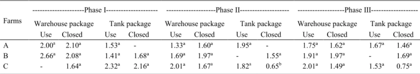
The fungal counts of the shrimp feed were similar in farms “A” and “B” at all the cultivation phases, regardless of the collection site and package type (closed or opened), as shown in table 1. The lowest fungal count was found in farm “C” from feed from a closed package stored in phase II tank. It
could be verified that the farms had acquired shrimp
feed from all phases of growth with previous fungal contamination, based on the counts of the closed package samples. Therefore, feed manipulation and feed storage method used in the farms did not interfere in the amounts of isolated fungi. The samples analyzed presented values below 3.0CFU g-1 (Tables
1 and 2). Fungal count was similar in the farms
Table 1 - Mean count of filamentous fungi and yeast (CFU/g) in feed supplied to shrimp at different growth stages in Piauí coastal area.
---Phase I--- ---Phase II--- ---Phase
III---Warehouse package Tank package Warehouse package Tank package Warehouse package Tank package
Farms
Use Closed Use Closed Use Closed Use Closed Use Closed Use Closed
A 2.00a 2.10ª 1.53ª - 1.33ª 1.60ª 1.95ª - 1.75ª 1.62ª 1.67ª 1.46ª
B 2.66ª 2.08ª 1.41ª 1.68ª 1.69ª 1.97ª - 1.55ª 1.91ª 1.97ª - 1.69ª
C - 1.64ª 2.32ª 2.16ª 2.01ª 1.67a 1.82a 0.65b 2.01ª 1.49ª 1.53ª 0.75ª
(P>0.05) when the rainy and dry season collections were compared (Table 2).
All the analyzed samples had counts under the proposed limits of 4.0log10 CFU-1 g-1 (GMP, 2008),
indicating a good microbiological quality. These results were similar to those obtained by CALVET et al. (2009), when identifying the toxigenic mycobiota in trout rations.
The prevalent fungi in feed used in shrimp farming in Piauí (Figure 1) belonged to the gender
Aspergillus and its teleomorphs (66.1%). Thirty-four
Aspergillus strains were identified; with A. flavus
(38.2%) being the most prevalent. Other important toxigenic species, such as two strains of A. niger
aggregated (Figure 2) were also isolated. These strains produced ochratoxin A when were qualitatively compared with standards by TLC.
Two strains of A. flavus produced
aflatoxins B1, B2, G1 and G2. The amount of AF B1
varied from 0.39 to 0.42ng g-1; AF B
2 from 0.18 to
0.27ng g-1, AF G1 1.78ng/g and AF G2 0.09ng g-1.
These strains can be classified as atypical A. flavus
(TRAN-DINH et al., 1999; PILDAÍN et al., 2004; PERRONE et al., 2007). This strain isolated presents all macroscopic and microscopic morphological characteristics of A. flavus differentiating morphologically A. parasiticus (KLICH, 2002).
Thus, the molecular and phylogenetic classification and identification of the strain becomes necessary,
because there are no reports related to this variety of fungi in Brazil. Possibly, these strains may be a new species into the section Flavi not described yet.
In general, all samples showed that
Aspergillus, the main toxicogenic fungi, were the
prevalent genera. The high incidence of A. flavus observed in shrimp feed indicates the possible
presence of aflatoxins (CAST, 2003). The isolated
fungi from stored feed in closed packages could be Table 2 - Mean count of filamentous fungi and yeast (CFU/g) in shrimp feed during the dry and rainy seasons.
---Rainy period (AT=27º C, RH=71.5%)--- ---Dryperiod (AT=30.9º C, RH=62.5%)---Warehouse package--- ---Tank package--- ---Warehouse package--- ---Tank package---Farms
Opened Closed Opened Closed Opened Closed Opened Closed
A 2.06ª 1.64ª 2.70ª - 1.52ª 1.85ª 1.25ª
-B 2.04ª 1.99ª 2.61ª 1.64ª 2.29ª 2.18ª - 1.98ª
C 2.01ª 1.59ª 2.17ª 1.56ª 1.60ª 2.09ª 1.48ª 2.11ª
a= Equal letters in the same line and column do not present P<0.05% difference; CFU g-1 = colony-forming units per gram in log 10.
AT=Average Temperature; RH=Relative Humidity.
caused by the use of contaminated raw materials, or yet, by processing, according to BINTVIHOK et al., (2003).
From the 108 samples analyzed, only
15 (13.88%) presented contamination by aflatoxin
B1at levels that varied from 0.25ng to 360ng g-1.
Two samples did not comply to the standard values recommended by the Brazilian legislation, with values above 50ng g-1 for aflatoxin B1 (BRAZIL,
1988). Aflatoxin B2, G1 and G2were not detected. The feeds used at different phases of cultivation, stored both in closed and opened packages, were found to present low fungi count in all the farms
analyzed, throughout the year. Aflatoxin B1 was the
only mycotoxin detected in the analyzed feed. Intake of mycotoxins by shrimp may lead to economic losses, as these toxins interfere in their metabolism, overloading the hepatopancreas. BOONYARATPALIN et al. (2001) fed ration
contaminated with aflatoxin to shrimp, confirming
the presence of these toxins in their muscles after four weeks.
The low incidence of aflatoxins B1
is probably due to the high feed turnover on the farms. This practice prevents the storage of feeds for a prolonged time, inhibiting fungi growth and multiplication and, consequently, the production of mycotoxins.
CONCLUSION
The samples of shrimp feed analyzed showed low fungal counts indicating good
microbiological quality. However, aflatoxin B1 was
detected in 15 samples, two out of 15 with values above the recommended by Brazilian legislation. It was noticeable the isolation of two strains
classified as atypical A. flavus Group II (Lineage
“S”), which produced aflatoxin B1 B2, G1 and G2
and were macroscopic and microscopic different of
A. parasiticus. The molecular identification of this
strains are in progress in order to differentiate from
A. parasiticus and var A. flavusparvisclerotigenus.
ACKOWLEDGEMENTS
This research was carried out with grants from Conselho Nacional de Desenvolvimento Científico e Tecnológico (CNPq), Universidade Federal do Piauí (UFPI), Universidade Federal Rural do Rio de Janeiro (UFRRJ), Fundação de Amparo a Pesquisa e ao Desenvolvimento Científico e Tecnológico do Maranhão (FAPEMA) (Brazil) and Universidade Nacional de Río Cuarto (UNRC) (Argentina).
REFERENCES
ABCC (ASSOCIAÇÃO BRASILEIRA DE CRIADORES DE CAMARÃO). Censo da produção anual de 2010. Online. Available from: <http://www.abccam.com.br/censo_2010>. Accessed: Sept. 24, 2011.
BARBOSA, T.S. et al. Mycobiota and mycotoxins present in finished fish feeds from farms in the Rio de Janeiro State, Brazil.
International Aquatic Research, v.5, p.3, 2013. Available from: <http://www.intaquares.com/content/5/1/3>. Accessed: Jul. 30, 2014. doi: 10.1186/2008-6970-5-3.
BINTVIHOK, A. et al. Aflatoxin contamination in shrimp feed and effects of aflatoxin addition to feed on shrimp production. Journal of Food Protection, v.66, n.5, p.882-885, 2003. Available from: <http://www.ncbi.nlm.nih.gov/pubmed/12747701>. Accessed: Apr. 12, 2007.
BOONYARATPALIN, M. et al. Effects of aflatoxin B1 on growth performance, blood components, immune function and histopathological changes in black tiger shrimp (Penaeus
monodon Fabricius). Aquaculture Research, v.32, Suppl.1, p.388-398, 2001. Available from: <http://onlinelibrary.wiley.com/ doi/10.1046/j.1355-557x.2001.00046.x/full>. Accessed: Apr. 12, 2007. doi: 10.1046/j.1355-557x.2001.00046.x.
BRASIL. Ministério da Agricultura. Portaria MA/SNAD/SFA n.07, de 09/11/88. Fixa padrões de tolerância para aflatoxinas em alimentos para consumo animal: matérias primas e rações, 1988.
Diário Oficial da União, p.21.968, 09 nov. 1988. Seção I.
CALVET, R.M. et al. Micoflora toxígena de rações de trutas.
Revista Higiene Alimentar, v.23, p.170-171, p.429-430, 2009.
CAST (COUNCIL FOR AGRICULTURAL SCIENCE AND TECHNOLOGY). Mycotoxins: risks in plant, animal and human systems. Ames, Iowa, 2003. Task Force Report n.139.
FAO. Fishery Information, Data and Statistics Unit. FishStat plus: universal software for fishery statistical time series. Version 2.3. Rome, 2006. Available from: <http://www.fao.org/fi/statist/ FISOFT/FISHPLUS.asp>. Accessed: Apr. 12, 2007.
GARCIA, D. et al. Predicting mycotoxins in foods: a review. Food Microbiology, v.26, p.757-769, 2009. Available from: <http:// www.sciencedirect.com/science/article/pii/S0740002009001397>. Accessed: Apr. 13, 2010. doi: 10.1016/j.fm.2009.05.014.
GEISEN, R. Multiplex polymerase chain reaction for the detection of potential aflatoxin and sterigmatocystin producing fungi.
Sistematic and Applied Microbiology, v.19, p.388-392, 1996. Available from: <http://dx.doi.org/10.1016/S0723-2020 (96) 80067-1>. Acesso em: Apr. 12, 2008. doi: 10.1016/S0723-2020 (96) 80067-1.
GMP (GOOD MANUFACTURING PRACTICES). Certification
scheme animal feed. Sector 2006. Appendix 1: Product standards; Regulations on Product Standards in the Animal Feed Sector, GMP 14. Available from: <http://www.bezpecna-krmiva.cz/soubory/ gmp_standard_08_EN.pdf>. Accessed: Dec. 01, 2014.
GOPINATH, R.; RAJ, R.P. Histological alterations in the hepatopancreas of Penaeus monodon Fabricius (1798) given aflatoxina B1 incorporated diets. Aquaculture Research, v.40,
p.1235-1242, 2009. Available from: <http://onlinelibrary.wiley. com/doi/10.1111/j.1365-2109.2009.02207.x/full>. Accessed: May 22, 2011. doi:10.1111/j.1365-2109.2009.02207.x.
GOPINATH, R. et al. Ultrastructural changes in the hepatopancreas of Penaeus monodon Fabricius 1798 given aflatoxin B1 diets. Aquaculture Research, v.43, p.32-43, 2012. Available from: <http://onlinelibrary.wiley.com/doi/10.1111/ j.1365-2109.2011.02798.x/pdf>. Accessed: Feb. 03, 2012. doi: 10.1111/j.1365-2109.2011.02798.x.
KLICH, M.A. A laboratory guide to the common Aspergillus
species and their teleomorphs. Australia: CSIRO - Division of
Food Processing, Australia, 2002. 116p.
NUNES, A.J.P. et al. Carcinicultura ameaçada: produtores sofrem com as mortalidades decorrentes do vírus da mionecrose infecciosa (IMNV). Panorama da Aquicultura, v.14, n.83, p.37-51, 2004.
PEREYRA, C.M. et al. Fungi and mycotoxins in feed intended for sows at different reproductive stages in Argentina. Veterinary Medicine International, v.2010 p.1-7, 2010. Available from: <https://www.scienceopen.com/document_file/b356bf0d-d98c- 4ad5-9ed7-1b0547d2c9a1/PubMedCentral/b356bf0d-d98c-4ad5-9ed7-1b0547d2c9a1.pdf>. Accessed: Apr. 12, 2012. doi:10.4061/2010/569108.
PERRONE, G. et al. Biodiversity of Aspergillus species in some important agricultural products. Studies in Mycology, v.59, p.53-66, 2007. Available from: <http://www.studiesinmycology.org>. Accessed: May 30, 2010. doi: 10.3114/sim.2007.59.07.
PILDAIN, M.B. et al. Analysis of population structure of Aspergillus flavus from peanut based on vegetative compatibility, geographic origin, mycotoxin and sclerotia production.
InternationalJournal of Food Microbiology, v.93, p.31-40, 2004. Available from: <http://www.elsevier.com/locate/ijfoodmicro>. Accessed: May 30, 2010. doi:10.1016/j.ijfoodmicro.2003.10.007.
PITT, J.I.; HOCKING, A.D. Fungi and food spoilage. 3.ed. London, New York: Springer Dordrecht Heidelberg, 2009. 524p.
ROCHA, I.P. Impactos sócio-econômicos e ambientais da carcinicultura brasileira: mitos e verdades, 2007. Available from: <http:www.abccam.com.br>. Accessed: Apr. 12, 2008.
TRAN-DINH, N. et al. Molecular genotype analysis of natural toxigenic and nontoxigenic isolates of Aspergillus flavus and
A. parasiticus. Mycologycal Research, v.103, n.11, p.1485-1490, 1999. Available from: <http://dx.doi.org/10.1017/ S0953756299008710>. Accessed: Oct. 12, 2012. doi: 10.1017/ S0953756299008710.