Bioaccumulation of animal adenoviruses in the pink shrimp
Roger B. Luz
1, Rodrigo Staggemeier
1, Rafael B. Fabres
1, Mayra C. Soliman
1,
Fernanda G. Souza
1, Raoni Gonçalves
2, Ivone V. Fausto
2, Caroline Rigotto
1,
Larissa S. Heinzelmann
1, Andréia Henzel
1, Juliane D. Fleck
1, Fernando R. Spilki
11Laboratório de Microbiologia Molecular, Universidade Feevale, Novo Hamburgo, RS, Brazil. 2Laboratório de Biologia e Microscopia, Universidade Federal do Rio Grande do Sul, Imbé, RS, Brazil.
Submitted: April 17, 2014; Approved: November 16, 2014.
Abstract
Adenoviruses are among the most promising viral markers of fecal contamination. They are fre-quently found in the water, sediment and soil of regions impacted by human activity. Studies of the bioaccumulation of enteric viruses in shrimp are scarce. The cities located in the northern coast of the lake systems in Southern Brazil have high urbanization and intensive farming rates, and poor sewage collection and treatment. One hundred (n = 100)Farfantepenaeus paulensispink-shrimp specimens and 48 water samples were collected from coastal lagoons between June 2012 and May 2013. Water samples were concentrated and the shrimp, mashed. After DNA extraction, samples were analyzed by real time polymerase chain reaction (qPCR) in order to detect and quantify viral genomes. Thirty-five percent of shrimp samples were positive for contamination, predominantly by avian ad-enoviruses. A total of 91.7% of water samples contained adenoviruses DNA, with the human form being the most frequent. Our results provided evidence of significant bioaccumulation of adenovirus-es in shrimp, showing the extent of the impact of fecal pollution on aquatic ecosystems.
Key words:adenovirus,Farfantepenaeus paulensis, pink-shrimp, water quality, Northern Coast of Rio Grande do Sul.
Introduction
Crustaceans plays a crucial role in consumer-resource relationships in aquatic ecosystems. Damage to these popu-lations may alter ecological functions and have a signifi-cant impact on the social, economic and health conditions of nearby communities (Seeliger and Odebrecht, 1996). Farfantepenaeus paulensis(Peréz-Farfante, 1967), known as the pink shrimp, is native to the Brazilian coast, and can be found along the entire coastline between northeastern Brazil and Argentina. Shrimp fishing, even at low levels, contributes significantly to the income of artisanal fisher-men living in local communities and helps to maintain re-gional trade networks (Reis and D’Incao, 2000; Cotrimet al., 2007).
Crustaceans, clams and oysters can bioaccumulate enteric viruses and bacteria from surrounding water and sediment (Beuretet al., 2003). Shrimp and other
crusta-ceans may also accumulate enteric viruses and bacteria through feeding on contaminated organisms, becoming pathogen vectors (DiGirolamoet al., 1972; Umeshaet al., 2008). Moreover, it was postulated that organisms in con-taminated water may carry viruses on their surface or bow-els, acting as mechanical vectors (Stentifordet al., 2009). Shrimp contamination by poliovirus types 1 and 2 has been reported in Venezuela (Boteroet al., 1996), while entero-virus contamination has been identified in India (Umeshaet al., 2008). Although viruses transmitted by humans or other animals are unlikely to be able to replicate in shrimp cells, the consumption of undercooked contaminated shrimp may pose a serious health risk.
Adenoviruses (AdVs) belong to the Adenoviridae family. Contamination generally occurs by waterborne or fecal-oral transmission, making AdVs a promising viral in-dicator of fecal contamination in surface and groundwater (Thurston-Enriquezet al., 2003; Rigottoet al., 2010;
Oli-DOI: http://dx.doi.org/10.1590/S1517-838246320140323
Send correspondence to F.R. Spilki. Laboratório de Microbiologia Molecular, Universidade Feevale, Novo Hamburgo, RS, Brazil. E-mail: [email protected].
veiraet al., 2012; Vecchia et al., 2012). AdVs are non-enveloped and have double-stranded DNA, and are there-fore more resistant to environmental degradation and water treatment than other contamination indicators (Tavareset al., 2005; Bofill-Maset al., 2006; Jianget al., 2007; Griffin et al., 2008). AdVs are the most prevalent human viruses in sewage and can be found in contaminated bodies of water throughout the year (Rigotto, 2010), although contamina-tion is more frequent in temperate regions (Tavareset al., 2005). The main sources of water contamination are sew-age leaksew-age or discharge and urban or rural runoff. AdVs are often found in urban rivers (Taniet al., 1995; Castig-nollesa et al., 1997; Chapron et al., 2000), or polluted coastal waters (Puiget al., 1994; Pinaet al., 1998; Jianget al., 2001; Rigottoet al., 2010), where they may persist for up to four months (Lipp and Rose, 1997; Jianget al., 2001). The annual risk of infection by consumption of contami-nated water is of 0.9% for one infectious unit in 100 liters of water (Mena and Gerba, 2008), and although most infec-tions are asymptomatic, AdVs can cause paralysis, menin-gitis, myocarditis, congenital heart defects, infectious hep-atitis, ocular infections and gastroenteritis (Pina et al., 1998; Lenaertset al., 2008). AdV levels can be used as in-dicators of water and food contamination and as a tool for the evaluation of fecal contamination patterns in different regions. AdVs cannot replicate outside the host cell and are highly species-specific, facilitating the identification of the sources of microbial contamination, and the implementa-tion of corrective or mitigating measures (Field and Samad-pour, 2007). In the southernmost state of Brazil, the pres-ence of AdVs has been detected in sewage-contaminated surface waters in the state capital (Vecchiaet al., 2012) and in contaminated groundwater in the countryside (Oliveira et al., 2012).
The Tramandaí, Armazém and Custódia Lagoons are part of the Tramandaí River basin, located in the Coastal Hydrographic Region of Rio Grande do Sul, in southern Brazil. The lagoon system is connected to the Atlantic Ocean by a small watercourse in the Tramandaí Lagoon. The Camarão River joins the Armazém and Custódia La-goons. The Tramandaí and Armazém Lagoons are classi-fied by the Luiz Roessler Environmental Protection Agency (Fundação Estadual de Proteção Ambiental Hen-rique Luiz Roessler; FEPAM) as brackish water type 2, while the Custódia Lagoon is classified as brackish water type 1. The latter can therefore be used for recreational or fishing activities provided fecal coliform organisms are ab-sent (FEPAM, 2000). Although the exact state of sanitation in the cities of Tramandaí, Imbé and Osório, which sur-round the lagoons, is unknown, the average shortfall in sewage treatment in Southern Brazil has been found to be as high as 82% (Clarke and King, 2006). At the time of writing, Imbé has no sewage collection system, and in Tramandaí, only 28% of the total sewage load is treated (SNIS, 2011). The disposal of untreated sewage into bodies
of water can modify the levels of dissolved nutrients in the water, induce eutrophication and reduce local diversity (Seeliger and Odebrecht, 2010). The tributaries of the la-goons are also contaminated by domestic and agricultural runoff, creating a potential health hazard for the 100 to 200 thousand people who live in the area (SEMA, 2010; IBGE, 2011). Water contaminated with urban sewage may contain several waterborne pathogens, including bacteria, protozoa and viruses. The consumption of contaminated water or food and recreation in contaminated waters represents an especially serious risk for children, the elderly and the immunocompromised (Fong and Lipp, 2005). The lack of sanitation and land use guidelines may pose a serious im-pact on water quality and aquatic ecosystems (FEPAM, 2000). Other areas in the basin are also used for agriculture and livestock production (mostly cattle, swine and chicken). The lagoons are also the habitat of several avian species and serve as a resting place for migratory birds.
As a result of the poor sewage treatment and intensive disposal of untreated wastewater in their surrounding area, Brazilian lagoons may represent an ideal location for the study of fecal pollution. The study of these lagoons is espe-cially relevant given the diversity of mammal, avian and aquatic species inhabiting the area. The aim of the present study was to investigate the potential bioaccumulation of adenovirus DNA in shrimp collected from the lagoon sys-tem.
Material and Methods
Sampling sites
Samples were collected from two shrimp fishing sites and ten water sampling sites in the Tramandaí, Armazém and Custódia Lagoons, located on the northern coast of the state of Rio Grande do Sul. Sites were delimited with the help of the Center for Coastal, Limnological and Marine Studies (CECLIMAR) of the Federal University of Rio Grande do Sul (UFRGS). Samples were collected by boat from the sites shown in Figure 1. CECLIMAR donated 50 F. paulensisshrimp collected from the Camarão River and Armazém Lagoons in April 2013, as well as 50 shrimp of the same species obtained from the estuary in 2011. Ten 500-milliliter (mL) samples of water were also aseptically collected at five time points between June 2012 and May 2013.
Sample preparation
The shrimp were fully macerated and stored at -80 °C until processing. Approximately 1 gram of macerated tis-sue was diluted in 1 mL of Eagle’s minimum essential me-dium (E-MEM), which was then vortexed and centrifuged for 10 min at 18,000 xg.
Water samples were concentrated by adsorption-elution, as described by Katayamaet al.(2002), with minor modifications. Briefly, 0.6 mg MgCl2were added to each
sample and pH was adjusted to 5±0.5. The samples were
filtered through negatively charged nitrocellulose mem-branes (0.45mm, 47 mm diameter; HA Millipore®,
Massa-chusetts, USA). Membranes were washed with 87.5 mL of 0.5 millimolar (mM) sulfuric acid (H2SO4) and 2.5 mL of 1 mM sodium hydroxide. The final extract was neutralized with 12.5 microliters (mL) of H2SO4,50 mM, and 12.5mL of Tris-EDTA 100x buffer. The filtrate was stored at -80 °C.
Molecular techniques
Viral DNA was extracted from concentrated water samples and the supernatant from the macerated tissue samples using a DNA/RNA extraction kit (Stratec®, Berlin, Germany), according to manufacturer’s instructions.
Two pairs of primers were used for quantitative poly-merase chain reaction (qPCR) assay and high resolution melting analysis (HRM). For the detection of human (HAdV), canine (CAV), bovine (BAV), avian (AvAdV)
AvAdV (80.5 °C) (Fig. 2). Genotyping was confirmed in a subset of samples by direct sequencing (data not shown).
Given the overlap between the melting peaks of dif-ferent AdV species, when more than one form of the virus was present, a separate reaction was used for the detection
of HAdV. This reaction involved VTB2-F
(5’-GAGACGTACTTCAGCCTGAAT-3’) and VTB2-R (5’-GATGAACCGCAGCGTTCAA-3’) primers at 20 pM, as described by Wolfet al.(2010), and was performed un-der the same time and temperature conditions as the re-mainder of the reactions described. However, in this procedure, the annealing temperature was lowered to 55 °C and the number of cycles was reduced to 40. HAdV had a specific melting peak of 86°C.
In each run, no template control (NTC) and negative control samples were used to exclude contamination. Stan-dard curves were used to determine the genome copies (gc) in each samples. Ten-fold serial dilutions of the control fragment from 10-1to 10-5starting at 6.01 x 10⁷gc/5
mL
(HAdV-5; VTB2 primer set) and 6.88 x 10⁸ gc/5 mL
(HAdV-2; ADV primer set)were used in each reaction as positive controls. The mean coefficient of determination (R2) of these reactions was 0.998, their efficiency was 98.7% and the analytical sensitivity was 8-12 genome cop-ies per 1mL/reaction. Both sets of primers target a highly
conserved region of the hexon gene. All reactions were run
in duplicate using the Platinum® SYBR® Green qPCR Super Mix-UDG (Life Technologies Corporation, Carlsbad, California 92008, USA) commercial kit. qPCR reactions was performed in a thermal cycler (iQ5 Bio-Rad, Biorad, Hercules, California 94547, USA) under the fol-lowing conditions: each 25mL reaction contained 12.5mL
of Mix, 5.5mL of ultrapure water (DNAse/RNAse free),
1mL of each primer (20 pM) and 5mL of the nucleic acid
extracted from each sample.
Results and Discussion
AdV contamination was detected in 35 (35%) of the 100 samples collected from both fishing sites. AvAdV was present in 17 samples (17%), BAV in 13 (13%), CAV in 7 (7%) and PoAdV in 2 (2%). An average of 5.8 x 10⁴ ge-nome copies (3.31 x 10²-8.18 x 10⁵) were present per gram of tissue. HAdV was not found in any tissue samples with either primer set. For reasons which still remain unknown, HAdV may not accumulate in pink shrimp tissue, or may not be detectable by the methods used in the present study. This result may also suggest that these bodies of water may be less contaminated by human feces than animal waste, since the geometric mean of genome copies of HAdV/L was lower than of other viruses.
AdVs were detected in 44 of the 48 (91.7%) water samples collected. Contamination was present in at least
718 Luzet al.
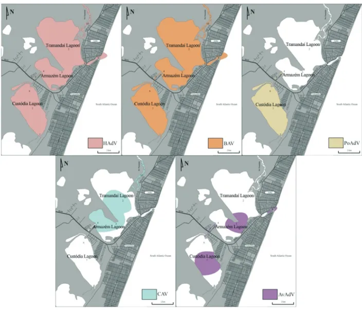
one sample drawn from every site. The ADV-F1 and ADV-R1 primers identified adenoviral DNA in 38 samples (79.2%), of which 23 (47.9%) were by BAV, 7 (14.6%) by HAdV, 5 (10.4%) by AvAdV, 4 (8.4%) by CAV and 4 (8.3%) by PoAdV. The VTB2-F and VTB2-R primers re-vealed the presence of HAdV DNA in 28 (58.3%) samples. When both primers were used, HAdV was detected in 64.6% (31 samples) of samples, with an average of 8.15 x 10⁵genome copies (3.95 x 10³-1.09 x 10⁹) per liter. HAdV and BAV were found in all sampling sites. CAV was pres-ent in the Tramandaí Lagoon, Armazém Lagoon and Tra-mandaí River. AvAdV was found in the Armazém Lagoon, Custódia Lagoon, Camarão River and at the point where the lagoons connect to the ocean. PoAdV was detected in the Camarão River and Custódia Lagoon only.
The results are summarized in Tables 1 through 4. A schematic representation of the viral distribution in the area studied is shown in Figure 3. Viral contamination was not
statistically associated with climate variability, and did not differ between sampling sites and times (data not shown).
ples, although detection rates and genome copies were lower in shrimp tissue than in water samples. The preva-lence of contamination by each type of AdV in shrimp tis-sue was as follows: AvAdV (17%), BAV (13%), CAV
(7%) and PoAdV (2%). AdV was detected in 91.7% of wa-ter samples, which were found to be contaminated by the human (64.6%), bovine (47.9%), avian (10.4%), canine (8.3%) and porcine (8.3%) forms of the virus. While animal
720 Luzet al.
Table 1- Adenovirus contamination in water; HAdV – human adenovirus; BAV – bovine adenovirus; CAV – canine adenovirus; AvAdV – avian adenovirus; PoAdV – porcine adenovirus.
Sampling site HAdV (%) BAV (%) CAV (%) AvAdV (%) PoAdV (%)
Site 1 - Tramandaí River Entrance 60 60 0 0 0
Site 2 – Pontal 80 80 20 0 0
Site 3 - Armazém Lagoon 100 60 20 20 0
Site 4 - Camarão River Entrance 40 60 20 0 0
Site 5 - Camarão River 75 25 0 50 25
Site 6 - Custódia Lagoon 60 20 0 0 40
Site 7 – Center of the Custódia Lagoon 40 20 0 20 20
Site 8 - Garibaldi Bridge 100 75 0 0 0
Site 9 - Barra Outfall 60 60 0 20 0
Site 10 - Tramandaí River Embranchment 40 20 20 0 0
Table 2- Mean genome copies of adenovirus per liter of water; HAdV – human adenovirus; BAV – bovine adenovirus; CAV – canine adenovirus; AvAdV – avian adenovirus; PoAdV – porcine adenovirus.
Sampling site HAdV BAV CAV AvAdV PoAdV
Site 1 - Tramandaí River Entrance 1.30 x 10⁵ 1.13 x 10⁶ 0 0 0
Site 2 – Pontal 8.66 x 10⁵ 2.18 x 10⁶ 3.33 x 10⁴ 0 0
Site 3 - Armazém Lagoon 9.02 x 10⁴ 1.37 x 10⁷ 1.02 x 10⁸ 2.82 x 10⁸ 0
Site 4 - Camarão River Entrance 8.88 x 10⁵ 6.68 x 10⁵ 1.25 x 10⁶ 0 0
Site 5 - Camarão River 1.32 x 10⁶ 4.95 x 10⁶ 0 6.46 x 10⁶ 5.53 x 10⁷
Site 6 - Custódia Lagoon 1.15 x 10⁶ 1.10 x 10⁶ 0 0 2.41 x 10⁶
Site 7 – Center of the Custódia Lagoon 1.84 x 10⁵ 5.18 x 10⁶ 0 1.09 x 10⁹ 1.94 x 10⁷
Site 8 - Garibaldi Bridge 1.69 x 10⁵ 2.50 x 10⁶ 0 0 0
Site 9 - Barra Outfall 2.55 x 10⁵ 2.73 x 10⁶ 0 3.30 x 10⁵ 0
Site 10 - Tramandaí River Embranchment 1.59 X 10⁵ 2.59 x 10⁵ 2.58 x 10⁵ 0 0
Table 4- Mean genome copies of adenovirus per gram of tissue; HAdV – human adenovirus; BAV – bovine adenovirus; CAV – canine adenovirus; AvAdV – avian adenovirus; PoAdV – porcine adenovirus.
Fishing site HAdV BAV CAV AvAdV PoAdV
Site 1 – Armazém Lagoon 0 2.95 x 10³ 6.05 x 10³ 1.23 x 10⁵ 0
Site 2 – Camarão River 0 1.03 x 10⁵ 1.18 x 10⁴ 3.81 x 10⁴ 0
Estuary 2011 0 1.73 x 10⁴ 1.37 x 10⁴ 2.60 x 10³ 5.92 x 10³
Table 3- Adenovirus contamination in shrimp; HAdV – human adenovirus; BAV – bovine adenovirus; CAV – canine adenovirus; AvAdV – avian adenovirus; PoAdV – porcine adenovirus.
Fishing site HAdV (%) BAV (%) CAV (%) AvAdV (%) PoAdV (%)
Site 1 – Armazém Lagoon 0 8 8 8 0
Site 2 – Camarão River 0 12 4 24 0
viruses pose no risk for penaeid shrimp or human health, they can affect other species and be indicative of proximity to a source of pollution which could also contaminate bod-ies of water with other non-specbod-ies-specific pathogens, such as bacteria, parasites and protozoa. All ecosystems re-quire clean water, and enteric infections can lead to serious economic, social and environmental consequences for live-stock or wild populations, rendering them more susceptible to other infections (Barardiet al., 2013). The avian AdVs detected in the present study may derive from poultry farms, since the cities surrounding the lagoons are esti-mated to produce approximately 15 thousand broilers (IBGE, 2010), or wild life. Of the 27 species of birds found in the lagoon system throughout the year, 10 use the lagoon as a food source (Ramos and Daudt, 2004), so that wild life contamination may have been partly responsible for our findings. All species of birds are susceptible to AvAdV contamination, and may shed the virus for more than 14 weeks after inoculation. Although most infections are sub-clinical, symptoms such as respiratory diseases, diarrhea, arthritis, inclusion body hepatitis, hydropericardium syn-drome and egg drop synsyn-drome have occasionally been found to occur (McFerran and Smyth, 2000). If not prop-erly treated, the waste produced by the 6,000 swine and 30,000 heads of cattle in the cities of Tramandaí, Imbé and Osório, all of which are located in the Tramandaí River ba-sin to the north of the Tramandaí lagoon (IBGE, 2010), may contaminate both surface and groundwater. In winter months, cattle can also be found in open or unfenced urban areas, which are often used for complementary grazing (Ramos and Daudt, 2004). Cattle infected by the BAV may exhibit conjunctivitis, pneumonia, enteritis and poly-arthritis (Flores, 2012). In swine, PoAdV may lead to diar-rhea or respiratory complications (Hammondet al., 2003). CAV may be found in the feces of both wild and domestic canids, has a worldwide distribution and may cause infec-tious canine hepatitis and infecinfec-tious canine tracheobron-chitis, depending on the viral type (Flores 2012).
Enteric viruses are found frequently and at high levels in bodies of water contaminated by urban sewage or live-stock waste. The prevalence of enteric viruses in surface water has been found to be very high in North America (Chapronet al., 2000) but somewhat lower in Brazil, where the rate of contamination by AdV ranges from 64.2% in the coastal region of the state of Santa Catarina (Rigottoet al., 2010) to 21.4% in the urban area of Porto Alegre (Vecchia et al., 2012). The primers used in the present study identi-fied HAdV DNA in 64.6% of water samples. This finding shows the impact of poor waste management in the cities surrounding the lagoons on the water quality of the region. Approximately 80% of homes in the Tramandaí River ba-sin have individual sewage disposal systems. In Osório and Tramandaí, solid waste repositories also contribute to water contamination, by leakage into rivers, streams and lakes. The maximum amount of HAdV DNA detected in water
samples exceeded 10 million genome copies per liter. This number is alarming, considering that the basin supplies wa-ter to a population ranging from 100 to 200 thousand, in ad-dition to domestic animals or wild life, and that AdV has an extremely low infectious dose (i.e.one infectious unit is al-ready considered harmful) (Haaset al., 1993). Fecal con-tamination can also influence the concentration of organic matter, nitrates and phosphates, which, in excess, can be harmful to aquatic flora and fauna and lead to eutrophi-cation (Seeliger and Odebrecht, 2010).
Further studies may wish to use amplicon sequencing to confirm the findings obtained with our pan-specific primers and perform phylogenetic analyses, especially of AvAdV, since the lagoons are home to many migratory bird species. Since no infectivity assay was performed, we can-not speak to the viability of the virus. However, AdV are known to be strong indicators of fecal contamination. Loca-tions where adenoviruses are found, regardless of viability, may also be contaminated by other forms of the virus (Oliveiraet al., 2012; Vecchiaet al., 2012; Hartmannet al., 2013), different viral agents (Pinaet al., 1998) or even other contaminants (Sidhuet al., 2013).
The present results emphasize the impact of untreated sewage and livestock waste on fragile aquatic ecosystems. There is a pressing need for more adequate biological and chemical markers of water quality, higher investments in sanitation and joint efforts from the government, industry and population to reduce the generation and inadequate dis-posal of waste.
Acknowledgments
RBL had a master’s scholarship from
FAPERGS/CAPES. FRS is a CNPq research fellow. Finan-cial support was provided by the Fundação de Amparo à Pesquisa do Estado do Rio Grande do Sul (FAPERGS) and Conselho Nacional de Desenvolvimento Científico e Tec-nológico (CNPq). This project was supported by CECLIMAR, UFRGS, Fundação Estadual de Pesquisa Agropecuária do Rio Grande do Sul (FEPAGRO Saúde Animal) and the Universidade Feevale.
References
Barardi CRM, Viancelli A, Rigotto Cet al.(2013) Monitoring vi-ruses in environmental samples. Int J Environ Sci Eng Res 3:62-79.
Beuret C, Baumgartner A, Schluep J (2003) Virus-Contaminated Oysters: a Three-Month Monitoring of Oysters Imported to Switzerland. Appl Environ Microbiol 69:2292-2297. Bofill-Mas S, Albinana-Gimenez N, Clemente-Casares Pet al.
(2006) Quantification and Stability of Human Adenoviruses and Polyomavirus JCPyV in Wastewater Matrices. Appl En-viron Microbiol 72:7894-7896.
Castignolles N, Petit F, Mendel I et al. (1998) Detection of adenovirus in the waters of the Seine River estuary by nested-PCR. Mol Cell Probes 12:175-180.
Chapron CD, Ballester NA, Fontaine JHet al.(2000) Detection of astroviruses, enteroviruses, and adenovirus types 40 and 41 in surface waters collected and evaluated by the information collection rule and an integrated cell culture-nested PCR procedure. Appl Environ Microbiol 66:2520-2525. Clarke R, King J (2006) O Atlas da Água. Publifolha, São Paulo. Cotrim DS, Miguel LA, Fernández-Baldor Á (2007) Sistemas
Pesqueiros e a Pesca Artesanal: O caso de Tramandaí/RS. Congresso Brasileiro de Sistemas de Produção. Sociedade Brasileira de Sistemas de Produção, Fortaleza.
DiGirolamo R, Wiczynski L, Daley Met al.(1972) Preliminary Observations on the Uptake of Poliovirus by West Coast Shore Crabs. Appl Microbiol 23:170-171.
Durand SV, Lightner DV (2002) Quantitative real time PCR for the measurement of white spot syndrome virus in shrimp. J Fish Dis 25:381-389.
Erali M, Voelkerding KV, Wittwer CT (2008) High resolution melting applications for clinical laboratory medicine. Exp Mol Path 85:50-58.
FEPAM - Fundação Estadual de Proteção Ambiental Henrique Luiz Roessler (2000) Diretrizes Ambientais para o Desen-volvimento dos Municípios do Litoral Norte. Cadernos de Planejamento e Gestão Ambiental n. 1. FEPAM, Porto Ale-gre.
Field KG, Samadpour M (2007) Fecal source tracking, the indica-tor paradigm, and managing water quality. Water Res 41:3517-3538.
Flores EF (2012) Virologia Veterinária: Virologia Geral e Doen-ças Víricas. UFSM, Santa Maria.
Fong TT, Lipp EK (2005) Enteric Viruses of Humans and Ani-mals in Aquatic Environments: Health Risks, Detection, and Potential Water Quality Assessment Tools. Microbiol Mol Biol Rev 69:357-371.
Griffin JS, Plummer JD, Long SC (2008) Torque teno virus: an improved indicator for viral pathogens in drinking waters. Virol J 5:1-6.
Haas CN, Rose JB, Gerba Cet al.(1993) Risk assessment of virus in drinking water. Risk Anal 13:545-552.
Hammond JM, Jansen ES, Morrissy CJet al.(2003) Protection of pigs against ‘in contact’ challenge with classical swine fever following oral or subcutaneous vaccination with a recombi-nant porcine adenovirus. Virus Res 97:151-157.
Hartmann NM, Dartscht M, Szewzyk Ret al.(2013) Monitoring of adenovirus serotypes in environmental samples by com-bined PCR and melting point analyses. Virol J 10:1-9. IBGE - Instituto Brasileiro de Geografia e Estatística (2010)
Produção da Pecuária Municipal 38:1-65.
IBGE - Instituto Brasileiro de Geografia e Estatística (2011) Censo Demográfico 2010.
Jiang SC, Chu W, He JW (2007) Seasonal Detection of Human Viruses and Coliphage in Newport Bay, California. Appl Environ Microbiol 73:6468-6474.
Jiang S, Noble R, Chu W (2001) Human Adenoviruses and Coli-phages in Urban Runoff-Impacted Coastal Waters of South-ern California. Appl Environ Microbiol 67:179-184. Katayama H, Shimasaki A, Ohgaki S (2002) Development of a
vi-rus concentration method and its application to detection of
enterovirus and Norwalk virus from coastal seawater. Appl Environ Microbiol 68:1033-1039.
Lenaerts L, De Clercq E, Naesens L (2008) Clinical features and treatment of adenovirus infections. Rev Med Virol 18:357-374.
Lipp FK, Rose JB (1997) The role of seafood in foodborne dis-eases in the United States of America. Rev Sci Tech 16:620-640.
McFerran JB, Smyth JA (2000) Avian adenoviruses. Rev Sci Tech OIE 19:589-601.
Mena KD, Gerba CP (2008) Waterborne Adenovirus. Rev Envi-ron Contam T 198:133-167.
Monteiro GS, Fleck JD, Kluge Met al.(in press). Adenoviruses of canine and human origins in stool samples from a free-living pampas foxes (Lycalopex gymnocercus) and crab-eating foxes (Cerdocyon thous) in São Francisco de Paula, Rio dos Sinos basin. Braz J Biol (in press).
Nunan LM, Poulos BT, Lightner DV (2000) Use of polymerase chain reaction (PCR) for the detection of infectious hypo-dermal and hematopoietic necrosis virus (IHHNV) in penaeid shrimp. Mar Biotechnol 2:318-328.
Oliveira LK, Fleck JD, Comerlato Jet al.(2012) Enteric viruses in water samples from Brazilian dairy farms. Agr Water Man-age 111:34-39.
Pina S, Puig M, Lucena Fet al.(1998) Viral Pollution in the Envi-ronment and in Shellfish: Human Adenovirus Detection by PCR as an Index of Human Viruses. Appl Environ Micro-biol 64:3376-3382.
Puig M, Jofre J, Lucena Fet al.(1994) Detection of adenoviruses and enteroviruses in polluted waters by nested PCR amplifi-cation. Appl Environ Microbiol 60:2963-2970.
Ramos LA, Daudt RB (2004) Avifauna urbana dos balneários de Tramandaí e Imbé, litoral norte do Rio Grande do Sul. Biotemas 18:181-191.
Reis EG, D’Incao F (2000). The present status of artisanal fisher-ies of extreme Southern Brazil: an effort towards commu-nity-based management. Ocean Coast Manage 43:585-595. Rigotto C, Victoria M, Moresco Vet al.(2010) Assessment of adenovirus, hepatitis A virus and rotavirus presence in envi-ronmental samples in Florianopolis, South Brazil. J Appl Microbiol 109:1979-1987.
Seeliger U, Odebrecht C (2010). O Estuário da Lagoa dos Patos -Um Século de Transformações. U. Seeliger. FURG, Rio Grande.
Seeliger U, Odebrecht C, Castello JP (1996) Subtropical Conver-gence Environments: The Coast and Sea in the Southwest-ern Atlantic. Springer, New York.
SEMA - Secretaria do Meio Ambiente (2010) Relatório Anual de Recursos Hídricos. Secretaria do Meio Ambiente, Estado do Rio Grande do Sul, Porto Alegre, 168 p.
Sidhu JPS, Ahmed W, Gernjak Wet al.(2013) Sewage pollution in urban stormwater runoff as evident from the widespread presence of multiple microbial and chemical source tracking markers. Sci Total Environ 463-464:488-496.
SNIS - Sistema Nacional de Informações sobre Saneamento (2011) Diagnóstico dos Serviços de Água e Esgotos. Secre-taria Nacional Ambiental do Ministério das Cidades, Brasí-lia, 164 p.
Stentiford GD, Bonami JR, Alday-Sanz V (2009). A critical re-view of susceptibility of crustaceans to Taura syndrome, Yellowhead disease and White Spot Disease and
tions of inclusion of these diseases in European legislation. Aquacult 291:1-17.
Tani N, Dohi Y, Kurumatani Net al.(1995) Seasonal distribution of adenoviruses, enteroviruses and reoviruses in urban river water. Microbiol Immunol 39:577-580.
Tavares TM, Cardoso DDP, Brito WMED (2005) Vírus entéricos veiculados por água: aspectos microbiológicos e de controle de qualidade da água. Rev Patol Trop 34:85-104.
Thurston-Enriquez JA, Haas CN, Jacangelo Jet al.(2003) Inacti-vation of Feline Calicivirus and Adenovirus Type 40 by UV Radiation. Appl Environ Microbiol 69:577-582.
Umesha KR, Bhavani NC, Venugopal MNet al.(2008). Preva-lence of human pathogenic enteric viruses in bivalve
mol-luscan shellfish and cultured shrimp in south west coast of India. Int J Food Microbiol 122:279-286.
Vecchia AD, Fleck JD, Comerlato Jet al.(2012) First description of Adenovirus, Enterovirus, Rotavirus and Torque teno vi-rus in water samples collected from the Arroio Dilúvio, Porto Alegre, Brazil. Braz J Biol 72:1-7.
Wolf S, Hewitt J, Greening GE (2010) Viral Muliplex Quantita-tive PCR Assays for Tracking Sources of Fecal Contamina-tion. Appl Environ Microbiol 76:1137-1149.
Associate Editor: Cynthia Canêdo da Silva