1. Department of Rheumatology, Hospital Vega-Baja, Alicante, Spain 2. Department of Emergency, Hospital Santa Ana, Granada, Spain; 3. Infectious Diseases Unit, Hospital General de Elche, Alicante, Spain 4. Systemic Autoimmune Diseases Unit, Hospital Universitario San Cecilio, Granada, Spain
5. Institute of Parasitology and Biomedicine López-Neyra, IPBLN-CSIC, Granada, Spain
6. Epidemiology, Genetics and Atherosclerosis Research Group on Systemic Inflammatory Diseases, Rheumatology Division, IDIVAL, Santander, Spain
IntroductIon
Rheumatoid Arthritis (RA) is a chronic multisystem di -sease with an estimated occurrence of 1 to 2 percent of the world population1. RA patients have an increased risk of cardiovascular (CV) events with high morbidi-ty and mortalimorbidi-ty as a result of rapid atherosclerosis2.
Interestingly, RA and atherosclerosis are both chro -nic inflammatory diseases sharing inflammatory biomarkers as well as a similar cellular activation pat-tern. The development of CV disease in RA patients has been associated with inflammation and autoimmunity. Considering the previously mentioned incidence of CV events in patients with RA, the identification of high risk RA individuals that may benefit from treatment, should be an important step in order to prevent overt CV disease. In this regard, in asymptomatic RA patients, several non-invasive surrogate markers have demon-strated the presence of subclinical atherosclerosis3,4. However, information on serological biomarkers of CVD in patients with RA is limited5.
Endothelial dysfunction itself is a process that in-volves genetic characteristics, cardiovascular risk fac-tors, and inflammation6. Endothelin-1 (ET-1)7which is mainly secreted by endothelial cells is a potent en-dogenous vasoconstrictor. It acts through two different types of receptors: ETA and ETB. ET-1 contributes to the development of inflammatory processes in the vas-cular wall, increasing superoxide anion production and cytokine secretion. It has been found to be associated with the activation of transcription factors such as nu-clear factor (NF)- B and also the expression of proin-flammatory cytokines8. In turn, these transcription fac-tors and proinflammatory cytokines stimulate ET-1 production9. Bellisai et al.10report that ET-1 increases the synthesis of TNF-a in macrophages and monocytes, which enhances the inflammatory response by stimulating the chemotaxis and phagocytosis of ma -crophages, monocytes and neutrophils. In different
Endothelin-1 serum levels in women
with Rheumatoid Arthritis
Alvarez-Cienfuegos A1, Cantero-Nieto L2, García-Gómez JA3,
Ríos-Fernández R4, Martin J5, González-Gay MA6, Ortego-Centeno N4
ACTA REUMATOL PORT. 2019;44:250-257
AbstrAct
Objective: The purpose of this study was to evaluate
serum Endothelin-1(ET-1) levels in female Rheumatoid Arthritis (RA) patients compared with healthy controls, examine possible associations between ET-1 with dif-ferent characteristic of the disease and investigate pos-sible associations between ET-1 with surrogate markers of cardiovascular disease (CVD).
Methods: This cross-sectional study was performed in
Vega-Baja Hospital, Orihuela (Spain) from November 2016 to May 2018. Sixty-three women with RA and sixty-five age and sex healthy controls were included in this study. Serum ET-1 was analyzed using ELISA.
Results: Serum levels of ET-1 in RA female patients
were higher than those in healthy controls (p 0.001). Serum le vels of ET-1 were positively associated with N-terminal pro-brain natriuretic peptide (NT-proBNP) (r = 0.27, p < 0.05) and with C-reactive protein (CRP) (r = 0.36, p < 0.05). ET-1 levels in women with RA were higher in smokers. Pre dnisone use was associated with lower ET-1 levels. No association with carotid intima media thickness was found.
Conclusions: we observed the presence of higher le
-vels of serum ET-1 in RA women patients compared with healthy controls. These increased levels of ET-1 are associated with inflammation and smoking and re-duced by prednisone intake.
Keywords: Endothelin-1; Rheumatoid Arthritis; CRP;
types of cells, increased production of reactive oxygen species (ROS) occurs via the NF- B, cyclooxygenase (COX) and nicotinamide adenine dinucleotide phos-phate (NADPH) oxidase-dependent pathways11-13.
The objectives of this study were to analyze ET-1 in a cohort of women with RA, examine possible associa -tions between ET-1 with different characteristic of the disease and investigate possible associations between ET-1 with surrogate markers of CVD.
mAterIALs And methods
PAtIent seLectIon
The study was performed in Vega-Baja Hospital, Ori-huela (Spain) from November 2016 to May 2018. We prospectively enrolled 63 consecutive women patients affected by RA and 65 healthy women who served as controls. All patients included in this study had nor-mal serum creatinine (Cr) levels, and met the 2010 American College of Rheumatology/European League Against Rheumatism (ACR/EULAR) criteria for RA14. Individuals with prevalent cardiovascular disease were excluded.
At the clinic visit, participants completed question-naires about their lifestyle characteristics, medical his-tory, and current medication used. Informed consent was obtained for all subjects, and the study was appro -ved by the Research Ethics Committee of Hospital Uni-versitario de Elche in Alicante, Spain, date: 22/11/2016, protocol number: pi35/2016 and con-ducted in accordance with the guidelines in the Dec-laration of Helsinki.
cArdIovAscuLAr Assesment
Disease severity was scored through disease activity score of 28 joints and joint damage was evaluated based on the Steinbrocker radiographic crite ria (I-IV). Current smokers were defined as those who reported having smoked ≥1 cigarette per day regularly during the year preceding the examination. Waist circumfer-ence, weight, and height were measured; and body mass index (BMI) was calculated as weight (kg)/height (m2). Blood pressure (BP) was measured twice in the left arm of the seated subject with a mercury column sphygmomanometer. The average of the 2 readings was used as the examination BP, and hypertension was defined as self-reported antihypertensive medication use, or a systolic BP ≥140 mm Hg, or a diastolic BP ≥90 mm Hg. Type 2 diabetes mellitus (T2DM) was
de-fined by self-reported use of insulin, or oral hypo-glycemic medications, or a fasting glucose level ≥126 mg/dl. Kidney function was assessed using the esti-mated glomerular filtration rate (eGFR) calculated by the CKD-Epi study equation15.
The CV risk was assessed using the Modified Sys-temic Coronary Risk Evaluation (mSCORE). The mSCORE was calculated using validated risk tables for both low and high risk populations16. For this study, the low risk table was used since Spain has been clas-sified as a low risk country for CVD. Carotid intima-media thickness (c-IMT) was measured by perform-ing carotid ultrasound examination in the common carotid artery and the detection of focal plaques in the extracranial carotid tree by manual technique using a commercially available scanner equipped with 7–12 MHz linear transducer as the patient was lying in the supine position with the neck rotated to the opposite side of examination as previously reported17. Carotid plaques were counted in each territory and defined as no plaque, unilateral plaque or bilateral plaques17. Val-ues of cIMT greater than 0.9 mm were considered ab-normal (cIMT thickening) and plaques were defined if the cIMT was greater than 1.5 mm18. In our study, an-kle-arm index was evaluated using a BIDOP model ES-100V3 vascular screening system (Hadeco, Inc, Kawasaki, Japan).
LAborAtory meAsurements
In all the cases, a fasting blood sample was taken in the morning, and was stored at -70°C until the assays were performed.
The sera were tested for creatinine, CRP, NT-ProB-NP and ET-1. Creatinine was determined by Jaffe method (Siemens Healthcare Diagnostic Inc. NY, USA). CRP was measured by turbidimetric im-munoassay (Siemens Healthcare Diagnostic Inc. NY, USA). NT-proBNP was quantified in heparinised plas-ma using a solid-phase two-site chemiluminescent im-munometric assay (Biomérieux, France). Serum ET-1 (Elabscience, USA) was measured by ELISA according to the manufacturer’s recommendations. Anti-citrulli-nated protein antibodies (ACPAs) were detected using a second-generation ELISA (ACPAs) kit (ORGENTEC Diagnostika GmbH. Mainz, Germany) while IgM RF was determined as part of routine analysis by turbidi-metric assay (Siemens Healthcare Diagnostic Inc. NY, USA) according to the manufacturers’ instructions. Fasting plasma glucose was measured in fresh speci-mens with a hexokinase reagent kit (Siespeci-mens
Health-cluded in our study as controls; mean age (SD) 52 ± 9 years. Most of them were also Caucasian (98.3%). LAborAtory resuLts
Laboratory tests of the patients and healthy controls in-cluded in the present study are shown in Tables I and II.
Forty-six (73.0%) and 45 (71.4%) of the 63 wom-en with RA were positive for rheumatoid factor and ACPAs, respectively. As expected, laboratory markers of inflammation found at the time of the study were high-er in women with RA than in controls (Table I). In this regard, the mean CRP in RA patients was 0.6 ± 0.8 mg/dl versus 0.2 ± 0.1 mg/dl in controls (p <0.001). Likewise, the mean ESR in the group of RA patients was 23.9 ± 15.8 mm/1sthour versus 11.3 ± 10.2 mm/1st hour in controls (p <0.001) (Table I). Patients with RA had lower uric acid levels than controls (3.9 ± 1.3 ver-sus 4.6 ± 1.3 mg/dl; p= 0.002). However, NT-proBNP levels were higher in patients with RA (79.8 ± 54.8 ver-sus 59.7± 38.4 pg/ml in controls; p= 0.01).
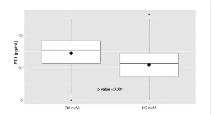
Interestingly, the serum ET-1 concentrations were significantly higher in the RA patients than those in the control group: [28.9 (0-50) vs. 21.7 (0-50), pg/ml; p
0.001] (Figure 1).
cArdIovAscuLAr dIseAse rIsk fActors
As shown in Table I, patients had a mean BMI of 26.6±5.6 kg/m2, waist circumference of 103.8± 13.2 cm, ankle-arm index of 1.1±0.1, cIMT of 0.7±0.1 mm, mSCORE 2.0 ± 2.3. Fourteen (22%) of them had a smoking history.
Healthy controls had a mean BMI of 25.9 ± 4.3 kg/m2, waist circumference of 83.1 ± 13.4 cm, ankle-arm index of 1.2 ± 0.2, cIMT of 0.6 ± 0.2 mm, mSCORE 1.8 ± 2.5. Fourteen (21.5%) of them had a smoking history.
reLAtIonshIP between et-1 LeveLs And cArdIovAscuLAr rIsk fActors or dIseAse feAtures In PAtIents wIth rheumAtoId ArthrItIs
Table III shows the correlation coefficients between 1 and other markers in patients with RA. Levels of ET-1 were significantly correlated with smoking (p= 0.020). ET-1 levels also showed a statistically significant positive correlation with CRP (r = 0.36, p= 0.004) and with NT-proBNP (r = 0.27, p= 0.036). In contrast, an inverse correlation between prednisone intake and ET-1 levels (p= 0.034). However, there was no correlation of ET-1 levels with age, BMI, ankle-arm index, cIMT, care Diagnostic Inc. NY, USA). Total cholesterol and
triglyceride levels were determined by fully enzymatic techniques. High-density lipoprotein (HDL) was de-termined after precipitation of apolipoprotein B (apoB)–containing lipoproteins with magnesium sul-fate and dextran sulsul-fate. Low-density lipoprotein (LDL) was calculated using the Friedewald formula. All oth-er routine soth-erum biochemical parametoth-ers woth-ere mea-sured at the Department of Clinical Chemistry, Vega-Baja Hospital.
stAtIstIcAL AnALysIs
Data were analyzed by statistical software SPSS 18 (Chicago, IL, USA) and with the program R version Rx64.3.5.0 (Vienna, Austria), using independent sam-ples t-test, Mann-Whitney U test, and Chi-square test when appropriate. Spearman’s coefficient and Pearson’s correlation were calculated as suitable to determine the correlation between the bio-chemical parameters. P-values of less than 0.05 were considered statistically significant. The quantitative data were shown as mean ± standard deviation (SD) and median (Q1–Q3) as suita ble. To test if we can admit that the distribution is normal we use the Shapiro-Wilk test. Linear regression was used to examine the cross-sectional associations of plasma ET-1 concentrations with CRP and NT-proBNP.
resuLts
chArActerIstIcs of the study subjects The main features of the 63 women with RA and 65 controls included in this study are shown in Table I. The mean age (SD) of the patients was 53 ± 8 years. The majority were Caucasian (90.5%). The mean disea -se duration was 8.5 ± 5.8 years. The mean di-sea-se ac-tivity score in 28 joints (DAS28) according to the ery-throcyte sedimentation rate (ESR) indicated low disea se activity 3.0 ± 1.3. The mean health assessment ques-tionnaire (HAQ) was 0.75 ± 0.67. The mean brocker s stage was 2.75 ± 1.17 and the mean Stein-brocker s class 1.87 ± 0.69. At the time of the study 32 (50.7%) patients were receiving biologic agents (9 eta -nercept, 9 certolizumab pegol, 7 tocilizumab, 6 adali-mumab and 1 rituximab). Most patients (73%) had re-ceived or were undergoing methotrexate therapy (mean weekly dose 11.5 ± 4.8 mg in patients on methotrexa -te) and 16 patients took prednisone with a median dai-ly dose of 6.5 ± 3.5 mg.
in-tAbLe II. serum et-1 And study PArAmeters of rA PAtIents And heALthy controLs
RA HC
mean ± SD mean ± SD p-value
ET-1, pg/ml 28.9 ± 12.6 21.7 ± 11.7 <0.001 Cholesterol, mg/dl 212.7 ± 41 211.8 ± 37.3 0.89 LDL-C, mg/dl 120.1 ± 29.2 129.7 ± 30.7 0.06 HDL-C, mg/dl 69.9 ± 19.4 63.3 ± 13.1 0.02 Triglycerides, mg/dl 112.9 ± 55.6 107.7 ± 53.9 0.59 Uric acid, mg/dl 3.9 ± 1.3 4.6 ± 1.3 0.002 NT-proBNP, pg/ml 79.8 ± 54.8 59.7 ± 38.4 0.01 Fe, mg/dl 77.7 ± 28.9 85.6 ± 32.3 0.14
SD: standard deviation, RA: rheumatoid arthritis, HC: healthy control, ET-1: endothelin 1, LDL: low density lipoprotein, HDL: high density lipoprotein, Fe: iron, NT-proBNP: prohormone brain natriuretic peptide.
tAbLe I. chArActerIstIcs of women wIth rheumAtoId ArthrItIs And heALthy controLs
RA HC
mean ± SD mean ± SD p-value
Age, years 53.1 ± 8.3 52.7 ± 9.7 0.80
Height, cm 160.8 ± 6.2 169.9 ± 7.1 <0.001
Body weight, kg 68.7 ± 14.5 67.2 ± 12 0.52
Body mass index, kg/m2 26.5 ± 5.6 25.9 ± 4.3 0.49
Waist circumference 103.8 ± 13.1 83.1 ± 13.4 <0.001
Ankle-arm index 1.1 ± 0.1 1.2 ± 0.2 <0.001
cIMT mm 0.7 ± 0.1 0.6 ± 0.2 <0.001
mSCORE 2 ± 2.3 1.8 ± 2.5 0.63
Duration of RA, years 8.5 ± 5.8 – –
DAS28-ESR 3 ± 1.3 – – HAQ 0.75 ± 0.67 – – Steinbrocker’s stage 2.75 ± 1.17 – – Steinbrocker’s class 1.87 ± 0.69 – – Smoking, n (%) 14 (22.2) 14 (21.5) 0.47 Hypertension, n (%) 10 (15.8) 11 (16.9) 0.87 Diabetes mellitus, n (%) 3 (4.7) 2 (3) 0.62 Dyslipidemia, n (%) 13 (20.6) 14 (21.5) 0.57 Prednisone, mg/day 6.5 ± 3.5 – – Methotrexate, mg/week 11.5 ± 4.8 – –
Biologic agent use, n (%) 32 (50.7) – –
RF positive, n (%) 46 (73) – – ACPAs positive, n (%) 45 (71.4) – – CRP, mg/dl 0.6 ± 0.8 0.2 ± 0.1 <0.001 ESR, mm/h 23.9 ± 15.8 11.3 ± 10.2 <0.001 Serum creatinine, mg/dl 0.58 ± 0.11 0.7 ± 0.2 <0.001 eGFR, ml/min 108.7 ± 28.7 99 ± 13.2 0.01
SD: standard deviation, RA: rheumatoid arthritis, HC: healthy control, DAS: disease activity score, ESR: erythrocyte sedimentation rate, CRP: C-reactive protein, eGFR: estimated glomerular filtration rate, RF: Rheumatoid Factor, ACPAs: Anti-citrullinated protein antibodies, HAQ: Health Assessment Questionnaire, cIMT: Carotid intima-media thickness, mSCORE: Modified Systemic Coronary Risk Evaluation.
disease duration, disease activity or ACPA or RF status (Table III).
In the linear regression model, higher log10 ET-1 concentrations were associated with higher CRP [ = 0.024 95% CI (0.041, 0.008); p= 0.004] and NT-proBNP [ = 1.173 95% CI (2.246, 0.098); p=0.032].
dIscussIon
RA is an inflammatory, systemic, autoimmune, chron-ic disease of unknown cause, characterized by physi-cal disability, progressive destruction of the joints and increased mortality, mainly due to CVD19,20.
ET-1 might play an important role in inflammatory processes and vasculopathy in connective tissue dis-eases (CTD) as a potent physiological vasoconstrictor, released after activation and/or damage of endothelial cells21. In various autoimmune diseases such as sys-temic lupus erythematosus (SLE)22, systemic sclerosis (SSc)23or RA24, increased plasma ET-1 levels have been found. Moreover, elevated ET-1 serum levels have been implicated in the pathophysiology of both vascular and fibrotic manifestations in SSc23. Increasing evidence suggests a potential central role of endothelial
dys-function in RA pathogenesis25-27, specifically in patients with high inflammatory activity28. Furthermore, some other studies suggest that chronic inflammation in the course of RA leads to endothelial function impairment, regardless of the disease activity29. Either by the prolif-eration of new blood vessels or by over expression of inflammatory mediators, the endothelial cells play a key role in the systemic disease process and further in-ternal organ damage26,30.
In our study, we found significantly elevated plasma ET-1 levels in women with RA compared with healthy controls. Clinical studies also reported elevated plasma levels of ET-1 in patients with RA31-33.
ET-1 can stimulate the production of pro-inflam-matory cytokines such as interleuk6, and CRP is in-duced by IL-6 during inflammation. In keeping with that, we observed a statistically significant positive cor-relation between ET-1 levels and CRP levels.
Different studies have evaluated the relationship be-tween ET-1 and CRP. Plasma ET-1 levels are found ele-vated and correlated with CRP in patients with inflam-matory pathologies such as acute ischemic stroke34, exacerbations of chronic obstructive pulmonary di -sease35, and acute myocardial infarction treated with direct coronary angioplasty36.
fIGure 1. Median serum concentration of Endothelin-1 (ET-1) in Rheumatoid Arthritis patients and healthy controls. 2 tailed Mann Whitney U test for unpaired sample. HC: healthy controls; RA: Rheumatoid arthritis.
monary epithelial cell line under stimulated or basal conditions. In line with these findings, we disclosed a negative correlation between ET-1 levels and the use of prednisone. With respect to this, patients from our se-ries who were on treatment with prednisone had low-er levels of ET-1.
A close relationship exists between the natriuretic peptides of the cardiovascular system and ET-1. Dif-ferent studies have shown ET-1 to be a potent stimula-tor of the synthesis and release of these peptides in car-diac tissues38,39. It has also been suggested that part of the vasodilatory action of these natriuretic peptides might be due to a reduction in the basal production of ET-140,41. The hypothesis has been advanced that con-tributing to the regulation of vascular tone, a feedback mechanism exists between them42,43. Interestingly, we disclosed a significant positive correlation between ET-1 levels and NT-proBNP levels in our patients with RA, which was independent of age.
liwi ska-Mosso et al.44 found that tobacco smoking has a direct effect on the endothelium, leading to an increased level of ET-1. Bossard et al.45observed a signi -ficant correlation of ET-1 with smoking. In agreement with these observations, we also found a significant asso ciation between ET-1 levels and smoking status in patients with RA.
In our study we could not confirm a correlation be-tween disease activity, disease duration or RF/ACPA seropositivity with ET-1 levels. It was also the case for cIMT or traditional CV risk factors.
There are several limitations in our study that should be considered. First, this study was a cross-sectional analysis that reflected the status of a population in a particular period. The cross-sectional design of this study does not allow drawing causal inferences. This study focused only on RA women; therefore, the find-ings of this study cannot be generalized to men with RA. However, it has a number of strengths derived from the monocentric design of the study with the inclusion of consecutive RA patients homogeneously evaluated and the careful analysis of data performed by a dedi-cated physician.
concLusIons
We observed the presence of higher levels of serum ET-1 in RA women compared with healthy controls. These in-creased levels of ET-1 are associated with inflammation and smoking and reduced by prednisone intake.
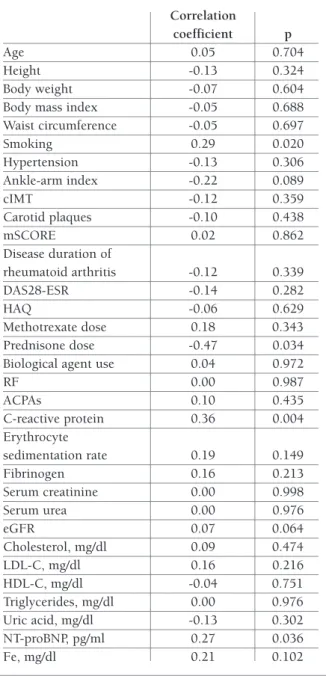
tAbLe III. correLAtIons between et-1 And study PArAmeters In rheumAtoId ArthrItIs PAtIents. Correlation coefficient p Age 0.05 0.704 Height -0.13 0.324 Body weight -0.07 0.604
Body mass index -0.05 0.688 Waist circumference -0.05 0.697 Smoking 0.29 0.020 Hypertension -0.13 0.306 Ankle-arm index -0.22 0.089 cIMT -0.12 0.359 Carotid plaques -0.10 0.438 mSCORE 0.02 0.862 Disease duration of rheumatoid arthritis -0.12 0.339 DAS28-ESR -0.14 0.282 HAQ -0.06 0.629 Methotrexate dose 0.18 0.343 Prednisone dose -0.47 0.034 Biological agent use 0.04 0.972
RF 0.00 0.987 ACPAs 0.10 0.435 C-reactive protein 0.36 0.004 Erythrocyte sedimentation rate 0.19 0.149 Fibrinogen 0.16 0.213 Serum creatinine 0.00 0.998 Serum urea 0.00 0.976 eGFR 0.07 0.064 Cholesterol, mg/dl 0.09 0.474 LDL-C, mg/dl 0.16 0.216 HDL-C, mg/dl -0.04 0.751 Triglycerides, mg/dl 0.00 0.976 Uric acid, mg/dl -0.13 0.302 NT-proBNP, pg/ml 0.27 0.036 Fe, mg/dl 0.21 0.102
cIMT: Carotid intimal medial thickness, m-SCORE: modified systematic coronary risk evaluation, DAS: disease activity score, HAQ: Health Assessment Questionnaire , RF: rheumatoid factor, ACPAs: Anti-citrullinated protein antibodies, eGFR: estimated glomerular filtration rate, LDL: low density lipoprotein, HDL: high density lipoprotein; Fe: iron
Calderon et al.37observed that dexamethasone and triamcinolone acetonide down-regulate the production and synthesis of ET-1 by a transformed human
pul-corresPondence to
Antonio Alvarez-Cienfuegos
Department of Rheumatology, Hospital Vega-Baja Crta. Orihuela-Almoradi, S/N
03314 Orihuela (Alicante), Spain E-mail: [email protected]
references
1. Gibofsky A. Overview of epidemiology, pathophysiology, and diagnosis of rheumatoid arthritis. Am J Manag Care 2012; 18: S295-302.
2. Castañeda S, Nurmohamed MT, González-Gay MA. Cardiovas-cular disease in inflammatory rheumatic diseases. Best Pract Res Clin Rheumatol 2016; 30:851-869.
3. Kerekes G, Soltész P, Nurmohamed MT, et al. Validated meth-ods for assessment of subclinical atherosclerosis in rheumatol-ogy. Nat Rev Rheumatol 2012; 8:224-234.
4. Corrales A, Parra JA, González-Juanatey C, et al. Cardiovascu-lar risk stratification in rheumatic diseases: carotid ultrasound is more sensitive than Coronary Artery Calcification Score to detect subclinical atherosclerosis in patients with rheumatoid arthritis. Ann Rheum Dis 2013; 72:1764-1770.
5. López-Mejías R, Castañeda S, González-Juanatey C, et al. Car-diovascular risk assessment in patients with rheumatoid arthri-tis: The relevance of clinical, genetic and serological markers. Autoimmun Rev 2016; 15:1013-1030.
6. Totoson P, Maguin-Gaté K, Nappey M, Wendling D, Demougeot C. Endothelial dysfunction in rheumatoid arthritis: Mechanis-tic insights and correlation with circulating markers of systemic inflammation. PLoS One 2016; 11: e0146744.
7. Kowalczyk A, Kleniewska P, Kolodziejczyk M, Skibska B, Gora-ca A. The Role of Endothelin-1 and Endothelin Receptor An-tagonists in Inflammatory Response and Sepsis. Arch Immunol Ther Exp 2015; 63:41-52.
8. Yeager ME, Belchenko DD, Nguyen CM, et al. Endothelin-1, the unfolded protein response, and persistent inflammation: role of pulmonary artery smooth muscle cells. Am J Respir Cell Mol Biol 2012; 46:14-22.
9. Virdis A, Schiffrin EL. Vascular inflammation: a role in vascular disease in hypertension?. Curr Opin Nephrol Hypertens 2003; 12:181-187.
10. Bellisai F, Morozzi G, Scaccia F, et al. Evaluation of the effect of bosentan treatment on proinflammatory cytokine serum levels in patients affected by systemic sclerosis. Int J Immunopathol Pharmacol 2011; 24:261-264.
11. Donate PB, Cunha TM, Verri WA JR, et al. Bosentan, an endo -thelin receptor antagonist, ameliorates collagen-induced arthri-tis: the role of TNF-a in the induction of endothelin system genes. Inflamm Res 2012; 61:337-348.
12. Kleniewska P, Piechota-Polanczyk A, Michalski L, et al. Influ-ence of block of NF-kappa B signaling pathway on oxidative stress in the liver homogenates. Oxid Med Cell Longev 2013; 2013:308358.
13. Piechota A, Goraca A. Influence of nuclear factor- B inhibition on endothelin-1 induced lung edema and oxidative stress in rats. J Physiol Pharmacol 2011; 62:183-188.
14. Aletaha D, Neogi T, Silman AJ, et al. 2010 Rheumatoid arthri-tis classification criteria: an American College of Rheumatolo-gy/European League Against Rheumatism collaborative initia-tive. Ann Rheum Dis 2010; 69:1580-1588.
15. National Kidney Foundation. K/DOQI clinical practice
guide-lines for chronic kidney disease: evaluation, classification, and stratification. Am J Kidney Dis 2002; 39:S1-266.
16. Baena-Díez JM, Subirana I, Ramos R, et al. Validity Assessment of Low-risk SCORE Function and SCORE Function Calibrated to the Spanish Population in the FRESCO Cohorts. Rev Esp Cardiol (Engl Ed) 2018; 71:274-282.
17. Corrales A, González-Juanatey C, Peiró ME, Blanco R, Llorca J, González-Gay MA. Carotid ultrasound is useful for the cardio-vascular risk stratification of patients with rheumatoid arthritis: results of a population-based study. Ann Rheum Dis 2014; 73:722-727.
18. Gonzalez-Juanatey C, Llorca J, Martin J, Gonzalez-Gay MA. Carotid intima-media thickness predicts the development of cardiovascular events in patients with rheumatoid arthritis. Semin Arthritis Rheum 2009; 38:366-371.
19. Goodson N. Coronary Artery disease in rheumatoid arthritis. Curr Opin Rheumatol 2002; 14: 115-120.
20. Kitas GD, Erb N. Tackling ischaemic heart disease in rheuma-toid arthritis. Rheumatology (Oxford) 2003; 42: 607-613. 21. Mayes MD. Endothelin and endothelin receptor antagonists in
systemic rheumatic disease. Arthritis Rheum 2003; 48: 1190--1199.
22. Yoshio T, Masuyama J, Mimori A, Takeda A, Minota S, Kano S. Endothelin-1 release from cultured endothelial cells induced by sera from patients with systemic lupus erythematosus. Ann Rheum Dis 1995; 54:361-365.
23. Vancheeswaran R, Magoulas T, Efrat G, et al. Circulating en-dothelin-1 levels in systemic sclerosis subsets—a marker of fi-brosis or vascular dysfunction? J Rheumatol 1994; 21:1838--1844.
24. Haq A, El-Ramahi K, Al-Dalaan A, Al-Sedairy ST. Serum and synovial fluid concentrations of endothelin-1 in patients with rheumatoid arthritis. J Med 1999; 30:51–60.
25. Brenchley PE. Antagonising angiogenesis in rheumatoid arthri-tis. Ann Rheum Dis 2001; 60:iii71-74.
26. Szekanecz Z, Szegedi G, Koch AE. Angiogenesis in rheumatoid arthritis: pathogenic and clinical significance. J Investig Med 1998; 46:27–41.
27. Koch AE. Angiogenesis as a target in rheumatoid arthritis. Ann Rheum Dis 2003; 62:ii60-67.
28. Van Doornum S, McColl G, Jenkins A, Green DJ, Wicks IP. Screening for atherosclerosis in patients with rheumatoid arthri-tis: comparison of two in vivo tests of vascular function. Arthri-tis Rheum 2003; 48:72-80.
29. Vaudo G, Marchesi S, Gerli R, et al. Endothelial dysfunction in young patients with rheumatoid arthritis and low disease ac-tivity. Ann Rheum Dis 2004; 63:31-35.
30. Sundy JS, Haynes BF. Pathogenic mechanisms of vessel damage in vasculitis syndromes. Rheum Dis Clin North Am 1995; 21:861-881.
31. Pache M, Schwarz HA, Kaiser HJ, et al. Elevated plasma en-dothelin-1 levels and vascular dysregulation in patients with rheumatoid arthritis. Med Sci Monit 2002; 8:CR616-619. 32. Kuryliszyn-Moskal A, Klimiuk PA, Sierakowski S, Ciolkiewicz
M. A study on vascular endothelial growth factor and endothe-lin-1 in patients with extra-articular involvement of rheuma-toid arthritis. Clin Rheumatol 2006; 25: 314-319.
33. Chen M, Li Z, Zhang Z, et al. Intervention of integrative medicine treatment has impact on serum levels of ET-1, TNF-a, MLT in RA-CVD. Saudi J Biol Sci 2018; 25: 959-964. 34. Giannopoulos S, Kosmidou M, Hatzitolios AI, Savopoulos CG,
Ziakas A, Karamouzis M. Measurements of endothelin-1, C-re-active protein and fibrinogen plasma levels in patients with acute ischemic stroke. Neurol Res 2008; 30: 727-730. 35. Kwon YS, Chi SY, Shin HJ, et al. Plasma C-reactive protein and
endothelin-1 level in patients with chronic obstructive pul-monary disease and pulpul-monary hypertension. J Korean Med Sci 2010; 25: 1487-1491.
36. Katayama T, Yano K, Nakashima H, et al. Clinical significance of acute-phase endothelin-1 in acute myocardial infarction pa-tients treated with direct coronary angioplasty. Circ J 2005; 69: 654-658.
37. Calderon E, Gomez-Sanchez CE, Cozza EN, et al. Modulation of endothelin-1 production by a pulmonary epithelial cell line. I. Regulation by glucocorticoids. Biochem Pharmacol 1994; 48: 2065-2071.
38. Winquist RJ, Scott AL, Vlasuk GP. Enhanced release of atrial na-triuretic factor by endothelin in atria from hypertensive rats. Hypertension 1989; 14:111-114.
39. Thibault G, Doubell AF, García R, Lariviere R, Schiffrin EL. En-dothelin-stimulated secretion of natriuretic peptides by rat atri-al myocytes is mediated by endothelin A receptors. Circ Res 1994; 74:460-470.
40. Levin ER. Endothelins. N Engl J Med 1995; 333:356-363. 41. Saijonmaa O, Ristimaki A, Fyhrquist F. Atrial natriuretic peptide,
nitroglycerine, and nitroprusside reduce basal and timulated endothelin production from cultured endothelial cells. Biochem Biophys Res Commun 1990; 173:514-520.
42. Kohno M, Horio T, Yokokawa K, et al. Atrial and brain natriuretic peptides: secretion during exercise in patients with essential hy-pertension and modulation by acute angiotensin-converting en-zyme inhibition. Clin Exp Pharmacol Physiol 1992; 19:193--200.
43. Kohno M, Yasunari K, Yokokawa K, Murakawa K, Horio T, Take-da T. Inhibition by atrial and brain natriuretic peptides of en-dothelin-1 secretion after stimulation with angiotensin II and thrombin of cultured human endothelial cells. J Clin Invest 1991; 87:1999-2004.
44. liwi ska-Mosso M, Milnerowicz S, Nabzdyk S, Kokot I, Nowak M, Milnerowicz H. The effect of smoking on endothelin-1 in patients with chronic pancreatitis. Appl Immunohistochem Mol Morphol 2015; 23:288-296.
45. Bossard M, Pumpol K, van der Lely S, et al. Plasma endothelin-1 and cardiovascular risk among young and healthy adults. Atherosclerosis 2015; 239:186-191.