From the School of Medicine, Federal University of Uberlândia1, School of Medicine of Ribeirão Preto, University of São Paulo2 and from the Pediatric Department, Children’s Institute, School of Medicine, University of São Paulo3.
IMMUNOGLOBULIN E-RHEUMATOID FACTOR IN
JUVENILE RHEUMATOID ARTHRITIS
Rosa Aparecida Ferreira1, Virgínia Paes Leme Ferriani2, Mônica Camargo Sopelete1, Deise Aparecida Oliveira Silva1, José Roberto Mineo1, Maria Helena Bittencourt Kiss3 and Carlos Henrique Martins Silva1
RHCFAP/3095
FERREIRA RA et al. - Immunoglonulin E-rheumatoid factor in juvenile rheumatoid arthritis. Rev. Hosp. Clín. Fac. Med. S. Paulo 57(5): 209-216, 2002.
OBJECTIVES: To determine the presence of immunoglobulin E-rheumatoid factor in patients with juvenile rheumatoid arthritis and to correlate it with clinical and laboratory parameters.
METHODS: A multicenter prospective study was carried out from January 1993 to January 1999 with the enrollment of 3 centers of pediatric rheumatology. Ninety-one children with juvenile rheumatoid arthritis diagnosed according to the American College of Rheumatology criteria were studied: 38 (42%) with systemic, 28 (31%) with pauciarticular, and 25 (27%) with polyarticular onset. Ages ranged from 2.1 years to 22.6 years (mean 10.5 ± 4.7), with 59 (65%) girls. The control group consisted of 45 healthy children. The detection of immunoglobulin E-rheumatoid factor was carried out utilizing an enzyme-linked immunosorbent assay. Associations of immunoglobulin E-rheumatoid factor with immunoglobulin M-rheumatoid factor (latex agglutination test), total serum immunoglobulin E, erythrocyte sedimentation rate, antinuclear antibody, and functional and radiological classes III or IV were analyzed.
RESULTS: Positive immunoglobulin E-rheumatoid factor was found in 15 (16.5%) of the 91 children with juvenile rheumatoid arthritis: 7 (18.5%) with systemic, 5 (18%) with pauciarticular, and 3 (12%) with polyarticular onset. A significant correlation was observed between immunoglobulin E-rheumatoid factor and total serum immunoglobulin E in the juvenile rheumatoid arthritis patients. No correlation was found between immunoglobulin E-rheumatoid factor and positive latex agglutination slide test, erythrocyte sedimentation rate, antinuclear antibody, or the functional and radiological classes III or IV in any disease onset group. In 4 out of 45 control children (8.9%), immunoglobulin E-rheumatoid factor was positive but with no correlation with total serum immunoglobulin E levels.
CONCLUSIONS: Immunoglobulin E-rheumatoid factor could be detected in 16.5% of juvenile rheumatoid arthritis patients, particularly in those with high levels of total serum immunoglobulin E, and immunoglobulin E-rheumatoid factor appears not to be associated with disease activity or severity.
DESCRIPTORS: Rheumatoid factor. Immunoglobulin E. Juvenile rheumatoid arthritis. ELISA.
INTRODUCTION
Rheumatoid factors (RF) are spe-cific autoantibodies directed against antigenic determinants on the Fc frag-ment of the IgG molecule1. However,
RFs are not unique to rheumatoid ar-thritis; they sometimes occur in nor-mal elderly individuals, healthy immu-nized individuals, and patients with other autoimmune diseases or chronic
infections2. Several studies have
shown that only 5% to 25% of chil-dren with juvenile rheumatoid arthri-tis (JRA) have positive latex aggluti-nation tests for RF3.
Children with JRA who are RF posi-tive are likely to be girls, to be older at disease onset, and to have many joints involved. It has been suggested that they have more roentgenographic evi-dence of bony erosion and an overall poorer prognosis than their rheumatoid factor-negative counterparts5. Some
clinical and immunogenetic associa-tions are well known, such as nodes, bony erosions, functional incapacities, and HLA Dw4 (DRB1*0401) and Dw14 (BRB1*0404) associations5,6.
In one-third of patients with JRA who present idiotypic expression with cross-reaction for RF, classical RF (IgM-RF) is not observed7. It is
possi-ble that in this group of patients, other RF isotypes are present8. With the use
of ELISA, an increased detection is observed in JRA, with IgM-RF being demonstrated in 22% to 35% of pa-tients, IgG-RF in 4% to 6%, and IgA-RF in 30% to 60%, depending on the type of disease onset9-11. The presence
of IgA-RF and IgM-RF has been ob-served in a varying percentage of chil-dren with JRA, mainly in the polyar-ticular onset type12,13. However, the
fre-quency and clinical and prognostic roles of the other RF isotypes (IgG and IgE) in JRA are still unknown14.
There is evidence suggesting that IgE-RF is associated with extra-articu-lar rheumatoid vasculitis and activity of the disease in rheumatoid arthri-tis15,16. We found 1 study about IgE-RF
in JRA in the literature17. These
inves-tigators observed that sera from 11/26 children with JRA had low concentra-tions of IgE-RF detected by the indi-rect immunofluorescence technique and suggested that IgE-RF may be in-volved in the pathogenesis of JRA and rheumatoid arthritis by eliciting type I and III reactions17.
The aims of the present study were to determine the presence and the lev-els of IgE rheumatoid factor (IgE-RF) in children with JRA and to correlate its presence with sex, age at onset,
dis-ease duration, activity of disdis-ease, erythrocyte sedimentation rate, antinu-clear antibody, total serum IgE, IgM-RF (latex agglutination), and func-tional and radiological classes III or IV.
PATIENTS AND METHODS
Patients
A prospective, multicentric study was carried out from January 1993 to January 1999. Ninety-one children (59 girls) with JRA diagnosed according to the American College of Rheumatol-ogy criteria18,6 were studied. Sera were
randomly obtained from patients who visited one of the following three Centers of Pediatric Rheumatology: Hospital de Clínicas da Universidade de São Paulo, São Paulo (51 cases), Hospital de Clínicas da Faculdade de Medicina de Ribeirão Preto da Universidade de São Paulo, Ribeirão Preto, São Paulo (28 cases), and Hos-pital de Clínicas da Universidade Fed-eral de Uberlândia, Minas Gerais (12 cases). The age of the patients ranged from 2.1 to 22.7 years (mean 10.5 ± 4.7). The type of disease onset was sys-temic in 38 children, pauciarticular in 28, and polyarticular in 25.
Forty-five healthy control children with no symptoms of connective tis-sue disease or acute or chronic infec-tions were selected from the pediatric outpatient clinic. The mean age of controls was 5.5 ± 3.3 years.
Inclusion criteria
All patients included had no clini-cal symptoms suggestive of allergic rhynitis, asthma, or atopic dermatitis. In addition, they had 3 samples of feces examined for helminths with negative results. None of the JRA pa-tients were receiving or had received gold salts or D-penicilamine.
METHODS
Serum was obtained from each pa-tient during the first appointment after obtaining fully informed consent from their parents or relatives. Approval for the study was also obtained from the 3 Institutional Ethics Committees. Serum samples were stored at –20°C.
The demographic and clinical char-acteristics of the JRA patients are shown in table 1 (sex, age at onset, dis-ease duration, and activity of disdis-ease). Serum was obtained from each patient for the following laboratory measure-ments: erythrocyte sedimentation rate (by the Wyntrobe method), antinuclear antibody (by the Hep 2 immunoassay method), and total serum IgE (measured by monoclonal antibody-based ELISA modified from a previously described radioimmunoassay (RIA) by Pollart19).
Values of total IgE ≥ 300 IU/mL were considered abnormally high (posi-tive)20. IgM-RF was determined by the
latex agglutination slide test using a commercially available kit (RapiTex RF, Hoechst, Marburg, Germany). Sam-ples with RF of 20 IU/mL or more were considered to be positive.
Active disease was defined by the presence of joint swelling or limitation of mobility with warmth, pain, or ten-derness in 1 or more joints. Severity was evaluated according to Steinbrocker’s functional and radio-logical classes21with classes III and IV
denoting major dysfunction.
Quantitative determination of the IGE rheumatoid factor by ELISA
IgE-RF was determined by a stand-ard ELISA technique according to SILVA22, with some modifications. The
samples were always analyzed in du-plicate.
washed in 0.01M phosphate buffered saline (PBS), pH 7.2, containing 0.05% Tween 20 (PBS-T) and blocked with PBS-T plus 1% bovine serum al-bumin (PBS-T-BSA) for 1 h at room temperature (RT). Washings in PBS-T were done between the steps of the re-action. The plates were incubated with serum samples diluted 1:5 in PBS-T, in duplicate, for 1 h at RT. Subsequently, peroxidase-rabbit IgG anti-human IgE, prepared as described by Wilson and Nakane23, at 1:500 in PBS-T was added
and incubated for 1 h at RT. Finally, enzymatic substrate consisting of 0.03% H2O2 and ortho-phenylenedi-amine in 0.1 M phosphate citrate buffer, pH 5.0, was added and incu-bated for 15 min at RT. Adding 2 N H2SO4 stopped the reaction, and the absorbancy was read at 492 nm in a plate reader (Titertek Multiskan, Flow Laboratories, USA). Two positive and 10 negative control sera were included in each assay. Positive sera were ob-tained from patients who had IgM-RF by the latex agglutination test and IgE-RF by previous ELISA tests in the standardization of the assays. Negative sera were obtained from healthy chil-dren.
The results were expressed as ELISA index (EI) and calculated as follows: EI = absorbancy of serum
samples / cut off, where cut off was de-termined as the mean absorbancy of the negative control serum plus 3 standard deviations. Samples present-ing EI values > 1 were considered posi-tive.
STATISTICS
Statistical analysis consisted of determinations of geometric means with 95% confidence intervals (CI) for measurements of total serum IgE. Comparisons between the ages of chil-dren with JRA were performed using the Student t test. This test was also
used to compare the geometric means of total serum IgE. The positive and negative samples were listed in contin-gency tables and analyzed by the χ2
and Fisher exact tests24. Significance
was accepted when P <0.05.
Statisti-cal Statisti-calculations were performed using a standard Statistical Package Social Science (SPSS) program.
RESULTS
IgE-RF was detected in 15/91 (16.5%) patients with JRA and 4/45 (8.9%) children from the control group (P >0.05) (Fig. 1A). Disease onset was
systemic in 7 (18.4%), pauciarticular in 5 (17.8%), and polyarticular in 3 (12%) (Fig.1B).
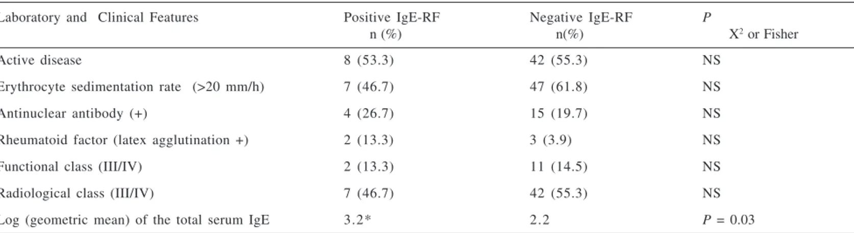
An attempt was made to compare IgE-RF-positive and negative patients in the JRA groups. Comparisons of the most significant clinical features be-tween the groups (with or without IgE-RF) are shown in table 2. There was no significant difference between the clinical and laboratory factors (disease activity, erythrocyte sedimentation rate, antinuclear antibody, or functional and radiological class III or IV) and the presence or not of the IgE-RF, ex-cept for the log of geometric mean of the total serum IgE, which was signifi-cantly higher (P = 0.03) in the group
of patients with positive IgE-RF (Ta-ble 2).
The presence or absence of IgE-RF in sera from JRA patients and control children was analyzed in comparison to total serum IgE (Fig. 2). There was a significant difference (P <0.05) in the
geometric mean of total IgE between JRA patients with positive (1420 IU/ mL) and negative (402 IU/mL) IgE-RF. In contrast, no statistically signifi-cant difference was found between positive (157 IU/mL) and negative (117 IU/mL) IgE-RF in the control group (P >0.05). In addition, the mean
of total IgE values for JRA patients
Table 1 - Clinical and laboratory features of the children with juvenile rheumatoid arthritis (n= 91) according to disease onset (systemic, pauciarticular, or polyarticular).
Clinical and Laboratory Features JUVENILE RHEUMATOID ARTHRITIS
Total Systemic Pauciarticular Polyartycular
(n = 91) (n = 38) (n = 28) (n = 25)
Females 59 (64.4%) 21 (55,3%) 17 (60.71%) 21 (84%)
Age (years) 10.5 (2.1 – 22.7) 11 (4.1 – 20.8) 9.9 (2.4-17.2) 10.3 (4.2-18.6)
Disease duration (years) 5.2 (0.2– 17.3) 6.2 (1.1-13.6) 4.4 (0.7-10.2) 3.7 (0.2-11.6) Age at onset (years) 5.2 (0.5 – 16.1) 4.7 (1.4-11.9) 5.5 (1.1-13.3) 6.7 (2.9-12.2)
Active disease 50 (55%) 21 (55.3%) 12 (42.8%) 17 (68%)
Erythrocyte sedimentation rate (>20 mm/h) 54 (59%) 26 (68.4%) 12 (42.8%) 16 (64%)
Anti nuclear antibody + 19 (21%) 1 (2.6%) 8 (28.6%) 10 (40%)
Rheumatoid factor (latex agglutination +) 5 (5.4%) ——— ———— 5 (20%)
Functional class (III/IV) 13 (14%) 11 (28.9%) ———— 2 (8%)
Radiological class (III/IV) 49 (54%) 26 (68.4%) 10 (35.7%) 13 (52%)
Table 2 - Laboratory and clinical features among 15 patients with positive FR-IgE with juvenile rheumatoid arthritis (JRA) and 76 patients with negative IgE-RF.
Laboratory and Clinical Features Positive IgE-RF Negative IgE-RF P
n (%) n(%) X2 or Fisher
Active disease 8 (53.3) 42 (55.3) NS
Erythrocyte sedimentation rate (>20 mm/h) 7 (46.7) 47 (61.8) NS
Antinuclear antibody (+) 4 (26.7) 15 (19.7) NS
Rheumatoid factor (latex agglutination +) 2 (13.3) 3 (3.9) NS
Functional class (III/IV) 2 (13.3) 11 (14.5) NS
Radiological class (III/IV) 7 (46.7) 42 (55.3) NS
Log (geometric mean) of the total serum IgE 3.2* 2.2 P = 0.03
NS = Not significant.
with IgE-RF was higher than that ob-tained for IgE-RF-positive control chil-dren, although the difference was not statistically significant (P = 0.055).
The geometric mean of total serum IgE for all JRA patients (495 IU/mL) was significantly higher than for the control group (121 IU/mL), P <0.0001
(Fig 3).
DISCUSSION
The pathogenic role of the rheuma-toid factor isotypes in JRA is not fully understood14. The importance of the
IgM isotype detected by agglutination methods is clear in terms of the classi-fication and prognosis of this disease, because this isotype defines the sero-positive polyarticular subtype, and it is related to progression to erosive dis-ease4. However, few studies are
avail-able on other RF isotypes in JRA14.
Only 1 investigation assessing the presence of IgE-RF in children with JRA was found in the literature17.
Eleven out of 26 children studied (42%) presented weakly positive reac-tions (undiluted sera) for IgE-RF when tested by indirect immunofluores-cence17.
In our study, IgE-RF was detected by ELISA in 15 (16.5%) of 91 children with JRA, and this detection was not Figure 1 - Levels of IgE-RF expressed in ELISA Index (EI). (A) Patients with JRA (n = 91) and
In our study, IgE-RF was detected in 4 (8.9%) control children, and at high concentrations in some of them, indicating a low specificity of this an-tibody. The children in this group were considered healthy, with no clinical history suggestive of atopy. However, it should be emphasized that in the patients with JRA studied here, the presence of IgE-RF was correlated with higher serum IgE concentrations, whereas this was not observed in the IgE-RF positive control group. These data seem to indicate that the stimuli responsible for IgE-RF production is different in healthy individuals and in patients with JRA.
The identification of IgE-RF in a group of patients with JRA stimulates the discussion of the role of this autoantibody and consequently of the IgE-dependent mechanisms involved in the pathogenesis of this disease. The prevalence of atopic disease in children with JRA is unknown. It has been re-ported that the incidence of IgE-spe-cific antibodies against pollen allergens in 40 children with JRA was similar to that expected for the normal popula-tion26. Some studies have suggested
that the prevalence of atopy among adults with rheumatoid arthritis is equal to or lower than that observed in the normal population, although high con-centrations of total serum IgE have been detected in these patients14,26. Recently,
Millauer in 1999, comparing patients with rheumatoid arthritis and patients with allergic diseases, observed that rheumatoid sera have high concentra-tions of total serum IgE and complexed anti-IgE, in addition to increased reac-tivity of specific IgE against only 1 al-lergen (Alternaria) and not against 9 other common allergens. Based on these findings, the authors concluded that the presence of large amounts of IgE probably does not result in an in-creased prevalence of allergic diseases in patients with rheumatoid arthritis, as previously mentioned14. On the other
Figure 2 - Levels of total serum IgE in JRA patients with positive IgE-RF (n = 15) and negative IgE-RF (n = 76), and control children with positive IgE-RF (n= 4) and negative IgE-RF (n = 41). The horizontal bars indicate the geometric means.
Figure 3 - Levels of total serum IgE expressed in IU/mL in the group of JRA patients (n = 91) and control children (n= 45). The horizontal bars indicate the geometric means.
related to the disease onset type. No correlation was observed between the presence of IgE-RF and any clinical or laboratory parameter. Previous studies have shown that low serum IgE-RF concentrations are detected in
hand, the presence of anti-IgE antibod-ies seems to be paradoxical. According to these authors, it is possible that anti-IgE antibodies may interfere with anti-IgE binding, thus preventing the sensitization of basophils and mast cells34.
Some interesting studies have dem-onstrated the presence of activated mast cells in experimental rheumatoid lesions27 and in synovial fluid of
pa-rheumatoid synovitis is uncertain. In conclusion, IgE-RF can be de-tected in some patients with JRA with higher total serum IgE levels. We be-lieve that the results reported here sug-gest an alteration of the humoral im-mune response in some children with JRA and stimulate future research lines aiming at the study of the IgE-depend-ent biological mechanisms involved in this disease.
tients with rheumatoid arthritis and JRA28, the increased expression of the
CD23 antigen in peripheral mononu-clear cells of patients with rheumatoid arthritis29,30 and JRA31, and the
repro-duction of an experimental model of arthritis by the inoculation of monomeric or complexed IgE32,33.
However, the value of these findings for the understanding of the physi-opathological mechanisms involved in
RESUMO RHCFAP/3095
FERREIRA RA e col. - Fator reuma-tóide-imunoglobulina E na artrite reumatóide juvenil. Rev. Hosp. Clín. Fac. Med. S. Paulo 57(5):209-216, 2002.
OBJETIVOS: Determinar os níveis séricos do fator reumatóide-imuno-globulina E na artrite reumatóide ju-venil e correlacioná-los com parâ-metros clínicos e laboratoriais.
MÉTODOS: Estudo multicêntrico prospectivo, realizado entre janeiro de 1993 a janeiro de 1999 com participa-ção de três centros de reumatologia pediátrica. Estudaram-se 91 crianças com o diagnóstico de artrite reumatóide
juvenil de acordo com os critérios do Colégio Americano de Reumatologia: 38 (42%) com a forma de início sistêmica, 28 (31%) pauciarticular e 25 (27%) poliarticular. A idade variou de 2,1 a 22,6 anos (média de 10,5 ± 4,7 anos) e 59 (65%) crianças eram do sexo feminino. O grupo controle constituiu-se de 45 crianças sadias. A detecção do fator reumatóide-imunoglobulina E foi realizada através de um ensaio imunoenzimático. Associações do fator reumatóide-imunoglobulina E com: fa-tor reumatóide-imunoglobulina M (látex), imunoglobulina E sérica total, VHS, FAN, classe funcional e radioló-gica III ou IV foram analisadas.
do-ença. Dos 45 controles, 4 (8,9%) tam-bém apresentaram fator reumatóide-imunoglobulina E positivo, mas não houve correlação com a imunoglo-bulina E sérica total.
CONCLUSÕES: O fator reuma-tóide-imunoglobulina E pode ser
detec-tado em 16,5% dos pacientes com ar-trite reumatóide juvenil, especialmen-te naqueles com níveis elevados de imunoglobulina E sérica total. O fator reumatóide-imunoglobulina E parece não se associar com atividade ou
gra-vidade da artrite reumatóide juvenil.
DESCRITORES: Fator Reuma-tóide. Imunoglobulina E. Artrite Reumatóide Juvenil. Ensaio imuno-enzimático.
REFERENCES
1 . SUTTON B, CORPER A, BONAGURA V et al. - The structure and origin of rheumatoid factors. Immunol Today 2000; 21(4): 177-182.
2 . TIGHE H & CARSON D A - Rheumatoid factor. In: KELLEY W N, HARRIS Jr. E D, RUDDY S et al. - Textbook of rheumatology. 5th ed. Philadelphia, Saunders, 1997. p.
241-249.
3 . PETTY RE, CASSIDY JT & SULLIVAN DB - Serologic studies in juvenile rheumatoid arthritis: a review. Arthritis Rheum 1977; 20 (suppl.): 260.
4 . SCHALLER JG - Juvenile rheumatoid arthritis. Pediatr Rev 1997; 18 (10): 337-349.
5 . EICHENFIELD AH, ATHREYA BH, DOUGHTY RA et al. - Utility of rheumatoid factor in the diagnosis of juvenile rheumatoid arthritis. Pediatrics 1986; 78 (3): 480 - 483.
6 . CASSIDY JT & PETTY RE - Juvenile rheumatoid arthritis. 3th
ed. Philadelphia, Saunder,1995. p. 133-223.
7 . ILOWITE NT, O‘REILLY ME, HATAM L et al. -Expression of the rheumatoid factor cross-reactive idiotype in JRA: association with disease onset subtype, disease activity and disease severity. Scand J Rheumatol 1992; 21: 51-54.
8 . ILOWITE NT, WEDGWOOD JF & BONAGURA VR - Expression of the major rheumatoid factor cross-reactive idiotype in juvenile rheumatoid arthritis. Arthritis Rheum 1989; 32 (3): 265-270. 9 . CASSIDY JT & VALKENBURG H A - A five year prospective study of rheumatoid factor tests in juvenile rheumatoid arthritis. Arthritis Rheum 1967; 10: 83-90.
10.WALKER SM, SHAHAM B, McCURDY DK et al. - Prevalence and concentration of IgM rheumatoid factor in polyarticular onset disease as compared to systemic or pauciarticular onset disease in active juvenile rheumatoid arthritis as measured by ELISA. J Rheumatol 1990; 17 (7): 936-940.
11.JARVIS JN, POUSAK T & KRENZ M - Detection of IgM rheumatoid factors by enzyme-linked immunosorbent assay in children with juvenile rheumatoid arthritis: correlation with articular disease and laboratory abnormalities. Pediatrics 1992; 90 (6): 945-949.
12.WALKER SM, McCURDY DK, SHAHAM B et al. -High prevalence of IgA rheumatoid factor in severe polyarticular-onset juvenile rheumatoid arthritis, but not in systemic-onset or pauciarticular-onset disease. Arthritis Rheum 1990; 33 (2): 199-204. 13.VARBANOVA BB, BALEVA M, NIKOLOV K et al. - Prevalence
of –IgM, -IgA and –IgG rheumatoid factors in seronegative polyarticular disease compared to pauciarticular disease in juvenile chronic arthritis as measured by ELISA. Adv Exp Med Biol 1999; 455: 61-68.
14.MILLAUER N, ZUERCHER AW, MIESCHER SM et al. - High IgE in rheumatoid arthritis (RA) patients is complexed with anti-IgE autoantibodies. Clin Exp Immunol 1999; 115 (1): 183-188.
15.MIZUSHIMA Y, SHOJI Y, HOSHI K et al. - Detection and clinical significance of IgE rheumatoid factor. J Rheumatol 1984; 11 (1): 22-26.
17.PERMIN G & EGESKJOLD EM - IgE anti-IgG antibodies in patients with juvenile and adult rheumatoid arthritis including Felty’s syndrome. Allergy 1982; 37 (6): 421-427.
18.BREWER EJ, BASS J, BAUM J et al. - Current proposed revision of JRA criteria. Arthritis Rheum 1977; 20 (2) (supll.): 195-199.
19.POLLART SM, CHAPMAN MD, FIOCCO GP et al. - Epidemiology of acute asthma: IgE antibodies to common inhalant allergens as a risk factor for emergency room visits. J Allergy Clin Immunol 1989; 83 (5): 875-882.
20.SILVA CHM - Contribuição para o estudo da imunoglobulina E (IgE) sérica total em crianças com artrite reumatóide juvenil. São Paulo, 1995. (Tese Doutorado em Pediatria - Faculdade de Medicina da Universidade de São Paulo).
21.STEINBROCKER O, TRAEGER CH & BATTERMAN RE -Therapeutic criteria in rheumatoid arthritis. JAMA 1949; 140: 659-662.
22.SILVA CHM -Fator reumatóide IgM detectado pelo teste de fixação do látex, reação de Waaler-Rose e ELISA em crianças com artrite reumatóide juvenil. São Paulo, 1990. (Dissertação Mestrado - Faculdade de Medicina, Universidade de São Paulo). 23.WILSON MB & NAKANE PK - Antibody conjugated to horseradish peroxidase. In: KANPP K & NOLUBAR GW (Eds.) - Immunofluorescence and related staining technique. Amsterdam, Elsevier Norths Holland Biomedical, 1978. p. 215-224.
24.TRIOLA MF - Introdução à estatística. 7ª. ed. Rio de Janeiro,
LTC (Livro Técnico e Científico E.S.A.), 1999.
25.MERETEY K, FALUS A, BÖHM U et al. - IgE class immune complexes in Felty’s syndrome: characterisation of antibody activities in isolated complexes. Ann Rheum Dis 1984; 43 : 246-250.
26.PESKETT SA, PLATTSMILLS TAE, ANSELL BM et al. -Incidence of atopy in rheumatic disease. J Rheumatol 1981; 8 (2): 321-324.
27.CAULFIELD JP, HEIN A, HELFGOTT SM et al. - Intraarticular injection of arthritogenic factor causes mast cell degranulation, inflammation, fat necrosis, and synovial hyperplasia. Lab Invest 1988; 59 (1): 82-95.
28.GRUBER B, BALLAN D & GOREVIC PD - IgE rheumatoid factors: quantification in synovial fluid and ability to induce synovial mast cell histamine release. Clin Exp Immunol 1988; 71 (2): 289-294.
29.CHOMARAT P, BRIOLAY J, BANCHEREAU J et al. - Increased production of soluble CD23 in rheumatoid arthritis, and its regulation by interleukin-4. Arthritis Rheum 1993; 36 (2): 234-242.
30.FERNÁNDEZ-GUTIÉRREZ B, HERNÁNDEZ-GARCÍA C, BAÑARES AA et al. - CD23 hyperexpression in rheumatoid arthritis: evidence for a B cell hyperresponsiveness to cognate and noncognate T-cell signals. Clin Immunol Immunopathol 1994; 72 (3): 321-327.
31.MASSA M, PIGNATTI P, OLIVERI M et al. - Serum soluble CD23 levels and CD23 expression on peripheral blood mononuclear cells in juvenile chronic arthritis. Clin Exp Rheumatol 1998; 16 (5) : 611-616.
32.DE CLERCK LS, STRUYF NJ & STEVENS, WJ - Activation of inflammatory cells by immune complexes containing IgE in serum and synovial fluid of patients with rheumatoid arthritis: a study using flow cytometric analysis. Ann Rheum Dis 1991; 50: 379-382.
33.DE CLERCK LS, STRUYF NJ, BRIDTS C H et al.-Experimental arthritis in rats induced by intra-articular injection of IgE aggregates: evidence of arthritogenic role of complexed IgE. Ann Rheum Dis 1992; 51: 210 - 213.
34.GRUBER BL - Immunoglobulin E, mast cells, endogenous antigens, and arthritis. Rheum Dis Clin North Am, 1991; 17 (2): 333-342.