UNIVERSIDADE DA BEIRA INTERIOR
Ciências da Saúde
Descoberta e desenvolvimento de inibidores da
17β-HSD-1 potencialmente úteis no tratamento
do cancro da mama
Experiência Profissionalizante na Vertente de Farmácia
Comunitária e Investigação
Aura Maria de Morais Vaz
Relatório de Estágio para obtenção do Grau de Mestre em
Ciências Farmacêuticas
Ciclo de Estudos Integrado
Orientador: Prof. Doutor Samuel Silvestre
Co-orientador: Prof
a. Doutora Luiza Granadeiro
Ao Bruno Abreu, porque o amor permanece ainda que separado por vales de ignomínia distância escoltada pela Saudade. Que elas se desvaneçam nesta nova fase da vida, e que no futuro impere enfim a partilha de uma vida preenchida de júbilo. Aos meus pais, avós e irmã Marta, que o amor de família seja sempre a força motriz da felicidade.
Agradecimentos
Em primeiro lugar, expresso um agradecimento profundo ao meu orientador, Professor Dr. Samuel Silvestre, por me ter introduzido ao mundo da química orgânica e farmacêutica e orientado com toda amizade, dedicação e continuidade, por todo o trabalho, confiança e paciência investidas em mim durante o trabalho para a obtenção do meu grau de mestre em Ciências Farmacêuticas. Agradeço também todo o apoio e amizade à minha co-orientadora Profa. Dra. Luiza Granadeiro e também agradeço à Profa. Dra. Carla Cruz pelo auxílio nas experiências de RMN.
Aos meus amigos e colegas de estudo e de trabalho no laboratório, com quem partilhei bons momentos de alegria e que nunca me abandonaram, sendo sempre um apoio nos momentos de maior trabalho e indecisão, muito agradeço! As saudades vão permanecer e ficarão sempre na minha memória com amizade! Ágradeço também especialmente à Tânia, Rita e Mafalda, por toda a amizade, paciência e apoio que partilhámos umas com as outras nesta difícil fase de escrita do Relatório de estágio!
Foi com muito apreço que no fim do meu percurso académico tive a oportunidade de contactar com o ambiente e vida diária da farmácia comunitária. Presto um marcado agradecimento a toda a equipa da Farmácia Sant’Ana, com quem criei bons laços de amizade, e um agradecimento muito especial à diretora técnica, Drª. Paula Bártolo por todo o apoio e elevada contribuição pessoal e científica que me proporcionou. Todos me acolheram com a maior atenção e simpatia, e me orientaram durante as horas de estágio, onde sempre me senti em casa.
Um especial e profundo agradecimento à minha família: pais, irmã, avós, tios e primos, e ao meu namorado Bruno, por todo o apoio e amor que me deram ao longo de todo o meu percurso académico e vida.
Finalmente, e não querendo esquecer ninguém, agradeço a todos os que, de uma forma ou outra, me ajudaram neste caminho!
Prefácio
Este Relatório de Estágio visa englobar e dar a conhecer ao público académico as áreas exploradas no estágio curricular de final de curso, para a obtenção do grau de Mestre em Ciências Farmacêuticas, sendo elas as áreas de Investigação (cuja escolha pessoal foi a vertente de Química Farmacêutica) e de Farmácia Comunitária, que no total perfizeram 800h de constante aprendizagem e enriquecimento pessoal e científico.
A perspetiva futura de um farmacêutico avizinha-se crítica, mas a força de espírito jamais poderá esmorecer em nós, futuros profissionais de saúde, com sonhos e ideais para além do impossível. Sonhemos sempre, lutemos sempre - juntos construiremos e receberemos um futuro melhor, um futuro digno e merecedor para todos nós.
Resumo
Capítulo I
O cancro da mama é o cancro mais comum entre as mulheres. Neste contexto, diversos estudos evidenciam que entre 40 a 60% de todos os cancros da mama são considerados hormono-dependentes, dependendo principalmente de 17β-estradiol para o seu desenvolvimento. No corpo humano, este estrogénio é originado maioritariamente a partir da estrona, um estrogénio com baixa atividade estrogénica, pela ação da enzima 17β-hidroxiesteroide desidrogenase tipo 1 (17β-HSD-1). Como esta enzima se encontra em elevadas quantidades em tecidos cancerígenos dependentes de estrogénios e tem elevada seletividade, a descoberta de inibidores da 17β-HSD-1, com o objetivo de minimizar a formação de estradiol, está a ganhar grande interesse nos dias de hoje.
Assim, baseado no conhecimento prévio de que vários D-homoesteroides têm interessantes propriedades inibitórias da 17β-HSD-1, neste trabalho pretende-se desenvolver vários derivados da estrona como potenciais inibidores da 17β-HSD-1 com elevadas propriedades inibitórias, baixa toxicidade e preparação sintética simples. Para este propósito foram realizados estudos computacionais, tais como o docking molecular e avaliação toxicológica in silico. A partir destes estudos, várias moléculas, em especial derivados da estrona funcionalizados em C2 e C3 e os seus homólogos com anel de D de 6 membros, lactonizado, foram seleccionados como potenciais novos inibidores da 17β-HSD-1.
Os resultados de docking realizados com o programa AutoDock Vina, revelaram que os referidos derivados lactonizados da estrona possuem elevados valores de afinidade para a enzima, quando comparados com os seus homólogos com anel D de 5 membros, e quando comparados com outros homólogos D-Homo, revelaram possuir iguais ou mesmo até superiores afinidades. Todos os compostos funcionalizados em C2 apresentaram valores de afinidade superiores quando comparados com o valor de afinidade da estrona, em especial substituintes hidrofóbicos ou com elevadas propriedades eletronegativas. Este tipo de funcionalização é vantajoso, já que se encontra descrito que diminui a estrogenicidade dos compostos. Adicionalmente descobriu-se também que substituições em C3, com grupos aceitadores em pontes de hidrogénio, como grupos éster ou heterocíclicos, bem como grupos lipofílicos, dão origem a compostos com elevados valores de afinidades. Os estudos de toxicidade in silico revelaram que as moléculas avaliadas são seguras em termos de efeitos mutagénicos, tumorigénicos, irritantes ou em efeitos a nível reprodutivo, exceto para as moléculas substituídas com C6-OH, que apresentaram risco a nível reprodutivo.
Neste momento estão ainda a ser realizadas sínteses químicas e a iniciar-se a avaliação biológica in vitro, com o objetivo de confrontar com os resultados obtidos nos cálculos computacionais e de efetuar estudos de relação-estrutura-atividade (REA) para este tipo de novos compostos inibidores da 17β-HSD-1.
Capítulo II
O capítulo II pretende resumir a minha participação no estágio profissional realizado na área da farmácia comunitária na Farmácia Sant’Ana – Covilhã. Nele encontram-se descritas todas as áreas de trabalho e funcionalidades, as funções desempenhadas pelo farmacêutico, entre outros diversos aspetos fundamentais para o funcionamento desta unidade de saúde dirigida para a comunidade.
Palavras-chave
Inibidores da 17β-HSD-1, cancro da mama, descoberta de fármacos, docking molecular, toxicidade in silico, farmácia comunitária
Abstract
1st ChapterBreast cancer is the most common cancer in women. In this context, several studies evidenced that 40 to 60% of all breast cancers are considered hormone-dependent, relying especially on estradiol for their development. In the human body, this estrogen is originated from estrone, an estrogen with low estrogenic activity, by the action of 17β-hydroxysteroid dehydrogenase type 1 (17β-HSD-1). As this enzyme is encountered in high amounts on estrogen-dependent cancer tissues and has high selectivity, the discovery of 17β-HSD-1 inhibitors, in order to minimize the formation of estradiol, is gaining high interest nowadays. Thus, based on previous knowledge that several D-homosteroids have interesting 17β-HSD-1 inhibitory properties, our group has been developing several new estrane derivatives as potential 17β-HSD-1 inhibitors with higher inhibitory properties, low toxicity and simpler synthetic preparation. For this purpose, computational studies, like molecular docking and in silico toxicological evaluation have been performing. From these studies, several molecules, mainly C2 and C3 functionalized estrone derivatives and their homologues with the lactonized D-ring have been elected as potentially novel 17β-HSD-1 inhibitors. Interestingly, docking results performed with AutoDock Vina revealed that the lactonized estrone derivatives have higher affinity values to the enzyme when compared to all their 5-membered D-ring homologues as well as equal or higher affinity values compared to the previously reported D-Homo derivatives. In silico toxicity tests also revealed that all molecules were safe regarding on mutagenic, tumorigenic, irritant or reproductive effects, except for C6-OH substituted molecules that presented risk on reproductive level. At the moment, chemical synthesis and in vitro biological evaluation are being performed.
2nd Chapter
This chapter aims to resume the professional traineeship realized in the field of Community Pharmacy, it’s presented in Chapter 2 an overview of the working areas and fields of this health unit managed for the society.
Keywords
17β-HSD type 1 inhibitors, breast cancer, drug discovery, molecular docking, in silico toxicity,
Índice
Capítulo I – Estágio em Investigação
Descoberta e desenvolvimento de potenciais inibidores da 17β-HSD-1 potencialmente úteis no tratamento do cancro da mama
1.1 Introduction 1
1.1.1 Breast Cancer and 17β-HSD-1 1
1.1.1.1 Current breast cancer therapeutic approaches 3
1.1.2 17β-HSD-1 4
1.1.2.1 Function and structure 4
1.1.2.2 Mechanism of action 5
1.1.2.3 Steroidal inhibitors of 17β-HSD-1 6
1.1.3 Drug design and discovery – the importance of computational
methods: molecular docking and in silico toxicity studies 13
1.1.3.1 (Q)SARs and in silico studies 13
1.1.3.2 Molecular Docking 14
1.1.3.2.1 Docking software 15
1.2 Objectives 17
1.3 Materials and methods 18
1.3.1 Computational studies 18
1.3.1.1 Software 18
1.3.1.2 Preparation of the ligands for molecular docking: 3D
molecular structures, minimizing energy and file formats 18 1.3.1.3 Choosing and preparing the macromolecule for docking
experiments 19
1.3.1.4 Grid-Box 19
1.3.1.5 Testing the accuracy of docking experiments 20
1.3.1.6 Docking results 21
1.3.1.7 In silico toxicity studies 22
1.3.2 Materials used on chemical synthesis 22
1.3.2.1 Reagents and solvents 22
1.3.2.2 Chromatography 22
1.3.2.3 Equipment and other devices 22
1.3.3 Experimental process 23
1.3.3.1 Baeyer-Villiger reaction of Estrone 23
1.3.3.2 Iodination reaction of Estrone with Sodium Iodide and Sodium
Chlorite 23
1.3.3.3 Iodination reaction of Estrone with Iodine and Copper (II) chorite di-hydrated (CuCl2.H2O)
24
1.3.3.5 Bayer-Villiger reaction of 2-nitroestrone 26
1.4 Results and Discussion 28
1.4.1 Computational studies 28
1.4.1.1 Molecular Docking studies 28
1.4.1.2 In silico toxicity studies 39
1.4.2 Chemical synthesis 39
1.5 Conclusions 43
1.6 Bibliography 44
Capítulo II – Estágio em Farmácia Comunitária
2.1 Introdução 51
2.2 Organização da farmácia Sant’Ana 52
2.2.1 Localização 52
2.2.2 Recursos humanos 52
2.2.3 Funções do Diretor Técnico, seus substitutos e adjuntos 53
2.2.4 Espaço físico da farmácia 54
2.2.5 Elementos interiores e exteriores distintivos da farmácia 55
2.2.6 Equipamentos gerais e específicos da Farmácia 55
2.2.7 Sistema informático 56
2.2.8 Gestão de qualidade 56
2.3 Informação e documentação científica 57
2.4 Medicamentos e outros produtos de saúde 58
2.4.1 Definições 58
2.4.2 Gamas de produtos de saúde disponíveis na farmácia 59
2.5 Aprovisionamento e Armazenamento 60
2.5.1 Aprovisionamento 60
2.5.1.1 Gestão de encomendas 60
2.5.1.2 Seleção dos fornecedores 60
2.5.1.3 Receção das encomendas 61
2.5.1.4 Preços 61
2.5.1.5 Prazo de Validade 62
2.5.1.6 Devoluções 62
2.5.1.7 Psicotrópicos, Estupefacientes e Benzodiazepinas 63
2.5.2 Armazenamento 64
2.5.2.1 Disposição dos medicamentos 64
2.5.2.2 Controlo de Temperatura e Humidade 65
2.6 Interação Farmacêutico-Utente-Medicamento 66
2.6.2 Uso racional de Medicamentos 66
2.6.3 Armazenamento domiciliário de medicamentos 67
2.6.4 Farmacovigilância 67
2.6.5 VALORMED 68
2.7 Dispensa de medicamentos 69
2.7.1 Medicamentos de uso humano 69
2.7.2 Receitas médicas 69
2.7.2.1 Elementos presentes na receita médica 69
2.7.2.2 Interpretação da receita médica 70
2.7.2.3 Quando se poderá dispensar um medicamento genérico na
receita médica? 71
2.7.2.4 Organização das receitas 72
2.7.3 Vendas suspensas e a crédito 72
2.7.4 Receitas médicas especiais – Estupefacientes e Psicotrópicos 72
2.7.5 Comparticipação de Medicamentos 73
2.7.5.1 Regimes de comparticipação 73
2.7.5.2 Medicamentos Genéricos e Preços de referência 74
2.8 Automedicação 75
2.9 Aconselhamento e dispensa de outros produtos de saúde 77
2.9.1 Produtos de dermofarmácia, cosmética e higiene 77
2.9.2 Produtos dietéticos para alimentação especial 77
2.9.3 Produtos dietéticos infantis 78
2.9.4 Fitoterapia e suplementos nutricionais (nutracêuticos) 79
2.9.5 Medicamentos de uso veterinário 80
2.9.6 Dispositivos médicos 80
2.10 Outros cuidados de saúde prestados na farmácia 82
2.10.1 Medição de Glicémia 82
2.10.2 Medição dos Triglicéridos e Colesterol 82
2.10.3 Medição da tensão arterial 83
2.10.4 Antropometria 83 2.10.5 Consultas de nutrição 83 2.10.6 Administração de vacinas 83 2.11 Preparação de medicamentos 85 2.11.1 Matérias-primas 85 2.11.2 Material de laboratório 86
2.11.3 Preparações magistrais e oficinais e Controlo de qualidade 87
2.11.4 Preço dos medicamentos preparados 88
2.11.5 Rotulagem e Validade 89
2.11.6 Várias preparações 89
farmacêuticas
2.11.6.2 Preparação do xarope de Propanolol 90
2.12 Contabilidade e Gestão da farmácia 91
2.12.1Colaboradores da Farmácia 91
2.12.2 Faturação e receituário 91
2.12.3 Documentos contabilísticos 92
2.12.4 Obrigações fiscais, preços e princípios gerais que os regem 94
2.13 Conclusão 95 2.14 Bibliografia 96 ANEXO I 97 ANEXO II 98 ANEXO III 100 ANEXO IV 104 ANEXO V 107
Lista de Figuras
Capítulo I
Figure 1.1 The evolution of a breast cell from normal to the neoplastic phase. 1 Figure 1.2 Partial representation of steroidogenesis, and substrates transformed
by 17β-HSD type 1. 3
Figure 1.3 17β-HSD type 1 complexing with 3β-diol(cyan). 5
Figure 1.4 Proposed mechanism of action of 17β-HSD type 1 in complex with
€estrone. 6
Figure 1.5 Examples of C3 substituted estrogens. 7 Figure 1.6 Examples of C6-substituted estrogens. 8 Figure 1.7 Example of C9-substituted estrogen. 8 Figure 1.8 Examples of C15-substituted estrogens. 9 Figure 1.9 Examples of C16-substituted estrogens. The compounds are examples
of hybrid inhibitors. 10
Figure 1.10 Examples of C17-substituted estrogens. 10 Figure 1.11 General structure of D-homo estrogens with substitutions on C2 and
C16. 11
Figure 1.12 Example of an estrogen with an heterocyclic E-ring, linked to a
pyridyl moiety. 11
Figure 1.13 Tibolone structure. 11
Figure 1.14 Comparison of D-homo estrone with the D-lactonized estrone (called
estrololactone) respectively. 12
Figure 1.15 Workflow for the use of drug and chemical toxicity databases and
models; starting from the source of data to the goal of predicting human health effects.
14
Figure 1.16 Grid Box for 3KMO.pdb. 20 Figure 1.17 Superimpose of the docked ligand (cyan) and the crystalized ligand 21
(green) in the binding pocket in affinity mode 4.
Figure 1.18 Schematic representation of Baeyer-Villiger reaction of estrone 23 Figure 1.19 Schematic representation of iodination reaction 23 Figure 1.20 Schematic representation of iodination reaction to afford both 2 and
4-iodoestrone 24
Figure 1.21 Schematic representation of nitration reaction 25 Figure 1.22 Scheme of Baeyer-Villiger reaction for 2-nitroestrone 26 Figure 1.23 Docked result for 17. 29 Figure 1.24 Docked C2-subsituted compounds 7, 26 and 21 in the crystal PDB#
3KM0 by AutoDock Vina. 30
Figure 1.25 Superimposal of docked results for C6β 27 (purple) and C6α 28
(golden). 31
Figure 1.26 Docked results for compounds 9 (magenta), 19 (yellow) and 44
(cyan). Notice the different types of interactions related to the importance of ring torsion.
32
Figure 1.27 Examples of moieties substituted on C3 proposed by our group. 33 Figure 1.28 Docking of the suggested compounds 58 and 60. 33 Figure 1.29 Resume of interactions of estrogen substitutions with 17β-HSD binding
pocket. 34
Figure 1.30 Synthetic pathway proposed to our work. The starting material is
Lista de Tabelas
Capítulo I
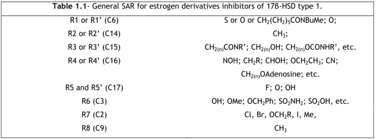
Table 1.1 General SAR for estrogen derivatives inhibitors of 17β-HSD type 1. 12
Table 1.2 Grid Parameters 20
Table 1.3 Results obtained for estrone derivatives 35
Table 1.4 Results obtained for estrone derivatives 36
Table 1.5 Results obtained for estrone D-lactonized ring derivatives 36
Table 1.6 Results obtained for estrone D-lactonized ring derivatives 37
Table 1.7 Results obtained for compounds 6 and 16 37
Table 1.8 Results obtained for docking of some D-homo derivatives 38
Table 1.9 Results obtained for estrone derivatives C3-substituted 38
Table 1.10 In silico toxicity results. N (drug conform) Y (undesired effect –
intermediate state)
39
Capítulo II
Lista de Acrónimos
Capítulo I
17β-HSD-1 17β- Hydroxysteroid Dehydrogenase type 1
3α-HSD 3α-Hydroxysteroid Dehydrogenase
3D Three-dimensional
ADMET Absorption, distribution, metabolism and excretion-toxicity
AKR Aldo-keto-reductase
AOM 5-α-androstan-3β
cLogP Calculated LogP
DHEA Dehydroepiandrosterone
DNA Deoxyribonucleic acid
EA Ethyl acetate
eq Equivalents
ER Estrogen Receptor
HTS High-Throughput Screening
ILS Iterated Local Search
MCPBA m-chloroperoxybenzoic acid or 3-chloroperoxybenzoic acid
MM2 Molecular Mechanics
mRNA Messenger Ribonucleic Acid
NADPH Nicotinamide adenine dinucleotide phosphate
NMR Nuclear Magnetic Resonance
OOP Out of plane
PDB Protein Data Bank
PE Petroleum Ether
Ph Phenyl
QSAR Quantitative structure-activity relationship
RMS Root mean square
SAR Structure-activity relationship
SDR Short-chain dehydrogenase reductase
THF Tetrahydrofurane
TLC Thin-layer chromatography
VS Virtual Screening
Capítulo II
ADME Assistência na Doença aos Militares do Exercito
ANF Associação Nacional de Farmácias
CEDIME Centro de Informação sobre Medicamentos
DCI Denominação Comum Internacional
DGF Direção Geral de Farmácia
FP Farmacopeia Portuguesa
IRC Imposto de Rendimento de pessoas Coletivas
IRS Imposto de Rendimento de pessoas Singulares
IVA Imposto sobre o Valor Acrescentado
MNSRM Medicamento Não Sujeito a Receita Médica
MSRM Medicamento Sujeito a Receita Médica
OTC Over-The-Counter
PIC Preço impresso na embalagem
PMA Preço Máximo Autorizado
PVF Preço de Venda à Farmácia
PVP Preço de Venda ao Público
SBC Sindicato de Bancários do Centro
Chapter I - Discovery and development
of 17β-HSD-1 inhibitors potentially
useful in breast cancer treatment
1.1 Introduction
1.1.1 Breast cancer and 17β-HSD-1
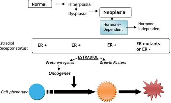
Breast cancer is one of the most common cancer diagnosed among women, and the third cause of death among them. 1,2,3 In Portugal, each year are detected about 4500 new cases of breast cancer, about 1500 women dying because of it.1 A high percentage of this cancer is hormone dependent - around 40 to 60% of all breast cancers and 75% of postmenopausal cases are hormone dependent, and have Estrogen Receptor (ER)-positive status, relying especially on estrogens for its growth, development and proliferation. For this reason, a good treatment approach may possibly depend on lowering estrogen levels in cancer tissues rather than in the hole organism. Breast cells suffer evolutions from the beginning of the formation of the tumor involving, for example, its ER status (Figure 1.1)3,4. In fact, a cancer that is hormone-dependent has a good prognosis concerning disease evolution and treatment, than one that is hormone-independent and has ER-mutated or non-functional. These non-functional receptors can explain why some women who are diagnosed having ER-positive tumors fail to respond to anti-estrogen therapy 3.
Figure 1.1 – The evolution of a breast cell from normal to the neoplastic phase. Adapted from Jorge R. Pasqualini et. al.3
Normal
Neoplasia
Hiperplasia Dysplasia Hormone-Dependent Hormone-Independent EstradiolReceptor status: ER + - ER + + ER + + ER mutants or ER -
ESTRADIOL
Proto-oncogenes Growth Factors
Oncogenes
Estradiol is a natural hormone present in the human body that presents the highest estrogenic activity – it is a ligand of the ER, and plays a critical role in the development of estrogen-dependent pathologies, including, besides breast cancer, a condition called endometriosis5,6. Meanwhile, estrone is an estrogen with less estrogenic activity, not directly related to the pathologies mentioned before 5.The binding of estradiol to the estrogen receptor triggers the formation of a complex that will interact with DNA, promoting the induction of biological responses at a cellular level, including cellular proliferation or protein synthesis (Figure 1.1). In breast carcinoma tissues, local estrogens are originated through two main pathways: one involves aromatase that functions as an androgen-estrogen converter and other uses steroid sulfatase, which converts estrone-3-sulphate (without estrogenic activity) to estrone 5,7,8. In this context, 17β-hydroxysteroid dehydrogenase type 1 (17β-HSD-1) is an enzyme involved in the last step in the synthesis of estrogens, being responsible for the final conversion of estrone, originated mainly by estrone sulfatase and also by aromatase, into estradiol (Figure
1.2)8,9. This enzyme is also responsible for other reductive conversions, like the transformation of androstenedione into testosterone and of DHEA into 5-diol, corresponding only to less than 10% of its total activity 10. This enzyme is not only overexpressed in breast cancer tissues, presenting high activity, but also has been found abundantly expressed in the granulosa cells of ovary, syncytiotrophoblasts of the placenta, liver and endometrium tissues, as well as in their tumors and in prostate tumors 5,11,12,13,14,15. In addition, it is also found in skin and fat tissue. It should be noted that estrogens, especially estradiol, in these tissues, have activities in the same cells where they are formed, acting in an intracrine way5,3. In almost 50% of breast cancer tissues and endometrial cancer cell lines, overexpression of 17β-HSD-1 mRNA has been reported, and also has been characterized as a prognostic marker for disease progression5,8.
Thus, suppressing the activity of this enzyme would result on reduced levels of estradiol formed, promoting a tumor regression, and, if we can talk about a forthcoming claim, the cure of this cancers16,25. Therefore, the inhibition of this selective enzyme, 17β-HSD-1, is an attractive approach for the design of new anti-tumor drugs, included in the so called steroidogenesis controllers.8,17 In fact, inhibition won’t probably affect the biosynthesis of other important steroid hormones, and thus the incidence of adverse effects, so common in chemotherapy, will be reduced. Also, the fact of this enzyme is overexpressed in this kind of tumors is interesting, because inhibition would cause a diminution of the estrogens more specifically in those places, instead of in the whole body 6. For these reasons, it is not surprising that this target is being highly studied by several research groups nowadays, and consequently a considerable amount of new promising compounds that can inhibit it have been found recently. Nevertheless, none of these compounds have been progressed to clinical trials11,18. It is crucial that inhibitors need to be designed carefully: since 17β-HSD-1 and 2 may have opposing action (type 2 converts estradiol into estrone), the former inhibitors must be screened for selectivity toward the later14,19.
Considering the importance of this enzyme it is important to know its structure, characteristics and inhibitors developed until now, as we are going to see further on this introduction.
Figure 1.2 – Partial representation of steroidogenesis, and substrates transformed by 17β-HSD type 1. Adapted from D. Poirier.17
1.1.1.1 Current breast cancer therapeutic approaches
There are several available treatment approaches for breast cancer, and those include: surgery (including lumpectomy, partial mastectomy and total mastectomy, and recently, conservative surgery1), sentinel lymph node biopsy followed by surgery, radiation therapy, chemotherapy, hormone therapy and targeted therapy 20. Surgery has a great impact in women life, especially concerning total mastectomy, causing nefarious moral consequences, hard post-surgical pain or loss of mobility of the upper members. Nowadays, surgery generally avoids total mastectomy, using instead less invasive procedures, as partial mastectomy and conservative surgery and is combined with radiotherapy and chemotherapy or/plus hormonal substitution, which has augmented the rate of survival on the women with this cancer. Chemotherapy consists in applying drugs that suppress or blocks cell proliferation, reducing tumor recurrence. The drugs available and approved for breast cancer treatment, as described by National Cancer Institute of USA 20, include:
Alkylating agents: Cyclophosphamide;
Antimetabolites: Methotrexate, fluorouracil and capecitabine (that is metabolized to fluorouracil) and gemcitabine;
aromatase aromatase 1,7 and 12 2 17β-HSD types ER specific effects 2 1 and 5
Topoisomerase II inhibitors: Antraciclines (doxorubicin, epirubicin) DNA intercalating cytotoxics: doxorubicin;
Citotoxics that interfere with tubuline: taxanes (paclitaxel, docetaxel); Tiroquinases inhibitors;
Others: estrogen receptors antagonists (Fulvestrant and toremifene), aromatase inhibitors (anastrozole, exemestane and letrozole), Ixabepilone and Lapatinib Ditosylate 2, 20.
Given the high percentage of hormone dependent breast tumors, composed by hormone-sensitive/dependent cells, hormone therapy is one of the most important approaches on breast cancer. This therapy consists in avoiding that cancer cells have access to estrogens needed for their development which can greatly help to stop or prevent the growth of the tumor 1. ER-positive breast cancers are usually treated with anti-estrogens, like tamoxifen. They bind ER instead of estrogen hormones blocking its effects. The main problem of tamoxifen is that it is not a pure anti-estrogen: depending on the location of ER, it presents estrogenic or estrogenic activity. Therefore, while in breast tissue it presents anti-estrogenic activity, in endometrium it presents anti-estrogenic activity, and then, although long-term adjuvant tamoxifen is beneficial, it can cause endometrium carcinoma 7,20,21. For this reason nowadays researchers try to find pure anti-estrogens, or other selective drugs25. These substances have, as the ones included on chemotherapy, systemic activity, affecting all cells of the organism 1.
We focus our attention on 17β-HSD-1 as being part of a development of a novel kind of treatment, called steroidogenesis control, consisting on the control of the production of endogen estrogenic compounds, such as estradiol. 17β-HSD-1 is not the only target, as we can find other enzymes involved on steroidogenesis, like other types of 17β-HDSs, 3α-HSD, 5α-reductase, aromatase and estrone sulfatase 8,22,23. Aromatase inhibitors are clinically available for treatment, and can be used by some postmenopausal women that have hormone-dependent breast cancer 20. As for aromatase and 17β-HSD-1, estrone sulfatase is responsible for maintaining high levels of estradiol in tumor cells – this enzyme catalyzes the hydrolysis of estrone sulfate to estrone, which is subsequently reduced to estradiol by 17β-HSD-1. The plasmatic concentration of estrone sulfate is greatly higher in women with breast cancer8.
1.1.2 17B-HSD-1
1.1.2.1 Function and structure
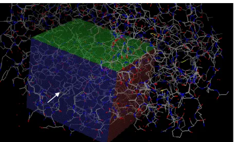
The human 17β-HSD-1 is a cytoplasmic reductive enzyme of the short-chain dehydrogenase/ (SDR) reductase and aldo-keto-reductase (AKR) (the only one being 17β-HSD-5) super-families (Figure 1.3). This enzyme catalyzes the stereo-specific oxido-reduction reactions of alcohols to carbonyls and, to date, 14 human 17β-HSDs have been identified 5,24,26. It is an homodimer, each subunit consisting of 327 amino acids, with a molecular weight between 34.5 and 34.9kDa, and it is dependent on NADH or NADPH as a cofactor 7,24,26,27. As seen before, it is a
reductive enzyme, 90% of its activity being responsible for the conversion of estrone into 17β-estradiol.
Figure 1.3 - 17β-HSD type 1 complexing with 3β-diol(cyan). Notice the cofactor NADPH (magenta) near the molecule and the presence of water molecules (red dots) on the binding pocket. This image was created with PyMol, from the crystal PDB# 3KM0, used in our work to perform the docking studies.
For some authors, the binding pocket of this enzyme might be a catalytic tetrad of Asn114, Ser142,Tyr155 and Lys159 26,28. Others consider that the tetrad sequence is composed by Tyr– X–X–X–Lys and a Ser residue, common to the dehydrogenases 15,25,27. For another’s it can be divided into three regions: the first recognizes the steroid phenolic A-ring and contains His221 and Glu282, which could form hydrogen bonds with O3 of the steroid: the second region binds to the central hydrophobic core of the steroid and contributes to the main thermodynamic force favoring the binding of substrates, and the third region is the catalytic region, that surrounds the D-ring and catalyzes the reduction of the carbonyl moiety to the hydroxyl present on estradiol, and contains Ser142, Tyr155 and Lys159 5.
1.1.2.2 Mechanism of action
The proposed mechanism of action is described in Figure 1.4. As can be seen
,
an hydrogen from NADPH attacks the C17 of estrone, forming an alcoholate stabilized by Tyr155 and Ser142. Then, Tyr155 transfers an hydrogen to estrone, being further reestablished by Lys159, which, in turn, is re-protonated by a water molecule 5,29,30. Lys159 is also responsible for the stabilization of the dinucleotide NADP+, by interacting with its ribose7.Figure 1.4 - Proposed mechanism of action of 17β-HSD type 1 in complex with estrone. Adapted from M. Neggry and Hartmann et. al. 29. The substrate E1 is represented in blue, the cofactor NADPH in red. The amino acids, which are either involved in the catalysis or are responsible for ligand/cofactor stabilization, are colored in black. The hydrogen bonds are in dashed lines while the proton transfers are highlighted with arrows.
1.1.2.3 Steroidal inhibitors of 17B-HSD-1
The knowledge of crystal structure of this enzyme greatly enhanced the design of several molecules that are promising 17β-HSD-1 inhibitors18. There are several structural classes of 17β-HSD-1 inhibitors and, as a major approach, we can divide the inhibitors or this enzyme as steroidal and non-steroidal 17,26,31. These molecules can also be grouped by their mechanism of action in three classes: the ones that compete with NADPH for it binding site, the ones that compete with the natural substrate, being mostly steroidal compounds, triggering reversible (the majority of the compounds seen below) or irreversible inhibition (mainly caused by substitutions on C16), and a third class of inhibitors, consisted on the so called hybrid compounds, combining moieties from NADPH and estradiol/estrone (this is, parts of both cofactor and substrate)26.
To test their inhibition capacity, compounds are assayed by different methods or strategies, presenting different values of IC50 or percentages of inhibition, depending on the method chosen. Therefore, it isn’t appropriated to directly compare the values obtained for the various compounds tested by different strategies, but one can have an idea of the capacity of inhibition provided by them. For this reason, a positive control must be included as a reference in all assays. The methods usually used to test the capacity of inhibition by the chemicals consist in:
Testing the compounds in homogenated HEK-293 transfected cells overexpressing 17β-HSD-1, in T-47D cells or transfected MCF-7 cells, by calculating of the amount of estrone transformed into estradiol;
Testing the compounds using the cytosolic fraction of human placenta (rich in 17β-HSD-1) by calculating the amount of estrone transformed into estradiol;
Directly measuring the transformation of estradiol into estrone in pure 17β-HSD-1 17. The estrogenicity of the compounds, can be evaluated by the study of proliferative activity on estrogen-sensitive cells, like T-47D, ZR-75-1 or MCF-7 cells 17. It is of remarkable importance to develop compounds with low estrogenic activity, in order to avoid cancer cell proliferation25.
Non-steroidal 17β-HSD-1 inhibitors that are being under study include phytoestrogens, which are most potent inhibitors among the naturally substances, but unfortunately are non-specific, gossypol and related compounds, thiophenepyrimidinone derivatives, 1-substituted 6-phenylnaphthalene derivatives, glycyrrhetinic acid derivatives, phenyl ketones, imidazoles and cinnamic acid ester derivatives, biphenyl mimics of estrone, thiophenepyrimidinones, (hydroxyphenyl) naphthols, bis( hydroxyphenyl) substituted arenes, flavonoids with OH in A-ring and heterocyclic substituted biphenylols 14,17,18,25,26. These compounds share common structural features, such as a phenolic ring and hydrophobic scaffolds, which will interact with the amino-acids present on the binding pocket of 17β-HSD-111. The major disadvantage of some natural phytoestrogen compounds, including flavonoids, is that they generally present unfavorable pharmacokinetic/ ADMET (absorption, distribution, metabolism, and excretion) characteristics and low-specificity due to cross-reactivity with other steroid metabolizing enzymes and steroid hormone receptors19,25.
In the group of steroidal inhibitors, we can make the following division – estrogen derivatives and non-estrogen steroids. Estrone and estradiol derivatives are largely described as 17β-HSD-1 inhibitors, although estrone derivatives present more inhibitory capacity than the estradiol ones, due to the carbonyl at C17 26. Next, general aspects of SAR (Structure Activity Relationship) are presented. Starting by the A ring, we can find:
The C2 substituted estrogens (this substitution is believed to reduce estrogenic activity 26, and also add some inhibition activity, especially with hydrophobic substitutions); generally includes small groups (see below) 17,26.
C3 substituted estrogens; also combined with substitutions on C2 and C15 and others, the moieties found are generally CH3, CH2Ph, SO2NH2 and SO3OH, the sulfamates being weak inhibitors 26; generally, in all inhibitors, the moiety present in C3 is the hydroxyl (OH).
In the B ring, we can find the following substitutions:
C6 substituted: some oxime derivatives and carbonyl moiety; they present some estrogenic activity, that is undesired for the treatment of this kind of cancers; the 6-substitution accompanies 16 and 17 6-substitutions, or can be alone; it was shown that the β configuration is preferable than the α to achieve better inhibition values 17,26.
Figure 1.6 – Examples of C6-substituted estrogens
In the C ring, the substitutions are unusual; however, we can find the follow:
C9 substitutions, described in the α plane, as a methyl group, along with some substitutions in C15, 16, 17 and others 17.
Figure 1.7 – Example of C9-substituted estrogen
D-ring is the most functionalized ring, presenting a variety of substitutions, combined or not with substitutions on the other steroid rings. In this group, another kind of new inhibitors is included: the estradiol hybrid derivatives, linking an adenosine moiety at C16, that projects into the NADPH binding site 5.
Substitutions at C15: there are described large alkyl spacers linked to C15 that bear polar moieties, like an amide, ester, carbonyl, hydrazone, alcohol, ether, urea, carbamate, retroamide, sufonyl urea, sulfamide, sulfamate, retrosulfonamide, retrocarbamate, retroester or a sulfonylcarbamate type side chain; can be found combined with substitutions on C2 and C3, or on the steroids with an E ring 13,17,26,32.
Figure 1.8 – Examples of C15-substituted estrogens
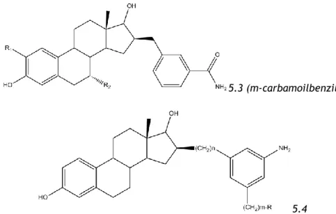
Substitutions on C16: one of the most common substitutions, that includes some irreversible inhibitors: ones with bromine in their composition of the chain, suicidal inhibitors (16-methylene substituted and with an α-hydroxyacetylenyc substitution) and alkylating inhibitors (processing a bromine in the end of an alkyl chain, working as a good leaving group). They are well tolerated, especially β-substitutions, and can present a dual activity: ER antagonists and inhibitors of 17β-HSD-117. We can find small or large substitutions on C16 - the large substitutions can interact with the co-factor (NADPH) at the active site of the enzyme, and the presence of a good leaving group at the end of the chain lead to good inhibition results. We can also find halogenated substitutions on this position (suicidal inhibitors). This group also includes different hybrid compounds that interact with the cofactor and are very potent inhibitors, although reversible, these hybrid structures can have large alkyl spacers (the optimal being 8 methylene groups) bound to C16, and to an the adenosine moiety (5.1), or a partial mimic of adenosine moiety, a meta substituted aniline (the simplified hybrid adenosine compounds(5.4), like m-carbamoylbenzyl or just carbamoyl substituent (5.3) in 16β-position), and other kind of compounds, whose spacers consists in amides linked to sulfamates or phenols (the peptide derivatives) (5.2) 17,26,33,34.
5.1
5.3 (m-carbamoilbenzil)
5.4
Figure 1.9 – Examples of C16-substituted estrogens. The compounds are examples of hybrid inhibitors.
Substitutions at C17: among others, include the 17-fluoro substituted estrogens- the Fluor can mimic the hydroxyl or carbonyl moieties because of it large electro-negativity and hydrogen-bond acceptor capacity, and is found combined with substitutions on C14, C15 and C16, as well as C2, and with double bonds on C8 and C9 26,11. Some substitutions including NO-R moiety are also described for estrogen derivatives 34. 17β-Formamido, 17β-benzamido as well as 17β-amino derivatives can be found in non-estrogenic molecules, and are associated with effective inhibition 17. The most common is to find the carbonyl moiety in C17, that mimics estrone, or the OH moiety, that mimics estradiol.
Figure 1.10 – Examples of C17-substituted compounds.
D-Homo derivatives: A new kind of inhibitors that present a 6-membered D-ring which proved to be very active 14,17,18,26. They are presented along with substitutions on C2 and in other places.
Figure 1.11 – General structure of a D-homo estrogens with substitutions on C2 and C16.
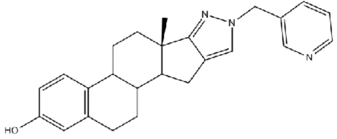
Other inhibitors present a heterocyclic E-ring, with 5 or 6 members, the bests presenting a pyridyl side-chain linked to it (Figure 1.12). 17,26,34
Figure 1.12 – Example of an estrogen with an heterocyclic E-ring, linked to a pyridyl moiety.
Finally, tibolone (a 19-nortestosterone steroid) (Figure 1.13) derivatives, whose metabolites have different binding affinities to estrogen, progesterone and androgen receptors, and enzymes involved in their metabolism.
Figure 1.13 – Tibolone structure.
Table 1.1- General SAR for estrogen derivatives inhibitors of 17β-HSD type 1.
R1 or R1’ (C6) S or O or CH2(CH2)5CONBuMe; O;
R2 or R2’ (C14) CH3;
R3 or R3’ (C15) CH2(n)CONR’; CH2(n)OH; CH2(n)OCONHR’, etc.
R4 or R4’ (C16) NOH; CH2R; CHOH; OCH2CH3; CN;
CH2(n)OAdenosine; etc.
R5 and R5’ (C17) F; O; OH
R6 (C3) OH; OMe; OCH2Ph; SO2NH2; SO2OH, etc.
R7 (C2) Cl, Br, OCH2R, I, Me,
R8 (C9) CH3
Further efforts need to be done, with the aim of reducing or abolishing the estrogen activity of these compounds, this is, the development of compounds with good inhibitory action, but with a low estrogenicity.
The promising compounds D-homo 2-substituted were the basis of our work. Moller et. al. studied on 2009 the importance and excellent results on 17β-HSD-1 inhibition relying in these compounds 18. The importance on 2-substitution is based on the fact that it is oriented to an unoccupied lipophilic sub-pocket, consisting of the side-chains of Val143, Met147, Phe259, Leu262 and Met279. In addition, 2-substitutions lower the intrinsic estrogenicity of the molecule. These molecules presented impressing results, with percentages of inhibition varying from 86 to 100% at 2µM, and IC50 in the nanomolar values (from 15nm to 126nm). Their homologues with a 5 membered D-ring, have inhibitions between 70 to 99% at 2µM, and IC50 from 35nm to 553nm. Basically, inserting a 6-membered D-ring on the estrogens, significantly improved the inhibitory properties and consequently, a lower amount of substance needed to totally inhibit the enzyme (lower IC50). In the synthesis, it was observed that the transformation of a 5-membered D-ring into a D-homo estrogen presented by the authors requires a complex synthesis pathway, with three steps and an unfavorable total yield. Our idea relies on building a 6-membered D-ring by a single step – the lactonization of D-ring by a Baeyer-Villiger (BV) reaction in 2-substituted estrogens (Figure 1.14). A lactone ring may, however, present a different activity compared to a D-Homo ring, due to the electronic density associated to the two oxygens. The lactones that we proposed to test and synthesize are called Westerfeld lactones, while are available a variant of this lactone, the Jacobsen’s lactone 37.
Figure 1.14 – Comparison of D-homo estrone with the D-lactonized estrone (Westerfeld’s lactone - estrololactone) respectively.
1.1.3 Drug design and discovery – the importance of computational
methods: molecular docking and in silico toxicity studies
By the last 30 years, drug development and research has grown and developed, partially thanks to the introduction of new computational methodologies in the field of chemistry 38. The so called chemoinformatics is a term introduced on 1998 by Dr. Frank K. Brown, and described by him as: “The use of information technology and management has become a critical part of the drug discovery process. Chemoinformatics is the mixing of those information resources to transform data into information, and information into knowledge for the intended purpose of making better decisions faster in the area of drug lead identification and organization”39,40.
With all the advances in the field of chemoinformatics being so important in the knowledge and global share of the information about several biological molecules and processes, serendipism is playing a less important role nowadays on drug discovery, although some examples may break this rule (e.g.: the discovery of sildenafil). The advance in the field of crystallography and NMR (Nuclear Magnetic Resonance) in molecular biology, and the creation of the Protein Data Bank (PDB), is very important to the success of some methodologies around the chemoinformatics. In fact, in PDB, thousands of proteins, most being involved and playing a major rule in pathologies, can be found and are accessible to anyone, especially for use in the docking methods, where a file of a macromolecule is needed to perform the tests38,39,40.
1.1.3.1 (Q)SARs and in silico studies
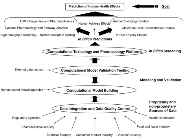
The basis of chemoinformatics relies on predicting the activity for some molecule: knowing which particular effects brings to a particular biologic system, we can then attempt to know and form a quantitative relationship between the biological activity and the structure of the chemicals – this leads us to the term QSAR – quantitative structure-activity relationship. QSAR quantizes features of the new chemical structure so that overall properties of the compound can be predicted based on the relationship between structure and activity computed using the knowledge derived from a training set. Together with SAR (which doesn’t accomplish the quantitative but only involves qualitative information) we can refer about (Q)SARs, which is involved in a series of techniques known as in silico approaches39. It is being applied on drug discovery including in toxicity prediction, risk assessment, lead optimization, prediction of oral bioavailability, blood brain barrier crossing, pharmacological target prediction, and even in other fields. These approaches are becoming relatively important in the pharmaceutical industry, especially identifying lead compounds for further tests relying in drug discovery 10,41,. The term in silico toxicology generally refers to a computational experiment, mathematical calculation, or scientific analysis of substances and organization of substance related data to assist in predictive toxicological and pharmacological profiling of chemical substances for understanding drug safety liabilities 41. All these in silico approaches work as
predictive or filtering tools. However, they need to be confirmed by experimental testing. (Figure 1.15).
Figure 1.15 – Workflow for the use of drug and chemical toxicity databases and models; starting from the source of data to the goal of predicting human health effects. Adapted from Luis G. Valerio Jr.41
1.1.3.2 Molecular Docking
In computational methods in drug discovery and development, we can find the Molecular Docking (referred here as Docking) method39. Docking consists on the search for spatial arrangements that fit two molecules together in energetically favorable configurations, and enables the prediction of the binding free energy of the complex (the binding affinity).Docking, thus, is ultimately interested in reproducing chemical potentials, which determine the bound conformation preference and the free energy of binding, as well as giving a three dimensional (3D) structure of the protein and the ligand molecule 38,42. Docking calculations explores enthalpic and entropic contributions, components of molecular interactions that lead to the final free energy of binding (ΔG). Enthalpic contributions includes electrostatic interactions (ionic bonds, charge-dipole and dipole-dipole interactions, (inductive interactions provided by intra-molecular contributions from the complex formed), dispersion forces (responsible for attractive interactions between non-polar molecule) and hydrogen bonds) and steric interactions (short-range repulsive forces), while entropic contributions are composed by translational and rotational energy (which means an
interference in the degrees of freedom of the ligand after the formation of the ligand-receptor complex), hydrophobic effect and solvent reorganization.43
Virtual Screening (VS) is a term directly connected to the Docking method. In contrast to High Throughput Screening (HTS), which consists in the search for hundreds or thousands of previously synthesized compounds of interest that bind to a target molecule in real conditions, VS performs this search by computational means, recurring to Docking, suggesting then computer hits 38. A great approach and use of VS is to perform a filter to a series of compounds, providing those who hit the target to be further synthesized and further evaluated by HTS assay. This attitude links to the concept of Green Chemistry, allowing less materials being wasted on the synthesis of compounds that were unlikely to have the wanted activity. Also, VS is a cheap and fast methodology 38,44.
To perform docking, usually the 3D structure of the target needs to be known and available by crystallography or NMR 38,44. This reveals a difficulty for some targets, as G-protein-coupled receptors, for which remains impossible, or remarkably difficult to crystalize or obtain structural information 38,42. Nevertheless, docking is, as described above, a very important methodology in drug design, as it can direct experimental efforts based on ligand-receptor binging predictions. In fact, a great number of drugs have been developed using docking methods at some stage of development. Thus, with the new advances on computer technology and the increase of the number of receptor structures in PDB and algorithm development allowing the creation of more accurate docking methods, evolving flexible ligands to complex with uncomplexed receptor structures, the future will promise that docking will be involved in even more drug design processes 42.
1.1.3.2.1 Docking software
In our work the docking software that has been used is the AutoDock Vina, an automated docking tool developed at The Scripps Research Institute, Molecular Graphics Laboratory, USA35. This software only performs docking of one single molecule at time, thus, it cannot perform VS tests. Unlike AutoDock 4.0 (the version available before the development of Vina), who uses as a docking method a genetic algorithm, to explore the full range of conformational flexibility and the rotational flexibility of selected receptor hydrogens, Vina uses an Iterated Local Search global optimizer 35,38,45. Iterated local search (ILS) is a method that generates a sequence of solutions generated by an embedded heuristic, leading to far better results than if one were to use repeated random trials of that heuristic. In this algorithm, a succession of steps consisting of a mutation and a local optimization are taken, with each step being accepted according to the Metropolis criterion. Mutation, or active site mutagenesis, is a measure of intrinsic binding energy based on the anchor principle, where the impact of a single amino acid substitution in the active site is measured in terms of catalytic efficiency or inhibitor binding46. The algorithm can be improved by iterations (the act of repeating a process usually with the aim of approaching a desired goal or target or
result) 46. That’s one of the functions that the user can decide: the higher the number of iterations, the longer is the search, but the results given are more accurate. Nevertheless, this new version gives more accurate and quick results comparing to the old version of AutoDock. Both treat the receptors as rigid, and the ligands as flexible molecules – this is remarkably important, given that the conformation a ligand adopts in solution is not necessarily the one it will adopt when bound to the target 38. Necessary to work, Vina requires a readable molecule and macromolecule saved in *.pdbqt file format, derived from *.pdb format with additional information: it stores the atomic coordinates, partial charges and AutoDock atom types47, and also information about the spatial search, thus requiring a grid box special coordinates, as we will describe further.
We can find different docking software, each one accomplishing specific docking and scoring methods, also known as docking and scoring functions, and different functionalities. Some of those are able to perform VS to hundreds or thousands of molecules presented. Respecting to the Scoring Function, it must obey to the thermodynamic and kinetic requirement – the complex must be the global minimum of the binding energy landscape, but at the same time this conformation must be accessible during the search 38. It is used to estimate the energy of a given protein-ligand arrangement, attempting to approximate the standard chemical potentials of the system, and it is the most important feature of a docking program 38,48.
1.2 Objectives
In this work, the objectives proposed were: to perform docking studies with AutoDock Vina to some estrogen derivatives into the macromolecule 17β-HSD-1, with the aim of finding compounds with high affinity to this enzyme, and also perform in silico toxicological studies with these molecules; to develop and synthesize some estrogen derivatives with special attention to the 2-substituted estrone derivatives and its lactonized D-ring homologues to be further evaluated in in vitro inhibitory assays.
We then expect that estrone derivatives with the 6-membered lactonized D-ring present higher affinity values in docking studies than the ones with a 5-membered D-ring. Also, we will confront the docking affinity studies and in silico toxicological studies of these lactonized compounds to their D-Homo homologues in order to find similarities or discrepancies on the results.
1.3 Materials and Methods
1.3.1 Computational studies
As previously referred, the docking experiment approached here is a semi-flexible docking, where the macromolecule was treated as a rigid body, while the ligands were treated like flexible bodies.
1.3.1.1 Software
The software used for the creation of the 3D (three-dimensional) molecules that would work as ligands in the docking studies was Chem3D v 12.0 software (Cambridge ChemBioOffice 2010).
For the preparation of the ligands (choose torsions and detecting aromatic carbons), macromolecule and construction of the Grid box, was used AutoDock Tools v 4.2 in MGM tools from The Scripps Research Institute.
The software used for adding the hydrogens to macromolecule, and to visualize the docking results including polar contacts was PyMol (The PyMol Molecular Graphics System) v 1.3 built to educational use.
The docking software used for the calculations was AutoDock Vina 35. It is a free new software as related before in our work, and it’s been used by some authors to perform docking searches nowadays.49,50
1.3.1.2 Preparation of the Ligands for molecular Docking: 3D molecular
structures, minimizing energy and file formats
3D structures of the molecules used in docking studies were constructed with the aim of Chem 3D, where the optimal conformational state was achieved with a modified version of Allinger’s MM2 force field. Molecular mechanics (force field) is a method for the calculation of molecular structures, conformational energies and other molecular properties, using concepts from classical mechanics35,53. It thus includes computation of Bond Stretching Energy, Angle Bending Energy, Torsion Energy, Non-Bonded Energy, Van Der Waals Energy (including cutoff parameters to improve the computational speed of calculations), Electrostatic Energy, charge/charge, charge/dipole and dipole/dipole contribution, Out of Plane (OOP) Bending, Pi Bonds and Atoms with Pi Bonds and Stretch-Bend Cross Terms43. From a pre-established optimized steroid nucleus geometry, the MM2 jobs were run until reached an gradient norm lesser than the minimum gradient norm (Root Mean Square [RMS] Gradient set of 0.010). The results of MM2 calculations were given by Total Energy in kcal/mol, and the molecules saved in *.pdb format with the geometry acquired by this calculations.
Additional conversion of the *.pdb format to *.pdbqt format was achieved in AutoDock Tools. The later provides the storage of the atomic coordinates, partial charges and AutoDock atom
types for further use on docking calculations by AutoDock Vina. This format also stores information about aromaticity and rotatable branches, which can be set on AutoDock Tools, if not performed automatically.
1.3.1.3 Choosing and preparing the macromolecule for docking experiments
Until 2010 there have been crystallized by X-Ray Diffraction several structures of 17β-HSD-1, which are available on Protein Data Bank (PDB) website 45. However, to choose the adequate structure which will lead to a reliable computational study, we have to consider some important information such as the crystal resolution, source of the enzyme, presence of water molecules (directly influenced by the resolution) and the ligands or co-factors crystallized within the macromolecule. The molecule chosen was proposed by M. Mazumdar et al. (PDB code of 3KM0), and comprises the 17β-HSD-1 in complex with 5-α-androstane-3-β, 17β-diol and NADPH, isolated from human placenta with a resolution of 2.30Å. This is the most recent crystal of 17β-HSD-1 added to PDB database until now (12-06-2012), and the fact of having an human origin is relevant for our studies.After choosing the most appropriate macromolecule, it was relevant to prepare it for the docking studies: add the Hydrogen atoms to residues and water molecules (performed with PyMol), remove the ligand that was co-crystallized with the macromolecule and define the Histidine charges to match the human physiologic environment (monoprotonated state38). The former is a very important step, as the crystal lacks all the hydrogen atoms. Also, the water molecules are very important to establish interactions between the ligand and the residues on the binding pocket, as they can turn some hydrogen acceptors residues to work as hydrogen donors, and the reverse38. So, regarding the importance of these moieties, the water molecules existent in the crystal, especially the ones near the pocket, were allowed to stay, giving higher affinity values. Before removing the ligand, we can take in account a precious information: the binding pocket location – crucial to the generation of the grid-box. Finally, AutoDock Tools performed corrections on amino-acids and global charges (gasteiger and Kollman charges), and merged the non-polar hydrogens. We allowed NADPH to stay, in order to better mimic the natural environment of the enzyme.
1.3.1.4 Grid-Box
The grid box is a way to identify the coordinates and spatial location of the binding pocket. The software AutoDock Tools generates a three-dimensional box within the macromolecule, which needs to be resized and placed over the desired pocket. Therefore, the presence of the ligand co-crystallized with the macromolecule in the pocket is very helpful for the accurate identification of the pocket. The software provides the values of space location of the center of the box and the box size (number of points in each dimension). Dimensioning the grid box to the approximate size of the pocket has only the purpose of reducing the time of calculation and number of iterations, thus assembling the computations more quickly and
accurately. The spacing of 1Å equals the distance of a C-C bond, and provides a bigger grid box without the need of too many points in each dimension. The information acquired after generating the grid box, is written down on the configuration parameters of the software AutoDock Vina for the macromolecule in use, so that the software knows in where in the molecule to perform the docking calculations.
Grid Box for 3KM0.pdbqt:
Figure 1.16 - Grid Box for 3KMO.pdb. This image was created with AutoDock Tools. Arrow indicates the location of the ligand co-crystallized on the binding pocket. Below is the information about spatial location of the gridbox.
Table 1.2. Grid parameters Spacing: 1Å
Dimension: Number of points in dimension: Center: Offset:
X 25 21.185 2.889
Y 20 -2.672 -3.583
Z 20 - 11.932 12.025
1.3.1.5 Testing the accuracy of docking experiments
To know if the software is performing accurate docking experiments there is a very simple but reliable test that consists in docking the ligand crystallized with the molecule51,52. If the docked molecule lies superimposed with the crystallized molecule, with no more than 2Å distance, the software is accomplishing good docking calculations 38,51,52. In this case, AOM (5-α-androstane-3-β) was identified and isolated from 3KM0, saved as a ligand with *.pdbqt format in AutoDock Tools, and then subject to a docking computation. Superimposing the results, one find that the best match lies on the fourth best affinity mode, with distances ranging from 1Å to 1.8Å. In the best affinity mode proposed by AutoDock Vina, the atoms that match have distances ranging from 3.4Å to 11Å.
Figure 1.17 - Superimpose of the docked ligand (cyan) and the crystalized ligand (green) in the binding pocket in affinity mode 4. This image was created with PyMol.
Nevertheless, the molecule fits in the same place where the ligand lies, in any affinity mode. As we can’t predict which docking affinity mode will match the real interaction mode, we will assume an average of the first four affinity values given by the software in our results, presenting also the best affinity mode value. The result presented by the fourth affinity mode proves us that the software is reliable on the docking results. It is important to mention, that in the majority of the docked results, the first affinity result presents the interactions with the molecule and adjacent amino-acids suggested by numerous authors as discussed before in the introduction: these include the interactions with Tyr155 and Ser142 with the carbonyl at C17 of estrone and its derivatives, and interaction with 3-OH and Hys221.
1.3.1.6 Docking results
The exhaustiveness selected of the calculations was the default (exhaustiveness=8). Results of docking in AutoDock Vina are presented in free energy of binding (ΔG) in kcal/mol, and clustered using a RMSD (root mean square deviation) cutoff value.RMSD values are calculated relative to the best mode and use only movable heavy atoms. Two variants of RMSD metrics are provided, rmsd/lb (RMSD lower bound) and rmsd/ub (RMSD upper bound), differing in how the atoms are matched in the distance calculation – the best configuration mode possesses rmsd/lb and rmsd/ub = 0. 47,53,58
As related by the developers of the software, the predicted free energy of binding is calculated from the intermolecular part of the lowest-scoring conformation, designated as 1, where the function g can be an arbitrary strictly increasing smooth possibly nonlinear function (1). C represents the contributions of intra-molecular or intermolecular interactions 47,48.
s1 = g(c1 − cintra1) = g(cinter1) (1)
The output values are given in order to the best configuration mode, and the distance between the best configuration mode are presented in armstrongs (Å) to preserve the ranking. Free energy of binding of the other low-scoring conformations uses cintra of the best binding mode:
1.3.1.7 In silico toxicity studies
The in silico toxicity studies were performed with the tools provided at The Organic Chemistry Portal (www.organic-chemistry.org) with OSIRIS Property Explorer. Molecules were designed directly on the program and then the values observed.
1.3.2 Materials used on chemical synthesis
1.3.2.1 Reagents and solvents
All synthesis were performed with reagents and solvents used as received. Estrone, used as substrate on the majority of the following reactions, as well as MCPBA (m-chloroperoxybenzoic acid), sodium bicarbonate (NaHCO3) and anhydrous sodium sulfate were purchased from Sigma-Aldrich. Sodium iodide (NaI) was acquired to Applichem, methanol was purchased to Normapur, glacial acetic acid and sodium chloride were purchased to Panreac, chloridric acid 37% (HCl) was acquired to Merck, sodium thiosulphate (Na2S2O2) was purchased to Fluka, Cooper(II) chloride di-hydrate was acquired to Riedel-de-Haën and nitric acid (HNO3) was purchased to Probus, S.A. Badalona.
All other solvents used were of adequate grade for chemical synthesis and purification: petroleum ether (PE), ethyl acetate (EA), diethyl ether and dichloromethane were acquired to Fisher Chemicals.
1.3.2.2 Chromatography
All chemical reactions were controlled by TLC (Thin Layer Chromatography), using commercial silica gel plates (silica gel 60 with a fluorescent indicator UV254) purchased to Mecrk/Macherey-Nagel. After the application of the compounds in the plates, elution was performed with an appropriate mixture of eluents in different proportions, as demanded by the kind of product/substrate, and then the plates were observed under UV light (254nm). They were posteriorly revealed by a solution of ethanol/sulphuric acid (95:5), followed by heating at 120ºC.
Column chromatography, in order to isolate and purify the products obtained on each reaction, were performed using Silica-gel 230-400 mesh, purchased from Merck, the eluent being described further for each reaction.
1.3.2.3 Equipment and other devices
The solvents were evaporated by using a rotary vacuum drier, from Heidolph. The UV254 revelator was model CN-15.LC.
RMN spectra were obtained by Bruker Avance III 600 spectrometer, and registered at 600 MHz for 1H RMN and at 151 MHz for 13C RMN, using CDCl