Joanna Melissa Ribeiro Gonçalves
Assessing Cadmium-based Quantum
Dots
Effects on the Gonads of the Marine
Mussel Mytilus galloprovincialis
UNIVERSIDADE DO ALGARVE
FACULDADE DE CIÊNCIAS E TECNOLOGIA
2017
Joanna Melissa Ribeiro Gonçalves
Assessing Cadmium-based Quantum
Dots
Effects on the Gonads of the Marine
Mussel Mytilus galloprovincialis
Mestrado em Biologia Marinha
(Especialidade em Ecotoxicologia)
Trabalho efetuado sobre a orientação da:
Professora Doutora Maria João da Anunciação Franco Bebianno
Doutora Nélia C. Costa Mestre
UNIVERSIDADE DO ALGARVE
FACULDADE DE CIÊNCIAS E TECNOLOGIA
2017
Assessing Cadmium-based QuantumDots Effects on the Gonads of the Marine Mussel Mytilus galloprovincialis
Declaração de autoria de trabalho
Declaro ser a autora deste trabalho, que é original e inédito. Autores e trabalhos consultados estão devidamente citados no texto e constam da listagem de referências incluída.
---
©Joanna Melissa Ribeiro Gonçalves
A Universidade do Algarve reserva para si o direito, em conformidade com o disposto no Código do Direito de Autor e dos Direitos Conexos, de arquivar, reproduzir e publicar a obra, independentemente do meio utilizado, bem como de a divulgar através de repositórios científicos e de admitir a sua cópia e distribuição para fins meramente educacionais ou de investigação e não comerciais, conquanto seja dado o devido crédito ao autor e editor respetivos
i
AGRADECIMENTOS
Em primeiro lugar gostaria de agradecer à Professora Maria João Bebianno por esta oportunidade de trabalhar na área que mais me interessa dentro de Biologia Marinha, pela experiência e os conhecimentos transmitidos, e a cima de tudo, pela a sua
compreensão dos meus horários devido ao trabalho, disponibilidade e orientação ao longo deste trabalho.
A seguir gostaria de agradecer ao Thiago Rocha por me ter deixado utilizar as amostras da sua tese de doutoramento e por todo o apoio que me deu mesmo estando do outro lado do mundo.
Gostaria de agradecer às pessoas que me acolheram e que tiveram a disponibilidade e interesse em transmitir me conhecimento. Um grande obrigado à Nélia Mestre e à Tainá Fonseca, por toda a disponibilidade, por toda a ajuda e por todos os conselhos que me deram ao longo deste ano.
Queria também agradecer ao Paulo Pedro pela ajuda prestada nas análises químicas. Um grande obrigado aos meus amigos, Beatriz Palhinhos, Rita Ferreira, João Pontes e Zé Patrício por toda a ajuda prestada no laboratório.
Aos meus pais, Victor e Celeste Gonçalves, pela a força e encorajamento ao longo da minha vida académica. Sem o vosso apoio, a todos os níveis, não teria chegado até aqui!
Por fim, gostaria de agradecer ao meu noivo, Antero Martins, por ter aturado todo o meu stress durante este ano, por me acalmar nos momentos que as coisas corriam menos bem, e por fim, por acreditar sempre em mim e nas minhas capacidades.
ii
ABSTRACT
Nanotechnology is found in many aspects of modern life, and its applications are continuously increasing. Engineered nanomaterials (ENMs) have helped improve the quality of life as well as economic growth. Quantum dots (QDs) are ENMs with unique optical and biofunctional properties, known to be useful in nanomedicine, biology and electronics. Of emerging concern is the fate of these ENMs, as they can enter the marine environment and prompt short-term and long-term effects on marine organisms. Tissue-specific responses in mussels have been studied, however knowledge on the effect of these ENMs in the gonads and possible effects on gametogenesis is scarce. The aim of this study was to assess the ecotoxicity of CdTe QDs (2 – 7 nm) in the gonads of the marine mussel Mytilus galloprovincialis, in comparison with its dissolved counterpart. Mussels were exposed to CdTe QDs and dissolved Cd for 14 days at 10µgCd.L-1. Cd accumulation, oxidative stress (superoxide dismutase - SOD, catalase -
CAT, glutathione peroxidase - GPx, glutathione S-transferase - GST) and oxidative damage (Lipid peroxidation – LPO), were analysed in the gonads looking for a distinction between male and female responses.
Results show that both Cd forms caused antioxidant responses to change in both male and female gonads, wherein QDs are more pro-oxidant when compared to dissolved Cd. In male mussels, QDs induced higher Cd accumulation than its dissolved counterpart, whilst both Cd forms showed high Cd accumulation in females. SOD, CAT, GPx and GST activities, in both male and female mussels, decreased after exposure to QDs, wherein females are the main sex affected. Females undergo oxidative damage after three days of exposure to QDs, shown by an 18.6-fold increase in LPO levels. Males also undergo oxidative damage due to exposure to QDs, with a gradual increase in LPO levels throughout exposure period.
Overall, QDs mediated toxicity is time and sex-dependent, wherein QDs effect on females is prominent after a short period of exposure, whilst in males, QDs toxicity is more noticeable after a longer time of exposure. Results predominately show that females are the sex most affected by CdTe QDs. Female gonads have twice as much lipid content compared to males, and these lipid contents are important sources of energy
iii
during gametogenesis and for embryo and larval development. Therefore, there is a rising concern on how gametes are affected and the potential changes at the cellular level that they may undergo, making it crucial to understand the effects of ENMs on gametogenesis, fertilization success, embryogenesis and larval development, as these may have serious impacts to population sustainability and ecosystem health.
Keywords: Engineered Nanomaterials, Cadmium Telluride (CdTe) Quantum dots, Mytilus
iv
RESUMO
A nanotecnologia trata da manipulação da matéria a nível atómico e molecular sendo que, atualmente as suas aplicações são diversas e contribuem para o melhoramento da qualidade de vida e a tendência para o aparecimento de novas aplicações revela ser promissor. Os nanomateriais manufaturados (ENMs) são componentes utilizados em produtos consumíveis, em tecnologia informática, equipamentos químicos e em medicina. Dentro dos ENMs existem os pontos quânticos (“quantum dots” – QDs) que são nanocristais semicondutores com propriedades óticas e biofuncionais, úteis para as mais variadas áreas como a medicina, a biologia ou a eletrónica. A nível da estrutura geral, um ponto quântico é constituído por um núcleo inorgânico com um material semicondutor (e.g. CdS ou CdTe), um invólucro inorgânico com um material semicondutor (e.g. ZnS) e com um gap de energia distante, à superfície é constituído por um revestimento orgânico aquoso (e.g. -ROOH). O invólucro dos QDs protege o núcleo de fenómenos como a oxidação ou a degradação, enquanto os ligandos à superfície aumentam a solubilidade e compatibilidade na água. As características abióticas do ambiente aquático tais como o pH, a força iónica, as radiações eletromagnéticas e UV, promovem diferentes transformações físico-químicas na superfície dos QDs, alterando o seu comportamento e o seu destino no meio aquático. Contudo, tem havido uma maior preocupação sobre o destino desses ENMs uma vez que poderão acabar no oceano e provocar efeitos, a curto e longo prazo, indesejáveis para os organismos marinhos sendo importante garantir que o potencial efeito tóxico seja adequadamente estudado e compreendido.
Os mexilhões são organismos sésseis e filtradores, com a capacidade de acumular contaminantes presentes no meio ambiente dentro dos seus tecidos, e são, portanto, reconhecidos como excelentes organismos para a biomonitorização. Os contaminantes que se acumulam nos tecidos podem causar stress e alterações na própria fisiologia dos mexilhões, fazendo com que seja possível a medição de parâmetros de avaliação do risco ambiental dos poluentes. Os mexilhões filtram água através das brânquias sendo as partículas comestíveis transportadas para o trato digestivo. A presença de contaminantes nestas partículas pode significar uma redistribuição diferente nos tecidos, incluindo nas gónadas.
v
A gametogénese é o processo de desenvolvimento das gónadas para produção de gâmetas, gâmetas esses libertados quando maduros na coluna de água onde ocorre a fertilização. Se por ventura, o desenvolvimento das gónadas for afetado pela presença de contaminantes acumulados nas gónadas, o sucesso da fertilização pode ser comprometido. Os contaminantes poderão ser responsáveis por afetar o desenvolvimento larvar e limitar a capacidade da população ser capaz de produzir uma geração futura viável, tendo impactos graves para todo o ecossistema.
O stress oxidativo ocorre quando um organismo se encontra num desequilíbrio de fatores pró-oxidantes e mecanismos antioxidantes devido à presença de radicais livres de oxigénio e outras espécies reativas de oxigénio (ROS). No ambiente aquático, os QDs têm a capacidade de gerar ROS, estando o tamanho do QD relacionado com a quantidade de ROS produzida. Para a manutenção da integridade celular de um organismo, a aniquilação de ROS é de grande importância. As enzimas antioxidantes como a superóxido dismutase (SOD), a catalase (CAT), a glutationa peroxidase (GPx) e a glutationa-S-transferase (GSTs), participam na remoção de ROS produzido. A SOD é responsável pela primeira linha de defesa antioxidante de um organismo, que catalisa o radical superóxido (·O2-) em peróxido de hidrogénio (H2O2) e oxigénio (O2). H2O2 que é
eliminado pela CAT, transformando H2O2 em O2 e H2O. A GPx usa a glutationa reduzida
(GSH) como cofator, e catalisa a redução de H2O2 em hidroperóxidos de água ou lípidos
nas suas respetivas formas estáveis de álcool, oxidando GSH a GSSG. GSTs são enzimas biotransformação de fase II que catalisam a transformação de vários compostos eletrofílicos em substâncias menos tóxicas, conjugando-as com glutationa (GSH). Apesar das enzimas antioxidantes terem a capacidade de proteger o organismo, os ROS produzidos aos níveis elevados podem desequilibrar o sistema de defesa antioxidante, levando à acumulação de subprodutos metabólicos que podem dar origem a danos oxidativos. A peroxidação lipídica (LPO) envolve a formação e propagação de radicais lipídicos, seguido dano oxidativo dos ácidos gordos polinsaturados (PUFA) e por isso, a determinação de LPO é uma abordagem para o estudo dos efeitos da exposição a xenobióticos.
O objetivo deste trabalho foi o de avaliar a ecotoxicidade de CdTe QDs nas gónadas do mexilhão Mytilus galloprovincialis, em comparação com Cd dissolvido. Os mexilhões foram expostos a CdTe QDs (2-7 nm) e Cd dissolvido, durante 14 dias, a uma
vi
concentração de 10 μgCd.L-1, onde foi analisada a acumulação de Cd, o stress oxidativo
(SOD, CAT, GPx, GST) e o dano oxidativo (LPO), tendo por objetivo comparar as possíveis diferenças nas respostas entre machos e fêmeas. As recolhas das amostras foram feitas após 3, 7 e 14 dias de exposição. Os resultados mostram que ambas as formas de Cd causaram alterações nas respostas antioxidantes das gónadas masculinas e femininas. Observou-se que os QDs são mais pró-oxidantes quando comparados com o Cd dissolvido. Nos machos, os QDs induziram maior acumulação de Cd, enquanto ambas as formas de Cd apresentaram elevada acumulação de Cd nas fêmeas, após 14 dias de exposição. As atividades de SOD, CAT, GPx e GST, em machos e fêmeas, diminuíram após a exposição a QDs, o que sugere que estes ENMs têm propriedades redox com capacidade de gerar ROS e stress oxidativo nos mexilhões. Embora não exista uma diferença significativa entre machos e fêmeas (p<0,05), as fêmeas mostraram ser mais afetadas pela exposição a QDs. As respostas antioxidantes dos machos e das fêmeas são dependentes do tempo e do tratamento, onde a resposta do sistema de defesa tornou-se cada vez mais inibitório ao longo do tempo de exposição a QDs. Em relação aos danos oxidativos nas gónadas, os resultados mostram que as fêmeas sofrem dano oxidativo após uma curta exposição aos QDs (3 dias), observando-se um aumento de 18,6 vezes no nível de LPO. Enquanto que nos machos existe um aumento gradual dos níveis de LPO ao longo do período de exposição, atingindo maiores níveis de LPO do que as fêmeas ao fim de 14 dias de exposição.
De uma forma geral, o efeito de QDs nas fêmeas é proeminente após um curto período de exposição, enquanto que nos machos, a toxicidade de QDs só é percetível ao longo do período de exposição. Analisando os resultados no seu conjunto, as fêmeas são o sexo maioritariamente afetado pela exposição a CdTe QDs. As gónadas femininas têm o dobro de conteúdos lipídicos em relação aos machos. Estes conteúdos lipídicos são fontes de energia importantes durante a gametogénese e para o desenvolvimento de embriões e larvas. Surge, portanto, uma preocupação crescente de como os gâmetas são afetados e as potenciais alterações a nível celular que podem sofrer, tornando-se crucial entender os efeitos que ENMs possam causar na gametogénese, sucesso de fertilização, embriogénese e desenvolvimento de larvas, pois podem ter graves impactos na sustentabilidade da população e na saúde do ecossistema.
vii
Palavras-chave: Nanomateriais manufaturados, pontos quânticos (CdTe QDs), Mytilus
viii
Table of Contents
AGRADECIMENTOS ... i ABSTRACT ... ii RESUMO ... iv FIGURE INDEX ... xLIST OF ABBREVIATIONS ... xii
CHAPTER 1. INTRODUCTION ... 1
1.1 Nanotechnology and Engineered Nanomaterials ... 1
1.2 Quantum dots and their applications as ENMs... 2
1.2.1 QDs characteristics and properties ... 2
1.2.2 QDs behaviour in the aquatic environment ... 4
1.2.3 QDs impacts on aquatic organisms ... 5
1.3 Bivalves as sentinel organisms ... 6
1.3.1 Selected model-system: Mytilus galloprovincialis ... 7
1.3.2 Gametogenesis ... 8
1.4 Oxidative stress ... 9
1.4.1 Antioxidant enzymes as biomarkers of oxidative stress ... 10
1.4.2 Oxidative damage ... 12
1.5 Objectives ... 13
CHAPTER 2. MATERIALS AND METHODS ... 14
2.1 QD characterization ... 14
2.2 Experimental design ... 14
2.3 Sex identification ... 15
2.4 Sample preparation for Cd accumulation and LPO determination. ... 15
2.4.1 Cd accumulation ... 15
2.5 Sample preparation for antioxidant enzyme activity analysis. ... 16
2.5.1 Total protein ... 16
2.5.2 Superoxide Dismutase ... 17
ix 2.5.4 Glutathione Peroxidase ... 18 2.5.5 Glutathione-S-Transferase ... 18 2.6 Lipid Peroxidation ... 19 2.7 Statistical Analysis ... 20 CHAPTER 3. RESULTS ... 21 3.1 Cd accumulation ... 21 3.2 Enzymatic Activity ... 22 3.2.1 Superoxide Dismutase ... 22 3.2.2 Catalase ... 24 3.2.3 Glutathione Peroxidase ... 25 3.2.4 Glutathione-S-Transferase ... 27 3.3 Oxidative damage ... 28 3.3.1 Lipid Peroxidation ... 28
3.4. Principal component analysis (PCA). ... 30
CHAPTER 4. DISCUSSION ... 32
CHAPTER 5. CONCLUSIONS ... 37
5.1. Future perspectives ... 38
x
FIGURE INDEX
CHAPTER 1. INTRODUCTION ... 1 Figure 1.1. Structure of a QD. Diameter of QD according to the QDs used in this study. (Image adapted from Rizvi et al., 2010) ... 3 Figure 1.2. M. galloprovincialis, Lamark 1819 (image taken from FAO, 2017) ... 7 CHAPTER 3. RESULTS ... 21 Figure 3.1. Cd concentration (mean ± std) (µg g-1 d. w.) in male and female gonads of
mussels M. galloprovincialis from controls (C), exposed to dissolved Cd (Cd2+) and CdTe
quantum dots (QDs) for 14 days. Different capital and lower-case letters indicate significant differences between treatments within the same time and for the same treatment between times, respectively, and * indicates significant differences between sex (p<0.05). ... 21 Figure 3.2. Comparison of SOD activity (mean ± std) (U.mg-1prot) between male and
female gonads of mussels M. galloprovincialis from controls (C), exposed to dissolved Cd (Cd2+) and CdTe quantum dots (QDs) for 14 days. Different capital and lower-case
letters indicate significant differences between treatments within the same time and for the same treatment between times, respectively, and * indicates significant differences between sex (p<0.05). ... 23 Figure 3.3. Comparison of CAT activity (mean ± std) (mmol min-1 mg protein-1) between
male and female gonads of mussels M. galloprovincialis from controls (C), exposed to dissolved Cd (Cd2+) and CdTe quantum dots (QDs) for 14 days. Different capital and
lower-case letters indicate significant differences between treatments within the same time and for the same treatment between times, respectively, and * indicates
significant differences between sex
(p<0.05).……….25 Figure 3.4. Comparison of GPx activity (mean ± std) (nmol.min-1.mg-1protein) between
male and female gonads of mussels M. galloprovincialis from controls (C), exposed to dissolved Cd (Cd2+) and CdTe quantum dots (QDs) for 14 days. Different capital and
xi
lower-case letters indicate significant differences between treatments within the same time and for the same treatment between times, respectively, and * indicates
significant differences between sex (p<0.05). ... 26 Figure 3.5. Comparison of GSTs activity (mean ± std) (nmol CDNB min-1.mg-1proteins)
between male and female gonads of mussels M. galloprovincialis from controls (C), exposed to dissolved Cd (Cd2+) and CdTe quantum dots (QDs) for 14 days. Different
capital and lower-case letters indicate significant differences between treatments within the same time and for the same treatment between times, respectively, and * indicates significant differences between sex (p<0.05). ... 28 Figure 3.6. Comparison of LPO levels (mean ± std) (MDA [nmol/mg prot]) between male and female gonads of mussels M. galloprovincialis from controls (C), exposed to dissolved Cd (Cd2+) and CdTe quantum dots (QDs) for 14 days. Different capital and lower-case
letters indicate significant differences between treatments within the same time and for the same treatment between times, respectively, and * indicates significant differences between sex (p<0.05). ... 29 Figure 3.7. Principal component analysis (PCA) of a battery of biomarkers (SOD, CAT, GPx, GST activities and LPO) in male (A) and female (B) gonads of mussels M. galloprovincialis from controls (C), exposed to dissolved cadmium (Cd) and CdTe Quantum Dots (QDs) for 14 days (p<0.05). ... 31
xii
LIST OF ABBREVIATIONS
ENMs – Engineered Nanomaterials QDs – Quantum dots
SPM – Suspended particulate matter NOM – Natural organic matter ROS – Reactive oxygen species SOD – Superoxide dismutase CAT – Catalase
GPx – Glutathione peroxidase GST – Glutathione-S-transferase GSH – Reduced glutathione GSSG – Glutathione disulfide LPO – Lipid peroxidation TP – Total proteins Cyt c – Cytochrome c
CDNB – 1-chloro- 2,4 -dinitrobenzene DAM – Daily assay mixture
DTT – 1,4-Dithiothreitol
EDTA – Ethylenediaminetetraacetic acid BHT – Butylated hydroxytoluene
1
CHAPTER 1. INTRODUCTION
1.1 Nanotechnology and Engineered Nanomaterials
Nanotechnology is a rapid developing key enabling technology that presents potential opportunities to improve all aspects of life. The European Committee for Standardization defines this technology as “the design, characterization, production, and
application of structures, devices, and systems controlling shape and size at the atomic scale” (CEN, 2017). Furthermore, the U.S. National Nanotechnology Institute (NNI)
determine nanotechnology as “the understanding and control of matter at the
nanoscale, at dimensions between 1 and 100 nanometres, where unique phenomena enable novel applications. Encompassing nanoscale science, engineering, and technology, nanotechnology involves imaging, measuring, modelling, and manipulating matter at this length scale.” (www.nano.gov). Whilst the U.S. Environmental Protection
Agency (EPA) describes nanotechnology as a “research and technology development at
the atomic, molecular, or macromolecular levels using a length scale of approximately one to one hundred nanometres in any dimension; the creation and use of structures, devices and systems that have novel properties and functions because of their small size and the ability to control or manipulate matter on an atomic scale” (EPA 100/B-07/001:
2007). The advancement of nanotechnology has led to engineered nanomaterials (ENMs), found to be useful across several commercial products, aiding towards the improvement of display technologies, electronics, nutrition, cosmetics, medical imaging and drug delivery as well as many other applications (Ju-Nam & Lead, 2008; Piccinno et al., 2012). In an environmental perspective, the EPA acknowledges that ENMs may have the potential to improve the environment via direct and indirect applications to detect, prevent and remove pollutants as well as to create cleaner industrial processes and environmental responsible products (EPA 100/B-07/001, 2007). However, there are unanswered queries regarding the impacts and possible toxicity that ENMs may bring upon the environment, and, is therefore, important to ensure that the potential risks are adequately understood.
2
1.2 Quantum dots and their applications as ENMs
There are two types of nanomaterials: organic and inorganic. The organic nanomaterials are those such as carbon nanotubes and fullerene derivatives, whilst the inorganic nanomaterials include nanoparticles based on metals (gold and silver), metal oxides (e.g. titanium dioxide, iron oxide, silicon dioxide, etc.) and semi-conductor nanoparticles known as quantum dots (QDs) (Hardman, 2006; Fadeel & Garcia-Bennett, 2010).
QDs are semi-conductor nanocrystals, with unique size dependent optical and electronical properties, transmitting these nanoparticles with high stability and a bright fluorescence (Alivisatos, 1996; Bao et al., 2007; Cao et al., 2007, Lu et al., 2008; Bao et al., 2010; Blickley & Guilio, 2010; Hsu et al., 2012). The U.S. EPA defines a quantum dot as “a closely packed semiconductor crystal comprised of hundreds or thousands of
atoms, and whose size is on the order of a few nanometres to a few hundred nanometres. Changing the size of quantum dots changes their optical properties.” (EPA 100/B-07/001:
2007). The unique properties QDs possess have shown to be beneficial in many applications. QDs have been found useful in electronics (e.g. LED, OLED, photovoltaic and lasers), nanomedicine (e.g. molecular profiling of cancer, antimicrobial agents, in
vivo tumour imaging and photodynamic therapy), analytical chemistry, pharmacy, and
molecular and cell biology (e.g. live-cell imaging, colocalization of genes/proteins, multicolour staining and flow cytometry) (Michalet et al., 2005; Deerinck et al., 2008; Ju-Nam & Lead, 2008; Tholouli et al., 2008; Kosaka et al., 2009; Rizvi et al., 2010; Byers & Hitchman, 2011; Zhang et al., 2012). Zhang and colleagues (2012) also highlight the potential of QDs in the application towards early stage diagnosis and development of disease- and patient-specific therapies.
1.2.1 QDs characteristics and properties
QDs physical, chemical and biological properties are strongly related to their wide range of applications. Alteration of QDs size, composition and surface coating, for example, can control the emission spectrum, enabling QDs to be tuned from visible and near-infrared to ultraviolet (UV) wavelengths (Rizvi et al., 2010). Quantum dot
3
confinement is observed when the size of the QD is smaller than that of the Bohr radius, as the particle size is too small to be comparable to an electron wavelength, leading to a transition from continuous to discrete energy levels (Michalet et al., 2005). As a result of quantum dot confinement, QDs are highly photostable as well as having a broad absorption, narrow and symmetric emission spectra, broad absorption cross-sections, and slow excited-state decay rates (Rizvi et al., 2010). Moreover, when QDs are coated or linked to functional groups (e.g -COOH), QDs solubility increases as well as giving QDs high-specific bio-activity (Michalet et al., 2005; Maysinger et al., 2007; Rizvi et al., 2010; Aye et al., 2013).
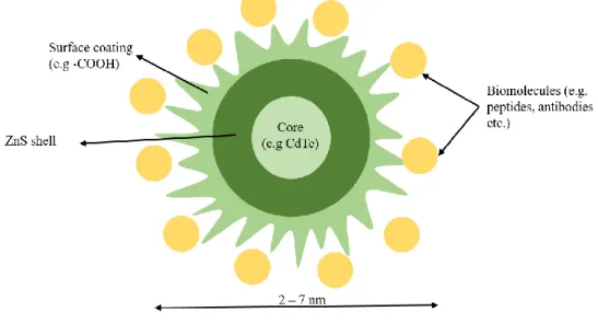
The general structure of a QD constitutes of an inorganic core semiconductor material, being the most common QD core used in biological and medical applications CdSe and CdTe (Smith et al., 2008), an inorganic shell of a distant band gap semiconductor material, such as ZnS, and further coated by an aqueous organic coating (ligands can be hydrophobic, hydrophilic or amphiphilic polymer) to which biomolecules, antibodies and/or drugs can be conjugated (see Fig.1) (Rizvi et al., 2010; Rocha et al., 2015b). The QDs shell protects the core from oxidation and degradation, while the surface ligands can increase the QDs water solubility and compatibility for applications in biological systems, enabling identification and action in specific biological targets (Maysinger et al., 2007; Smith et al., 2008; Rizvi et al., 2010)
Figure 1. Structure of a QD. Diameter of QD according to the QDs used in this study. (Image adapted from Rizvi et al., 2010)
4
1.2.2 QDs behaviour in the aquatic environment
Abiotic characteristics of the aquatic environment, such as pH, ionic strength, visible and UV electromagnetic radiation, promote different physicochemical transformation of QDs surface, therefore altering its behaviour and fate within the aquatic environment. Different processes influenced by these abiotic factors are for example, surface coating alterations, homo- and hetero-aggregation/agglomeration, disaggregation/deagglomeration, advection, diffusion, oxidation, NOM stabilization, bioturbation, settling, resuspension, interaction with macromolecules and/or organisms and biological transformation (Blickley & Giulio, 2010; Fabrega et al., 2011; Sousa & Teixeira, 2013; Corsi et al., 2014; Dale et al., 2015). The main physicochemical transformation of QDs is aggregation/agglomeration (Rocha et al., 2017). Morelli et al. (2012) and Rocha et al. (2014) acknowledge that QDs sediment more easily and are highly aggregated in seawater than in Milli-Q water. QDs – hetero-aggregation are a consequence of the interaction between dissimilar QDs, natural organic matter (NOM), suspended particulate matter (SPM) or biopolymers from aquatic biota, other ENMs or pollutants, being these processes dependent on collision frequency and attractive/repulsive forces at the QDs surface (Grasso et al., 2002).
The increase in wide applications of these ENMs also increase their release into the aquatic environment. This rises concern about the potential toxicity it may bring upon the aquatic environment as well as the interactions, bioaccumulation, and transfers within the aquatic food webs (Bouldin et al., 2008; Lewinski et al., 2011; Jackson et al., 2012; Farrell & Nelson, 2013; Ma & Lin, 2013; Lee & An, 2014) and potential toxicity that it may bring upon offspring (Hsu et al., 2012; Duan et al., 2013; Jong et al., 2013; Blickley et al., 2014). Most QDs are cadmium-based (Cd-based), being that the ecotoxicology related to QDs are mainly due to the release of Cd2+ ions from the
QDs core and/ or generation of free radicals or reactive oxygen species (ROS) (Gagné et al., 2008; Peyrot et al., 2009; Morelli et al., 2012; Tang et al., 2013; Katsumiti et al., 2014). The U.S. EPA and the International Agency on Cancer, classify Cd as human carcinogen (IARC, 1993; EPA, 1999), and is described as a non-essential metal and classified as a priority substance in the field of water policy by the European Water Framework Directive (Directive 2008/105/EC). The Ambient Water Quality Criteria for cadmium by
5
the U.S. EPA for 2016 are for estuarine/marine and freshwater, 33 µg/L and 1.8 µg/L, respectively (EPA 81 FR 19176, 2016). However, Cd-based QDs concentrations in the environment are unavailable, therefore, it is necessary to thoroughly study their potential ecotoxicological effects.
1.2.3 QDs impacts on aquatic organisms
Ecotoxicological impacts of Cd-based QDs in aquatic organisms have been noted in several studies using in vivo and in vitro exposure (Gagné et al., 2008; Peyrot et al., 2009; Katsumiti et al., 2014; Buffet et al., 2014; Rocha et al., 2014, 2015a), although the mechanisms of QDs-mediated toxicity are unclear, as it is dependent on the QDs size, shape, chemical composition, surface coating and the exposure conditions (Rocha et al., 2017). The QDs toxicity is mainly due to extra- and intra-cellular release of Cd2+ and
related to QDs dissolution (Peyrot et al., 2009; Domingos et al., 2011; Katsumiti et al., 2014), whilst the major mode of action of Cd-based QDs is associated to the oxidative damage caused by the production of free radicals or reactive oxygen species (ROS) (Gagné et al., 2008; Buffet et al., 2014). Because of quantum dot confinement, QDs can promote the delocalization of an electron from the valence band to the conduction band, generating an electron-hole pair (Ribeiro et al., 2012), making QDs efficient energy donors. This enables QDs to generate ROS in aqueous solutions, when exposed to UV and visible electromagnetic radiation (Ipe et al., 2005), being that the type and quantity of ROS generated is dependent on QDs composition as well as the fact that QDs photoactivation is reliant to size (Ipe et al., 2005; Ribeiro et al., 2012; Santana et al., 2015). The band gap energy, for example, of CdTe QDs is between 1.8 and 2.4 eV for valence band and conduction band, respectively, is potentially enough to reduce O2 and
to oxidise H2O molecules producing ROS such as the superoxide anion (·O2-) and the
hydroxyl radical (·OH-) (Santana et al., 2015). In aquatic organisms, substantial ROS
production, generated by the exposure to xenobiotics, such as QDs, can induce oxidative stress, meaning that ROS can bind to antioxidant defence mechanisms and cause abnormal activity or complete dysfunction (Blickley & Giulio, 2010).
6
1.3 Bivalves as sentinel organisms
Regarding aquatic nanotoxicity, bivalve species have a higher capacity to concentrate Cd-based QDs, from the water, inducing possibly tissue and cellular damage. Concerning sentinel organisms, bivalves have several characteristics that make them extremely important, and therefore, used extensively in ecotoxicology (Viarengo & Canesi, 1991; Livingstone, 1993; Canesi et al., 2012; Rocha et al., 2015b):
(i) Bivalves are sessile, filter-feeding organisms that have the capability to accumulate many contaminants present in the water, empowering measurements of stressor levels in their tissues, and therefore being a good indicator of the health of the surrounding environment;
(ii) These species are also easily collected and preserved under well-defined laboratory conditions, and have extensive background information about their biology and response to a wide range of environmental conditions;
(iii) They have a wide geographical distribution and are found at different latitudes, meaning that they are adapted to a variety of environmental parameters and therefore tolerant with respect to a wide range of environmental alteration. Their vast distribution enables comparisons between different areas;
(iv) Bivalves are found in high densities in quite stable populations, permitting repeated sampling and time-integrated indication of environmental contamination over a sample area;
(v) Many bivalve species are commercially used worldwide, and this increases their importance as sentinel organisms, as the information is crucial to understand the potential transfer of ENMs, such as Cd-based QDs, to humans.
Bivalve molluscs have been recognised as a unique target group for nanotoxicity (Canesi et al., 2012; 2015) after the first paper concerning possible hazards related with ENMs and their toxic effects towards aquatic organism (Moore, 2006).
7
Previous studies, using bivalves as sentinel organisms, have looked at tissue specific nanotoxicity by Cd-based QDs mainly assessing the effects in the gills and the digestive gland (Gagné et al., 2007; Peyrot et al., 2009; Rocha et al., 2014, 2015a, 2015c, 2016), as mussels take up water through their gills, filtering edible particles, and transport them to the digestive tract. In the presence of contaminants, such as Cd-based QDs, these can be redistributed differently in different tissues, including the gonads.
1.3.1 Selected model-system: Mytilus galloprovincialis
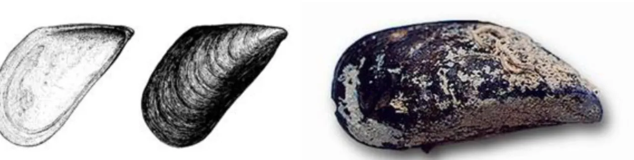
The marine mussel, Mytilus galloprovincialis (Lamark, 1819) (Figure 1.2.), commonly known as the “Mediterranean mussel”, was selected as model-system in this thesis to assess the toxicity of CdTe QDs in the gonads. As mentioned above, bivalve molluscs have been recognised as a unique target group for nanotoxicity (Canesi et al., 2012; 2015). Several studies have used M. galloprovincialis as a sentinel organism to assess the toxicity of QDs (Canesi et al., 2010; Montes et al., 2012; Gomes et al., 2012; 2013; Barmo et al., 2013; Hull et al., 2013; Katsumiti et al., 2014; Ruiz e al., 2015; Rocha et al., 2015b; 2016; 2017).
Figure 1.2. M. galloprovincialis, Lamark 1819 (image taken from FAO, 2017).
The mussel M. galloprovincialis is economically valuable and used in aquaculture, being mainly cultured in coastal waters of Galicia (NW Spain), and found to be produced in some Southern Mediterranean countries, The Russian Federation, Ukraine and South Africa, as well as cultured in China (FAO 2004-2017). In 2014, Europe produced 632 000 tonnes of bivalves, being the major producers Spain (223 000 tonnes), France (155 000 tonnes) and Italy (111 000 tonnes), whilst China produced 5 times more than Europe in the same year (12 million tonnes) (FAO, 2016). Within the bivalves, the production of
8
the marine mussel M. galloprovincialis was recorded as 116 262 tonnes in 2014, acknowledging that this number does not include the values produced by Spain or China (FAO 2004-2017). However, it is known that Spain and China produced 201 025 and >663 000 tonnes, respectively, of M. galloprovincialis in 2002 (FAO 2004-2017). This specie is native to the Mediterranean coast and the Black and Adriatic Seas, and has been registered as an invasive species by global invasive species database (GISD, 2015), notably to their ability to grow faster than native species and their tolerance to air exposure. M. galloprovincialis, as a filter feeder, mainly feeds on phytoplankton and organic matter, where it is capable of filtering 5 litres of seawater per hour with a size of 5 cm in length (FAO 2004-2017). This specie is sessile, meaning it lives attached to substrates (rocks and piers) by byssal threads secreted by the foot. Reproduction is gonochoristic, where males and females spawn simultaneously. Fertilization takes place externally, in the water column, and fertilized eggs then develop into free swimming larvae (FAO 2004-2017).
1.3.2 Gametogenesis
Gametogenesis is the process of gonad development towards the production of gametes that are then spawned when mature into the water column where the fertilization takes place. If gonads development is negatively affected by the presence of contaminants and/or if the contaminants are accumulated in the gametes, these can compromise the fertilization success, impair the larval development and limit the population capacity to produce a viable next generation, with major impacts to the whole ecosystem. The nanotoxicological effects of Cd-based QDs on the gonads of mussels have not been yet studied, however, some reviews of the effect Cd-based QDs on the reproductive system of other marine organisms are available (Lei et al., 2011; Hsu et al., 2012; Blickley et al., 2014). Lei and colleagues (2011) show that QDs (MAA-CdSe/ZnS QDs; ̴3.4±0.2 nm) at certain concentrations (0.5µM, 5µM and 10µM) influence the survival of zebrafish embryos, and Blickley and colleagues (2014) also demonstrate the presence of QDs (CdSe/ZnS QDs; 2-6 nm) in the eggs of the fish
Fundulus heteroclitus. Meanwhile, Hsu and colleagues (2012) suggest that QDs
9
Caenorhabditis elegans. Given that QDs have been assessed in affecting the
reproduction of the marine organisms mentioned above, it is therefore crucial to understand the nanotoxicity effects that QDs may have on the gonads of mussels and the outcomes of potential toxicity in relation to gametogenesis and fertilization success.
1.4 Oxidative stress
Oxidative stress is when an organism is experiencing an imbalance of pro-oxidant factors and antioxidant mechanisms (Blickley & Giulio, 2010), which is caused by the presence of oxygen free radicals and other reactive oxygen species. Reactive oxygen species (ROS) such as the superoxide anion (·O
2-), hydroxide anion (·OH), atomic oxygen
(12O2) and hydrogen peroxide (H2O2) (Kelly et al., 1998; Blickley & Giulio, 2010;
Girón-Pérez et al., 2013), are produced during normal metabolism. Lushchak (2011) demonstrates that there are numerous studies whereby prove a beneficial use of ROS by the biological system. ROS participate in enzymatic reactions, in the electron transport system in the mitochondria, in gene expression, signal transduction, nuclear transcription activation factor and in antimicrobial action of neutrophils and macrophages (Bayir, 2005). However, during metabolic processes, a small proportion (2-3%) of ROS may escape the protective shield (Davies, 1995), being this proportion supported by antioxidant enzymes as well as substances induced by xenobiotics (Bayir, 2005). Exposure to contaminants have been found to increase oxidative stress (Sohal et al., 2002) and to be responsible for the substantial production of ROS generated (Torres et al., 2008; Lushchak, 2011), which in consequence can bind to antioxidant defence mechanisms causing abnormal activity or complete dysfunction (Blickley & Giulio, 2010). In environmental toxicology, this matter has gained significant attention. The balance between pro-oxidant factors and antioxidant defences enable an assessment of oxidative damage influenced by diverse groups of chemical pollutants (Valavanidis et al., 2006).
10
1.4.1 Antioxidant enzymes as biomarkers of oxidative stress 1.4.1.1. Superoxide dismutase (SOD)
Superoxide dismutase (EC 1.15.1.1) is the enzyme responsible for the first antioxidant defence mechanism against ROS within an organism (Alscher et al., 2002; Li et al., 2009). SOD catalyses the conversion of the superoxide radical (·O
2-) into hydrogen
peroxide (H2O2) and oxygen (O2) (see Eq.1.). SOD’s presence is crucial to remove ·O2
-radicals formed as phospholipid membranes are impermeable to ·O
2- (Takahashi &
Asada, 1983). There are three forms of SOD’s based on the metal co-factor used. Iron SOD (Fe SOD) is one group found mostly in chloroplasts, whilst another form of SOD, manganese SOD (Mn SOD), is mostly found in the mitochondria and peroxisome (Alscher et al., 2002). Fe SOD and Mn SOD are present in both prokaryotic and eukaryotic organisms (Alscher et al., 2002). The third group is the copper-zinc SOD (Cu/Zn SOD) which has been mostly found in eukaryotes within the cytosol, although they can be found in other compartments (Alscher et al., 2002).
∙ O2−+ ∙ O 2
−+ 2H+ H
2O2+ O2
The activation of these specific SOD isoforms can serve as biomarkers in organisms experiencing pollutant-induced ·O
2- level increase (Barros et al., 2005; Murthy et al.,
2005). Thereby enabling measurements of SOD activity in the cytosolic fraction, given by the Cu/Zn SOD isoform, as well as the mitochondrial factor (Mn SOD).
1.4.1.2. Catalase (CAT)
Catalase (EC 1.11.1.6) is an enzyme and the primary antioxidant defence component which eliminates hydrogen peroxide (H2O2) (see Eq.2.).
SOD
Equation 1. Chemical reaction that SOD catalyses. Two superoxide radicals
(·O
11
Hydrogen peroxide (H2O2) is a non-radical ROS, that can penetrate through all
biological membranes and inactivate directly some enzymes (Alti et al., 2006). H2O2 also
reacts with metals, such as ferrous iron salts, and through Fenton-like redox cycling reactions to produce the hydroxyl radical (.OH) (Schaich, 1992), which has been
considered the most toxic free radical of biological and toxicological importance due to its potent oxidative capacity on lipids of biological membranes, proteins of enzymes and DNA (Richter, 1987; Stadtman and Levine, 2000; Jackson and Loeb, 2001; Valavanidis et al., 2006). Therefore, CAT activity is of extreme importance in breaking down H2O2 in
biological systems to avoid oxidative damage.
1.4.1.3. Glutathione Peroxidase (GPx)
Glutathione peroxidase (GPx) (EC 1.11.1.9) is an antioxidant enzyme with peroxidase activity that protects the organism from oxidative damage induced by peroxides (ROOH), such as H2O2 and lipid hydroperoxides (products of lipid
peroxidation) (Regoli & Principato, 1993; Júnior et al., 2001). Using reduced glutathione (GSH) as co-factor, GPx catalyses the reduction of H2O2 into water or lipid
hydroperoxides into their corresponding stable alcohol forms by oxidizing GSH into its oxidized form (GSSG) (see Eq.3.) (Vidal-Liñán et al., 2010).
Equation 3. GPx catalyses the reduction of H2O2 into two water molecules or lipid
hydroperoxides into their stable alcohol forms through oxidising two GSH molecules into its GSSG oxidised form.
2GSH GSSG GPx ROH/ 2H2O ROOH/ H2O2 2H2O2 2H2O + O2 CAT
12
Under natural conditions, in other words organisms that are not exposed to stress or to toxic agents, GPx is found to be among the most important antioxidant defences. The reduction of peroxides by GPx provides an efficient protection against oxidative damage and free radicals (Regoli & Principato, 1993).
1.4.1.4. Glutathione-S-transferase (GST)
Glutathione-S-transferase (GST) are a superfamily of multifunctional proteins (Van der Oost et al., 2003; Frova, 2006; Lee et al., 2007), being mainly involved in the detoxification of xenobiotics, as well as an antioxidant defence against ROS (Frova, 2006). GSTs catalyse nucleophilic attacks by reduced glutathione (GSH) on non-polar compounds which contain an electrophilic carbon, nitrogen or sulphur atom (Van der Oost et al., 2003; Hayes et al., 2005). Apart from their transferase activity, some isoforms of GST also express selenium-independent peroxidase activity for organic hydroperoxides (Hayes & Strange, 1995). GSTs, as phase II detoxifying enzymes, catalyse the transformation of a broad variety of electrophilic compounds, into less toxic substances, by conjugating them with glutathione (GSH) (Van der Oost et al., 2003). The biotransformation’s of xenobiotics is important as they change the biological activity and enhance the excretion of toxic compounds, thus preventing cell damage (Pereira et al., 2013). Given that GST can be induced or inhibited as a response to xenobiotics, these multifunctional proteins are widely used as biomarkers to assess the exposure of pollutants in aquatic organisms (Van der Oost et al., 2003; Amado et al., 2006).
1.4.2 Oxidative damage
Oxidative stress, known to cause damage to cellular macromolecules (Kelly et al., 1998; Matés, 2000), is defined by the imbalance of pro-oxidants and antioxidant mechanisms caused by the release of ROS, such as the superoxide anion (·O
2-), hydroxide
anion (·OH), atomic oxygen (1
2O2) and hydrogen peroxide (H2O2) (Kelly et al., 1998;
Blickley & Giulio, 2010; Girón-Pérez et al., 2013). Exposure to xenobiotics has been found to increase ROS production (Winston, 1991; Kelly et al., 1998; Livingstone, 2001; Pandey et al., 2003; Banni et al., 2005; Valavanidis et al., 2006; Farombi et al., 2007; Taylor & Maher, 2010; Girón-Pérez et al., 2013; Patil & David, 2013; Irinco-Salinas & Pocsidio,
13
2014) and despite organism’s protective antioxidant enzymes and their non-enzymatic co-factors, ROS produced at higher levels can overwhelm the antioxidant defence system, leading to accumulation of metabolic by-products and consequently oxidative damage (Hook et al., 2014).
1.4.2.1 Lipid Peroxidation (LPO)
LPO involves the formation and propagation of lipid radicals (Dianzani and Barrera, 2008; Girón-Pérez et al., 2013) ensuing in oxidative damage of polyunsaturated fatty acids (PUFA) (Repetto et al., 2012). Cell membranes are composed of PUFA, and therefore are a primary target for ROS attack, leading to a decrease in the membranes fluidity inclusive of cell membrane destruction (Gadjeva et al., 2005; Repetto et al., 2012; Girón-Pérez et al., 2013). Considering the accumulation of metabolic by-products, LPO is a chain reaction with membrane phospholipids, triggered by oxygen radicals or ROS (Repetto et al., 2012; Girón-Pérez et al., 2013).
For this reason, the determination of LPO is a great approach into understanding the effects of exposure to xenobiotics, as it reflects the action of ROS over biological lipids (Repetto et al., 2012). LPO is therefore used as a biomarker, in aquatic organisms, to quantify oxidative damage caused by pollutants.
1.5 Objectives
The aim of this study is to assess the effect of CdTe QDs (2 – 7 nm) in the gonads of the marine mussel Mytilus galloprovincialis, in comparison with its dissolved counterpart Cd2+. To accomplish the aim, Cd accumulation, oxidative stress (SOD, CAT,
GPx, GST) and oxidative damage (LPO) were used to assess the toxicity mediated by CdTe QDs, primarily looking for a distinction between male and female responses.
14
CHAPTER 2. MATERIALS AND METHODS
2.1 QD characterization
All relative information on QD characterization can be found in Rocha et al. (2014, 2015a, 2015c). Orange CdTe QDs were aquired from PlasmaChem GmbH (Berlin, CAS# 1306- 25-8) with 99.9% of declared purity, particle size of 2-7 nm, an emission wavelength at 590 ± 5 nm and a core coated by carboxyl groups (-COOH). A QD stock solution was made using Milli-Q water (100 mg.L-1), sonicated for 30 min (Ultrasonic bath VWR International, 230 V, 200 W, 45 KHz frequency) and kept in constant shaking. Dissolved cadmium stock solution was prepared in the same manner using Cadmium nitrate (Cd(NO3)2.4H2O) (Merck), but not sonicated. Suspensions of QDs in Milli-Q water and natural seawater (S = 36.3) were characterized by a combination of analytical techniques (Transmission Electron Microscopy, TEM; Dynamic Light Scattering, DLS; Electrophoretic Light Scattering, ELS) as described in Rocha et al. (2014, 2015a).
2.2 Experimental design
All the details on the experimental design and characterization of QDs can be found in Rocha and colleagues (2015c). Briefly, mussels M. galloprovincialis were collected from the Ria Formosa Lagoon (Portugal) and acclimated during 14 days.After acclimation, fifty mussels per treatment were placed into tanks (3 tanks per treatment) where they were exposed to 10 µgCd.L-1 of CdTe QDs (2-7 nm) and 10 µgCd.L-1 of Cd2+,
their soluble counterpart, simultaneously with a control group, that was kept in clean seawater, for 14 days. Mussels from each experimental condition were collected at the beginning of the experiment and after 3, 7 and 14 days of exposure. After sampling, mussels were dissected and gonads were then immediately frozen in liquid nitrogen and stored at -80 ºC until further use. Cd accumulation and the biomarkers of oxidative stress (SOD, CAT, GPx, GST) and of oxidative damage (LPO) induced by CdTe QDs and their dissolved counterpart were analysed in male and female gonads of M. galloprovincialis.
15
2.3 Sex identification
Sex identification was achieved by observation of an aliquot of a sample under an optical microscope (Compound Light Microscopy) at a magnification of 400×, for the presence of oocytes or spermatozoa. Samples were then stored at -80°C, with their respectful identification of male or female, for further analysis.
2.4 Sample preparation for Cd accumulation and LPO determination.
For the quantification of Cd concentration and LPO, samples were homogenized using the following process. Firstly, 15 ml falcon tubes were weighed with and without the sample, thus allowing to obtain the samples weight. 5 ml of Tris HCl [0.02M] and 50µl of butylated hydroxytoluene (BHT) was added to the samples and weighed again. In an ice bath, the sample was homogenized using an IKA Homogenizer (Ultra-Turrax T-25 model), for 2 minutes. The homogenized samples were weighed as well as the previously decontaminated centrifuge tubes. 3 ml of the homogenates were placed in the weighed centrifuge tubes and weighed again, and the remaining homogenates (1) were placed at 80ᵒC for 24 to 48 hours for further analysis of Cd concentration. The weighed 3 ml of homogenates were placed for 45 minutes to centrifuge at 30 000g and 4ᵒC. 500 µL of the resulting supernatant was removed and placed into microcentrifuge tubes and immediately frozen at -80ᵒC for quantification of LPO later, as well as 300 µL for the determination of total proteins.
2.4.1 Cd accumulation
For determination of Cd concentrations, gonad sample homogenates (2ml, Tris HCL [0.02 M] + 50µl BHT) were placed into previously weighed DigiTUBEs and weighed again. The homogenates were then dried at 80ᵒC, for 24 to 48h, digested with HNO3 and
analysed with a graphite furnace atomic absorption spectrometry (AAS AAnalyst 800 – PerkinElmer), as described by Rocha and colleagues (2016). Total Cd concentration are expressed as µg g-1 of dry weight tissue.
16
2.5 Sample preparation for antioxidant enzyme activity analysis.
Tissues were defrozen, weighed in previously weighed 15 ml falcon tubes, and suspended in 5 ml of 20 mM Tris-Sucrose buffer (0.5 M sucrose, 0,075 M KCl, 1 mM DTT, 1 mM EDTA, pH 7.6). Samples were further homogenized, in an ice bath, using a homogenizer (Ultra-Turrax T-25, IKA) for about 1 min.
Afterwards, the homogenate was transferred into previously weighed Nalgene centrifuge tubes and centrifuged for 15 minutes at 500g and 4ᵒC. Resulting supernatants were transferred into new weighed centrifuge tubes, for an additional centrifugation (12000g, 45 minutes, at 4ᵒC). Once the centrifugation has come to an end, the resulting supernatant (cytosolic fraction) was preserved in microcentrifuge tubes and stored at -80ᵒC for subsequent enzymatic activity analysis such as superoxide dismutase (SOD), catalase (CAT), glutathione peroxidase (GPx) and glutathione-S-transferase (GST) and total protein (TP) analysis.
2.5.1 Total protein
Total protein concentrations were measured in the cytosolic fraction following the colorimetric procedure of Bradford method (Bradford, 1976), using Bovin Serum Albumin as a standard (1 mg ml -1). The Bradford assay relies on the binding of Coomassie
Brilliant Blue G-250 dye to proteins (Bradford, 1976). The dye exists in three forms: cationic (red), neutral (green), and anionic (blue) (Compton & Jones, 1985). Under acidic conditions, the dye is mainly in the doubly protonated red cationic form (Amax = 470 nm).
On the other hand, when the dye binds to protein, it is converted to a stable unprotonated blue form (Amax = 595 nm) (Groth et al., 1963; Reisner et al., 1975). The
absorption of this blue protein-dye form was determined at 595 nm, using a microplate reader (Multimode microplate readers Infinite ® 200, Pro-Tecan). A standard curve was obtained and used to determine the sample concentration. Total protein, expressed as mg.g-1, was calculated using the following formula:
TP (mg.g-1) = 𝐶𝑜𝑛𝑐. (𝑚𝑔. 𝑚𝑙−1)× 𝑉𝑜𝑙.𝑇𝑟𝑖𝑠 (𝑚𝑙) 𝑊 𝑡𝑖𝑠𝑠𝑢𝑒 (𝑔)
17
2.5.2 Superoxide Dismutase
SOD activity was determined through a spectrophotometric method developed according to the method by McCord and Fridovich (1969). The activity was expressed in Units (U), wherein 1 U of activity corresponds to the amount of sample required to cause a 50% inhibition in the reduction of cytochrome c (Cyt c) by the anion superoxide (·O
2-)
generated by the xanthine/hypoxanthine system. Xanthine oxidase (Shmarakov & Marchenko, 2008; Kelley et al., 2010) is known to be responsible for the production of
·O
2- in the cytosol and peroxisomes of cells. Hence the incorporation of the
Xanthine/Hypoxanthine system within the method for the determination of SOD activity. The resoluteness of SOD was predicated on the enhancement of the absorbance engendered by the generation of Cyt c red, whose absorbance was quantified at 550nm. Results are expressed as U.mg-1protein, and calculated using the following formula:
SOD Activity (U.mg-1prot) = %𝐼 50 × 3 𝑉𝑠𝑎𝑚𝑝𝑙𝑒 × 1.4 ×1𝑐𝑚 𝑝𝑟𝑜𝑡𝑒𝑖𝑛𝑠 (𝑚𝑔) 2.5.3 Catalase
Following the method described by Greenwald (1985), the quantitative determination of CAT activity is based on the measurement of the consumption of hydrogen peroxide (H2O2) using a spectrophotometric assay at a wavelength of 240nm.
Blanks were comprised by 3000 µL of CAT buffer (KH2PO4 80 mM, K2HPO4 80 mM, pH
7.5) in quartz cuvettes, placed in duplicate. Then, 1900 µL of CAT buffer, 1000 µL of H2O2
and 100 µL of sample were added to a cuvette and placed in the spectrophotometer. The consumption of H2O2 over one minute of reaction was addressed by the decline in
absorbance (dAbs/min). Results are expressed as mmol min-1 mg protein-1 as following:
CAT (mmol/min/mg prot)
= (
(∆𝐴40)∗(𝑉𝑜𝑙 𝑠𝑎𝑚𝑝𝑙𝑒3 ) 𝑝𝑟𝑜𝑡 (𝑚𝑔 𝑚𝑙)⁄
)
18
2.5.4 Glutathione Peroxidase
Glutathione peroxidase (GPX) is an antioxidant enzyme induced by peroxides (ROOH), including H2O2, and uses reduced glutathione (GSH) as co-factor.
GPx activity was determined in a microplate reader, based on the method adapted from McFarland et al. (1999). In the microplate, 20 µL of blank (Tris-Sucrose buffer; 0.5 M sucrose, 0,075 M KCl, 1 mM DTT, 1 mM EDTA, pH 7.6) and 20 µL of sample were introduced into the wells in duplicate. 200 µL of DAM (3 mM GSH, 0.25 mM NADPH, 0.67 U/ml GR) solution was added to each well and incubated at 28ᵒC for two minutes. Then, 50 µL of substrate, Se-independent GPx (1 mM cumene hydroperoxide), was added to each well and placed inside the microplate reader (Infinite ® 200, Pro-Tecan). At 28ᵒC, the absorbance of each well of the microplate was read for 5 minutes, at 340 nm. Results were obtained through the decrease of the absorbance of nicotinamide adenine dinucleotide phosphate (NADPH) at 340 nm (Ԑ 340 (NADPH) = 0.005598 µM-1 cm-1), that
was consumed during the regeneration of reduced glutathione (GSH). GPx activity is expressed in mmol min-1mg prot-1, and calculated by the following formula:
GPx activity (mmol min-1 mg prot-1) =
(
(∆𝐴𝑏𝑠𝑠𝑎𝑚𝑝𝑙𝑒−∆𝐴𝑏𝑠𝑏𝑙𝑎𝑛𝑘) × 𝑉𝑡𝑜𝑡𝑎𝑙 ×1000 6.22 ×0.7485 ×𝑉𝑠𝑎𝑚𝑝𝑙𝑒 ×[𝑝𝑟𝑜𝑡𝑒𝑖𝑛 𝑚𝑔/𝑚𝐿])
2.5.5 Glutathione-S-Transferase
GST activity was determined in a microplate assay using the method described by McFarland et al. (1999) which was based firstly on Habig and Jakoby (1981). The activity is analysed using 1-chloro- 2,4- dinitrobenzene (CDNB) and reduced glutathione (GSH) as substrates and the change in absorbance measured at 340 nm every 30 seconds throughout 5min. 25 µL of Tris-Sucrose buffer (0.5 M sucrose, 0,075 M KCl, 1 mM DTT, 1 mM EDTA, pH 7.6) were added for blank, and for each sample to the wells, in replicates. After the preparation of DAM (60 mM CDNB, 0.2 M Tris-Sucrose) and GSH (10 mM) mixtures, 200 µL of the mixture was added to each well and the microplate was placed
19
inside the microplate reader (Infinite ® 200, Pro-Tecan) and absorbance of each well of the microplate was read for 3 minutes, at 340nm. GST activity is expressed as µmol CDNB min-1 mg prot-1, and calculated by the following formula:
GST activity (µmol CDNB min-1 mg prot-1) =
(
(𝐴𝑏𝑠𝑠𝑎𝑚𝑝𝑙𝑒−𝐴𝑏𝑠𝑏𝑙𝑎𝑛𝑘) ×𝑉𝑡𝑜𝑡𝑎𝑙 ×𝐷𝐹 9.6 ×0.6135 ×𝑉𝑜𝑙𝑠 ×[𝑝𝑟𝑜𝑡𝑒𝑖𝑛 𝑚𝑔/𝑚𝐿])
2.6 Lipid Peroxidation
Lipid peroxidation (LPO) was measured by the presence of its terminal products such as malondialdehyde (MDA) and 4-hydroxynonenal (Solé et al., 2010; Patil & David, 2013). MDA is a qualified biomarker of toxic effect as it can define the stress level of an individual caused by ROS (Erdelmeier et al., 1998). To quantify LPO, a colorimetric method based on the method elaborated by Erdelmeier et al. in 1998 was used. Using the extraction put aside for LPO during sample preparation (see section 2.4), LPO was determined in the soluble fraction. Before any processing, three reagents must be prepared (R1 – 1methyl-2-phenylindone, R1 diluted (18ml R1 + 6ml methanol), and R2 –
methanesulfonic acid, as well as standard solutions of malonaldehyde bis dimethyl acetal at three concentrations (A - 30mM; B - 10mM; C - 20µM). Then a water bath is pre-heated to 45ᵒC, and the samples defrosted and maintained the whole time in an ice bath. In the microcentrifuge tubes for LPO kept earlier, 200µl of each concentration of standard solution was added as well as 650µl of R1 diluted, and then shaken using a
vortex. 150µl of R2 was then added under the fume hood, and mixed well with the tube
closed. It was then incubated for 60 minutes in the water bath at 45ᵒC. The samples were then centrifuged at 15 000g for 10 minutes to obtain a clear supernatant. A microplate reader (Infinite ® 200, Pro-Tecan) was used and the samples were transferred from their respective microcentrifuge tubes into a microplate. The microplate was kept on top of ice whilst filling the wells. To each well, 150µl of standard solution and 150µl of the resulting supernatant of the sample from the centrifuge was added, to ensure 4 replicates of each. Then the plate was introduced into the equipment and the
20
absorbance was measured at 586 nm. MDA results are presented as units of nmol.mg-1
protein, and is calculated by the following formula:
MDA (nmol.mg-1 protein) =
(
𝐴𝑏𝑠−𝑏𝑎 (𝜇𝑚𝑜𝑙/𝑙)×𝑣𝑜𝑙𝑢𝑚𝑒 𝑇𝑟𝑖𝑠 (𝑚𝑙) 𝑊𝑒𝑖𝑔ℎ𝑡 𝑡𝑖𝑠𝑠𝑢𝑒 (𝑔)
𝑇𝑜𝑡𝑎𝑙 𝑝𝑟𝑜𝑡𝑒𝑖𝑛 (𝑚𝑔/𝑔)
)
2.7 Statistical Analysis
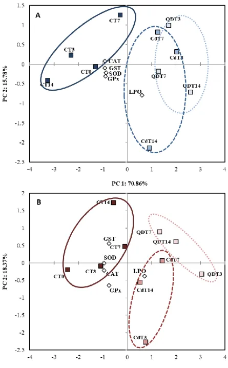
In order to understand the results of the experiment, three statistical tests have been implemented. The significant differences between treatments, time and sex was evaluated by a Two-Way ANOVA at a 95% confidence level (p<0.05), and a Tukey Post-hoc allowed pairwise comparisons among experimental conditions (p<0.05). These statistical analyses were performed on R software (R Core Team, 2017). A Principal Component Analysis (PCA) was also used to evaluate the relationship between the different treatments [unexposed mussels and Cd-exposed mussels (QDs and dissolved Cd)] and the analysed variables [antioxidant enzymes (SOD, CAT, GST activities) and oxidative damage (LPO)] in both male and female gonads and along the exposure period (14 days). The PCA was performed to differentiate sex specific responses towards both Cd forms. Results were considered significant when p < 0.05. PCAs were performed on Statistica 7.0 software (Statsoft Inc., 2005, USA).
21
CHAPTER 3. RESULTS
3.1 Cd accumulation
Cd content in the gonads of unexposed mussels did not change over time (p>0.05) however significant differences were registered between sex, whereby females show 43-fold higher Cd content than male mussels (p<0.05). Cd concentration in male and female mussels increased significantly after 14 days of exposure to QDs (males: 490.7-fold; females: 9-fold) and dissolved Cd (males: 194.9-fold; females: 10.8-fold) when compared to unexposed mussels (p<0.05; Fig 3.1.).
After 14 days of exposure, the Cd accumulated by male gonads exposed to QDs is significantly different from both dissolved Cd-exposed and controls (2.5-fold and 23.2-fold, respectively; p<0.05). On the other hand, Cd accumulation in female gonads is higher in those exposed to dissolved Cd than QD-exposed (1.2-fold), being significantly different from the Cd accumulated by Cd-exposed males, though not significantly
Figure 3.1. Cd concentration (mean ± std) (µg g-1 d. w.) in male and female gonads of mussels M.
galloprovincialis from controls (C), exposed to dissolved Cd (Cd2+) and CdTe quantum dots (QDs) for 14 days. Different capital and lower-case letters indicate significant differences between treatments within the same time and for the same treatment between times, respectively, and * indicates significant differences between sex (p<0.05).
22
different from females exposed to QDs. Contrarily, males have a much higher Cd accumulation in the gonad from QDs than from the readily available Cd form. However, no significant differences were found between males and females exposed to QDs after 14 days, whilst significant differences were found between sexes in mussels exposed to dissolved Cd (p<0.05).
3.2 Enzymatic Activity
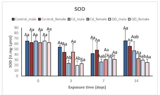
Antioxidant enzymes activities of unexposed mussels did not change over time (p>0.05; Fig. 3.2 – 3.5), with exceptions in GPx (males and females, p<0.05) and GST (males, p<0.05). A decrease in all antioxidant enzymes activities was registered in both male and female gonads after exposure to CdTe QDs and dissolved Cd (Fig. 3.2 – 3.5). SOD, CAT, GPx and GST activities changed after exposure to QDs, suggesting that these ENMs have potential redox properties with the capacity to generate ROS and oxidative stress in marine mussels. No significant differences in enzymatic activities were found between male and female mussels (p>0.05), though significant differences were registered in relation to the treatment and time of exposure within male and female gonads, separately (p<0.05).
3.2.1 Superoxide Dismutase
In mussels exposed to QDs, SOD activity in male gonads decreased after 3 days of exposure by 3.3-fold (p<0.05; Fig 3.2), increasing slightly after 7 and 14 days of exposure (1.9-fold and 2.2-fold, respectively) although still maintaining low values of SOD activity when compared to controls, suggesting an inhibition of SOD. Similarly, in mussels exposed to dissolved Cd, the pattern of SOD activity in male gonads was similar to that found in QDs, showing a decrease after 3 days of exposure (2.8-fold), however after 7 and 14 days of exposure SOD activity increased (2.5-fold and 1.4-fold).
Significant differences were registered in male gonads after 3 days of exposure in both dissolved Cd and QD treatments when compared to unexposed mussels (p<0.05; Fig 3.2), being SOD activity in mussels exposed to QD 1.2-fold smaller than SOD activity represented by mussels exposed to dissolved Cd.
23
On the contrary, there were no significant differences in female gonads exposed to both treatments (p>0.05; Fig 3.2). However, when comparing to males, females exposed to dissolved Cd showed an increase in SOD activity (1.9-fold) and a similar level of decrease after 14 days of exposure (1.5-fold), rather than the significant decrease in SOD activity represented by male gonads after 3 days exposure. Female gonads exposed to QDs presented a similar pattern to the male gonads, a decrease in SOD activity after 3 days of exposure (2.7-fold) with a slight increase in SOD activity after 7 and 14 days of exposure (2-fold and 2.5-fold, respectively). In dissolved Cd exposed mussels, females represent higher SOD activity than males after 3 and 7 days of exposure (1.9-fold and 1.2-fold, respectively; Fig 3.2) whilst after 14 days of exposure, males show higher SOD activity than females (1.5-fold). However, in QD-exposed mussels, females only show higher SOD activity than males after 3 days of exposure (1.2-fold), representing lower SOD activity after 7 and 14 days of exposure (1.1-fold and 1.2-fold, respectively). No significant changes were observed when comparing SOD activity between male and female gonads (p>0.05; Fig 3.2).
Figure 3.2. Comparison of SOD activity (mean ± std) (U.mg-1prot) between male and female gonads of mussels M. galloprovincialis fromcontrols (C), exposed to dissolved Cd (Cd2+) and CdTe quantum dots (QDs) for 14 days. Different capital and lower-case letters indicate significant differences between treatments within the same time and for the same treatment between times, respectively, and * indicates significant differences between sex (p<0.05).
24
Overall, after 14 days of exposure, SOD activity did not increase when compared to unexposed mussels. In fact, SOD activity decreased suggesting an inhibition of SOD, possibly due to a high generation of ·O
2-. QD-exposed mussels represent lower SOD
activity than in dissolved Cd-exposed mussels, whereby male gonads showed to be more affected by the generation of ROS due to dissolved Cd after 3 days of exposure than female gonads. However, after 7 and 14 days of exposure, females showed lower SOD activity than the males, suggesting that a longer time of exposure of female mussels to QD may possibly cause SOD activity to be increasingly inhibited.
3.2.2 Catalase
In QD-exposed mussels, CAT activity decreased after 3 days of exposure, in males, and maintained low values until the end of exposure (14 days), however a significant difference is only observed after 14 days (p<0.05; Fig 3.3). Males exposed to dissolved Cd on the other hand show a significant decrease in CAT activity after 3 days of exposure (2.3-fold, p<0.05), and an increase after 7 and 14 days of exposure (1.5-fold and 1.2-fold, respectively; p>0.05) maintaining values below those shown by unexposed mussels.
On the other hand, in female gonads exposed to dissolved Cd, CAT activity remained similar to those of unexposed mussels (p>0.05; Fig 3.3) and females exposed to QD show a significant difference after 3 days of exposure to both unexposed and dissolved Cd-exposed mussels (p<0.05). Until the end of the exposure period to QDs (14 days), female gonads maintained the low CAT activity shown just after 3 days, indicating significant differences between the two Cd forms in females (p<0.05).
CAT inhibition in mussels exposed to QDs is observed in both male and female gonads, being there no significant differences between the two sexes (p>0.05; Fig 3.3), thus suggesting that exposure to QD affects both sexes likewise. However, only males exposed to dissolved Cd showed significant differences (p<0.05; Fig 3.3) after 3 days of exposure increasing nearer to unexposed mussels CAT activity after 7 and 14 days.
![Figure 3.6. Comparison of LPO levels (mean ± std) (MDA [nmol/mg prot]) between male and female gonads of mussels M](https://thumb-eu.123doks.com/thumbv2/123dok_br/18055528.863372/44.892.134.762.526.851/figure-comparison-lpo-levels-mean-female-gonads-mussels.webp)