Two new species of harvestmen (Opiliones, Eupnoi,
Neopilionidae) from Waitomo, New Zealand
Christopher K. Taylor1, Anna Probert2
1 Dept of Environment and Agriculture, Curtin University, GPO Box U1987, Perth, WA 6845, Australia
2 School of Biological Sciences, University of Auckland, Private Bag 92019, Auckland Mail Centre, Auckland 1142, New Zealand
Corresponding author:Christopher K. Taylor (chris.taylor@curtin.edu.au)
Academic editor:Adriano Kury | Received 12 March 2014 | Accepted 6 August 2014 | Published 14 August 2014 http://zoobank.org/0A3E6FDD-E27D-49DF-92FD-04D0987C9CC0
Citation: Taylor CK, Probert A (2014) Two new species of harvestmen (Opiliones, Eupnoi, Neopilionidae) from Waitomo, New Zealand. ZooKeys 434: 37–45. doi: 10.3897/zookeys.434.7486
Abstract
Two new species of harvestman (Opiliones: Neopilionidae: Enantiobuninae) are described from the Waito-mo region of the North Island, New Zealand, Forsteropsalis bona sp. n. and F. photophaga sp. n. Both have been collected within caves in the region, where predation on glow-worms Arachnocampa luminosa has been previously recorded for one or both species (misidentiied as ‘Megalopsalis tumida’). However, both are regarded as troglophiles rather than strict troglobites due to the presence of specimens outside the cave systems, and the absence of troglobitic adaptations. Megalopsalis tumida (Forster, 1944) is identiied as a junior synonym of Forsteropsalis fabulosa (Phillipps & Grimmett, 1932).
Keywords
Taxonomy, arachnids, cave biota
Introduction
he Waitomo region of the North Island, New Zealand, has become internation-ally renowned as a tourist attraction owing to its extensive cave systems. he primary reason for their fame is their large population of glow-worms Arachnocampa luminosa
www.zookeys.org
Copyright Christopher K. Taylor, Anna Probert. This is an open access article distributed under the terms of the Creative Commons Attribution License (CC BY 4.0), which permits unrestricted use, distribution, and reproduction in any medium, provided the original author and source are credited.
(Keroplatidae). hese luminescent ly larvae construct silken nests on the roof of the cave from which they hang sticky threads to capture lying insects attracted to their light (Richards 1960). hey are themselves predated upon by harvestmen (Opiliones), which are able to avoid entanglement by the glow-worms’ threads and pull the larvae from their nests (Richards 1960; Meyer-Rochow and Liddle 1988).
Richards (1960) recorded two species of harvestmen feeding on glow-worms at Waitomo. One, Hendea myersi cavernicola Forster, 1954 (Triaenonychidae), was originally described from Waitomo. he second species was identiied by both Rich-ards (1960) and Meyer-Rochow and Liddle (1988) as Megalopsalis tumida (Forster, 1944) (Neopilionidae). Richards (1960) recorded this species feeding only on ma-ture glow-worm gnats; Meyer-Rochow and Liddle (1988) recorded it also feeding on pupae and late-instar larvae. Megalopsalis tumida is a junior synonym of
For-steropsalis fabulosa (Phillipps & Grimmett, 1932) (see below), and was originally
described from near Wellington. Examination of specimens collected from caves in the Waitomo region revealed the presence of two species of Neopilionidae, both of them described as new below. Which of these was the species mentioned by Richards (1960) and Meyer-Rochow and Liddle (1988) is unknown. Forsteropsalis bona sp. n. is the more similar to F. fabulosa, but a photograph of ‘Megalopsalis tumida’ in Meyer-Rochow and Liddle (1988) may show F. photophaga sp. n. It is not impos-sible that the two were confused.
While Meyer-Rochow and Liddle (1988) regarded Hendea myersi cavernicola as a true troglobite, and did not ind it outside the cave entrance, the collection of small numbers of ‘M. tumida’ outside the cave led them to regard it as troglophilic rather than troglobitic. Similar habits were inferred by Taylor (2013) for Megalopsalis
sufu-giens Taylor, 2013, described from caves in Western Australia. Further epigean
speci-mens are recorded herein for Forsteropsalis bona, while specimens collected in the cave were only a short distance from the entrance. Forsteropsalis photophaga has not yet been conclusively recorded outside the caves, but troglophily is also suggested for this spe-cies by the absence of strong adaptations for troglobitism.
Methods
Taxonomic descriptions
Forsteropsalis bona sp. n.
http://zoobank.org/32F7E0A3-1CE0-41EF-B7AA-48DDAE2A529A
Figure 1
Holotype male. WO. Lucky Strike Cave, Te Kuiti, on wall near entrance, 14 Feb 1959, K. A. J. Wise (MONZ).
Paratypes. WO. 2 males, Waitomo Valley, in stream crevice outside cave entrance (shady), specimens intertwined and sluggish, 30 Mar 1959, L. G. Watson (MONZ); 1 male, Weir Cave, Stubbs Farm, Waitomo, ca. 2 m from cave entrance within a 15 cm radius of Arachnocampa luminosa larvae, 23 Aug 2010, A. Probert (NZAC).
Etymology. From the Latin bonus, good, in contrast to the related Forsteropsalis fabulosa.
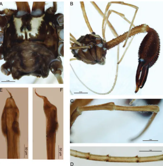
Male (n=4). Total body length 4.8–6.6; prosoma length 2.5–2.8, width 4.0–4.2. Prosoma (including ocularium) unarmed (Fig. 1A); ground colour in alcohol orange-brown with longitudinal yellow stripes on either side of ocularium (live coloration very dark brown [almost black] with orange-yellow stripes; appendages also black). Ozopores elongate, with small lanking lobes. Opisthosoma grey-yellow. Mouthparts cream-coloured; medial side of pedipalpal coxa with dense array of sharp denticles; cer-vix unarmed. Coxae yellow. Chelicerae (Fig. 1B): Segment I length 6.2–8.4; segment II 9.4–10.4. Elongate; segment I orange with lighter yellow patch at distal end, segment II dark orange-brown. Segment I denticulate, with denticles concentrated along dorsal, proventral and retroventral margins. Segment II massively inlated, evenly denticulate. Cheliceral ingers elongate, widely bowed apart; setae present on distal half of mobile inger. Pedipalps: Femur length 5.4–5.7; patella 2.2–2.4; tibia 2.8–3.1; tarsus 5.8–6.5. Distinctly elongate, yellow. Femur dorsally denticulate on proximal two-thirds; re-mainder of pedipalp unarmed. Setae sparse except for small concentration at prodis-tal end of patella; microtrichia present on tarsus and disprodis-tal half of tibia; prodorsal end of patella with distinct protrusion but without deinite inger-like apophysis (Fig. 1C). Tarsal claw without ventral tooth-row. Legs: Legs I femur length 8.6–9.9, patella 1.9–2.5, tibia 8.3–9.8; leg II femur 14.3–17.2, patella 2.2–2.8, tibia 14.4–17.8; leg III femur 7.5–8.7, patella 1.7–2.3, tibia 5.3–8.2; leg IV femur 8.5–10.7, patella 1.8–2.7, tibia 10.2–10.8. Femora sparsely denticulate, particularly in proximal half; remainder of legs unarmed. Distitarsus I with strong ventral tooth at distal end of each of irst ive or six pseudosegments (Fig. 1D). Tibia II with nine to ifteen pseudosegments; tibia IV with two pseudosegments. Penis (Fig. 1E–F): Shaft subquadrate; tendon long. Bris-tle groups relatively long, posterior brisBris-tle group with longest brisBris-tles reaching dorsal margin in lateral view. Glans short, subtriangular in ventral view, narrowing rapidly in lateral view.
Comments. Females of this species are currently unknown. Forsteropsalis bona
and enormous, sub-globose cheliceral segment II with widely bowed cheliceral ingers (Taylor 2011). In these features it strongly resembles F. fabulosa, and would key out to either F. fabulosa or F. tumida in the key to Forsteropsalis species provided by Taylor (2011). hese two species are synonymised below. Forsteropsalis bona can be distin-guished from F. fabulosa by the form of the pedipalpal patella: F. fabulosa has a distinct inger-like prodistal apophysis on the patella (Phillipps and Grimmett 1932: Fig. C p. 732), while the patellar apophysis is almost absent in F. bona (Fig. 1C). Forsteropsalis
fabulosa also has denticles both dorsally and ventrally on the pedipalpal femur, while
F. bona has denticles dorsally only.
An interesting feature of Forsteropsalis bona is the presence of a strong ventrodistal tooth on the end of each of the proximal pseudosegments of the distitarsus. his tooth sits between the two spinose setae generally present on each tarsal pseudosegment in all Enantiobuninae (Fig. 1D). Such a feature has not previously been recorded for this subfamily, though it is also present in F. fabulosa (specimens from MONZ, details giv-en in Taylor 2011). his may represgiv-ent a distinct synapomorphy of these two species. he glans of both Forsteropsalis fabulosa (Taylor 2011) and F. bona is relatively short compared to other Forsteropsalis species, and converges in shape on that of the Australian genus Megalopsalis (Taylor 2011, 2013). Nevertheless, the remaining fea-tures of these two species support a direct relationship with other New Zealand species of Pantopsalis and Forsteropsalis, and with Forsteropsalis in particular. hese features include dorsal papillae on the glans (Taylor 2011), setae on the mobile inger of the chelicera (absent in Megalopsalis except M. caeruleomontium; Taylor 2011, 2013), and an array of denticles on the medial side of the pedipalpal coxa (Fig. 3A; Taylor 2011).
Forsteropsalis fabulosa (Phillipps & Grimmett, 1932)
Macropsalis fabulosa Phillipps & Grimmett, 1932: 731–733, ig. p. 732.
Megalopsalis fabulosa (Phillipps & Grimmett) – Forster 1944: 186–187, igs 10–11
(misidentiication of Forsteropsalis inconstans).
Megalopsalis tumida Forster 1944: 188–189, igs 4–6 syn. n.
Forsteropsalis fabulosa (Phillipps & Grimmett) – Taylor 2011: 51, igs 99–101, 2012: 49.
Forsteropsalis tumida (Forster) – Taylor 2011: 60–61, igs 124–127.
Comments. Forsteropsalis fabulosa and F. tumida were distinguished in Taylor (2011) solely by the degree of dilation of the second cheliceral segment, with the latter supposedly more inlated in F. tumida than in F. fabulosa. A number of species of
Forsteropsalis and its sister genus Pantopsalis are now known to vary in cheliceral dilation (Taylor 2004, 2011, 2013, and see Forsteropsalis photophaga below). Forsteropsalis fabulosa
and F. tumida were both described from the Wellington district, and there seems to no
longer be any justiication for separating them as diferent species. Forsteropsalis tumida
is therefore regarded herein as a junior synonym of F. fabulosa (syn. n.).
Forsteropsalis photophaga sp. n.
http://zoobank.org/633102AB-D0FD-4F91-8A38-CFDF1ECDF08F
Figure 2
Holotype male. WO. Waitomo, Gardners Gut Cave System, 200 yards from Zweihöllen entrance, 25 June 1977, W. L. Blundell (MONZ).
Cave, Taumatamaire Rd, Waitomo County, 50 ft, 16 May 1966, K. A. J. Wise (MONZ); 2 males, Stubbs Farm, Waitomo, on rocky cave substrate, February 2013, G. Holwell et al. (NZAC); 2 males, Mangapohue Cave, Stubbs Farm, Waitomo, on rocky cave substrate, 21 Oct 2013, A. Probert & D. Townsend (NZAC).
Etymology. From the Greek phos, light, and phagein, to eat, in reference to this species’ predation of the glow-worm Arachnocampa luminosa.
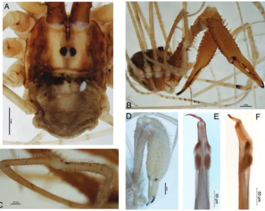
Male (n=7). Total body length 3.5–6.1; prosoma length 1.9–2.1, width 2.5–3.9. Prosoma (including ocularium) unarmed except for few black setae (Fig. 2A); ground colour orange-brown with longitudinal yellow stripes on either side of ocularium (live colouration light to mid-brown with pale yellow stripes). Ozopores elongate, with small lanking lobes. Opisthosoma grey-brown. Mouthparts cream-coloured; medial side of pedipalpal coxa with array of sharp denticles; cervix with single pair of denticles laterally. Coxa I orange; remaining coxae and venter of opisthosoma yellow. Chelicerae
and retrolateral rows of elongate denticles and some scattered median denticles proximally. Second segment mildly to notably inlated, sub-conical, evenly denticulate with longitudinal rows of more elongate denticles dorsally and retrolaterally. Cheliceral ingers elongate, slightly bowed apart; setae present along central third of mobile inger. Pedipalps: Femur length 4.6–6.5, patella 2.8–3.2, tibia 2.2–2.8, tarsus 4.8–5.7. Distinctly elongate; yellow. Median side of coxa with array of sharp denticles. Femur with few denticles dorsally in proximal half; remainder of pedipalp unarmed. Patella, tibia and proximal half of tarsus densely covered with plumose setae; microtrichia present over entirety of patella, tibia and tarsus; patella with small, rounded, prodistal apophysis (Fig. 2C). Tarsal claw without ventral tooth-row. Legs: Leg I femur length 8.1–11.0, patella 1.9–2.2, tibia 8.4–10.7; leg II femur 14.0–17.7, patella 1.9–2.5, tibia 16.0–19.0; leg III femur 7.1–9.4, patella 1.6–1.9, tibia 7.6–9.8; leg IV femur 9.0–12.2, patella 1.8–2.2, tibia 10.3–12.4. Yellow. Proximal half of femur I with few scattered dorsal denticles; remainder of legs unarmed. Tibia II with 12 pseudosegments; tibia IV with three pseudosegments. Penis (Fig. 2E–F): Shaft subquadrate; tendon long. Bristle groups relatively long, posterior bristle group with longest bristles reaching dorsal margin in lateral view. Glans relatively long, subrectangular in ventral view, remaining relatively deep to distal end but with dorsodistal end rounded.
Comments. Females of this species are currently unknown. he holotype of
Forsteropsalis photophaga when irst examined had a parasitic mite attached to the
opisthosoma (Fig. 2A). his mite is a representative of the Microtrombidiidae, a family that has not previously been recorded as parasitic on Opiliones; a more detailed description is currently being prepared by C. Taylor.
he genera Pantopsalis and Forsteropsalis have hitherto been regarded as well distinguished by the morphology of the cheliceral ingers (crescent-shaped in
Pantopsalis vs bowed in Forsteropsalis), pedipalpal patellar apophysis (hypersetose and rounded in Pantopsalis, sparsely setose and triangular in Forsteropsalis) and penile bristle groups (shorter in Pantopsalis than in Forsteropsalis) (Taylor 2004, 2011). he current species blurs this distinction: in its hypersetose and rounded pedipalpal apophysis it resembles Pantopsalis, but its elongate cheliceral ingers and penile bristle groups are more characteristic of Forsteropsalis. It also possesses an array of denticles on the medial side of the pedipalpal coxa as found in Forsteropsalis species (Fig. 3A; Taylor 2011). We therefore assign it to the latter genus herein. A hypersetose, rounded patella is also present in the female of F. grimmetti, though the male of that species possesses a more typical Forsteropsalis-type patella (Taylor 2011). It is possible that the hypersetose patella is in fact a symplesiomorphy of Pantopsalis and Forsteropsalis, with
F. photophaga being a basal member of the latter genus.
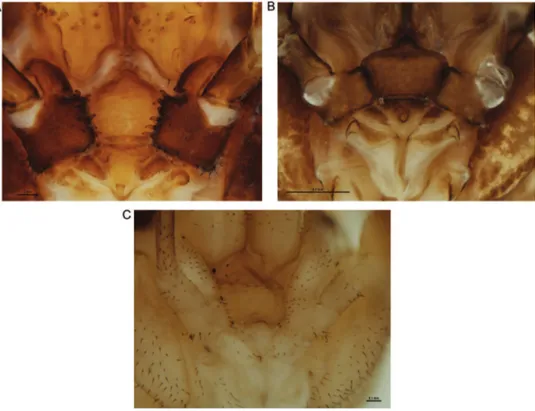
Figure 3. Mouthparts of selected New Zealand Neopilionidae, showing morphology of pedipalpal coxae. AForsteropsalis chiltoni, with medial denticles B Pantopsalis albipalpis, with sclerotised medial lange overhanging cervix CMangatangi parvum, with unarmed, simple coxae.
Acknowledgements
he authors would like to thank Phil Sirvid, Te Papa Tongarewa, for arranging the loan of specimens. Anna Probert was assisted in collecting specimens by Daniel Townsend, Greg Holwell and Christina Painting. Access to caves on Stubbs Farm was provided by Alister and Ann Stubbs. Christopher Taylor’s position at Curtin University is funded by Chevron Australia.
References
Crosby TK, Dugdale JS, Watt JC (1998) Area codes for recording specimen localities in the New Zealand subregion. New Zealand Journal of Zoology 25: 175–183. doi: 10.1080/03014223.1998.9518148
Forster RR (1944) he genus Megalopsalis Roewer in New Zealand with keys to the New Zea-land genera of Opiliones. Records of the Dominion Museum 1(1): 183–192.
Hickman VV (1957) Some Tasmanian harvestmen of the sub-order Palpatores. Papers and Proceedings of the Royal Society of Tasmania 91: 65–79.
Meyer-Rochow VB, Liddle AR (1988) Structure and function of the eyes of two species of opilio-nid from New Zealand glow-worm caves (Megalopsalis tumida: Palpatores, and Hendea myersi cavernicola: Laniatores). Proceedings of the Royal Society of London Series B (Biological Sci-ences) 233: 293–319. doi: 10.1098/rspb.1988.0023
Phillipps WJ, Grimmett RER (1932) Some new Opiliones from New Zealand. Proceedings of the Zoological Society of London 1932: 731–740.
Richards AM (1960) Observations on the New Zealand glow-worm Arachnocampa luminosa (Skuse) 1890. Transactions of the Royal Society of New Zealand 88(3): 559–574. Taylor CK (2004) New Zealand harvestmen of the subfamily Megalopsalidinae (Opiliones:
Monoscutidae) – the genus Pantopsalis. Tuhinga 15: 53–76.
Taylor CK (2011) Revision of the genus Megalopsalis (Arachnida: Opiliones: Phalangioidea) in Australia and New Zealand and implications for phalangioid classiication. Zootaxa 2773: 1–65. Taylor CK (2012) Clariication of the type status of Macropsalis fabulosa Phillipps & Grimmett
1932. Zootaxa 3194: 49–50.