Triggering Receptor Expressed on Myeloid
Cells (TREM)-2 Impairs Host Defense in
Experimental Melioidosis
Tassili A. F. Weehuizen1,2*, Tijmen J. Hommes1,2, Jacqueline M. Lankelma1,2, Hanna K. de Jong1,2, Joris. J.T.H. Roelofs3, Alex F. de Vos1,2, Marco Colonna4, Tom van der Poll1,2,5, W. Joost Wiersinga1,2,5
1Center for Infection and Immunity Amsterdam (CINIMA), Academic Medical Center, Amsterdam, the Netherlands,2Center for Experimental and Molecular Medicine (CEMM), Academic Medical Center, Amsterdam, the Netherlands,3Department of Pathology, Academic Medical Center, Amsterdam, the Netherlands,4Department of Pathology, Washington University in St. Louis, St. Louis, Missouri, United States of America,5Department of Medicine, Division of Infectious Diseases, Academic Medical Center, Amsterdam, the Netherlands
*t.a.weehuizen@amc.uva.nl;w.j.wiersinga@amc.uva.nl
Abstract
Background
Triggering receptor expressed on myeloid cells (TREM) -1 and TREM-2 are key regulators of the inflammatory response that are involved in the clearance of invading pathogens. Melioidosis, caused by the "Tier 1" biothreat agentBurkholderia pseudomallei, is a common form of community-acquired sepsis in Southeast-Asia. TREM-1 has been suggested as a biomarker for sepsis and melioidosis. We aimed to characterize the expression and function of TREM-1 and TREM-2 in melioidosis.
Methodology/Principal Findings
Wild-type, TREM-1/3 (Trem-1/3-/-) and TREM-2 (Trem-2-/-) deficient mice were intranasally infected with liveB.pseudomalleiand killed after 24, and/or 72 h for the harvesting of lungs, liver, spleen, and blood. Additionally, survival studies were performed. Cellular functions were further analyzed by stimulation and/or infection of isolated cells. TREM-1 and TREM-2 expression was increased both in the lung and liver ofB.pseudomallei-infected mice. Strik-ingly,Trem-2-/-, but notTrem-1/3-/-, mice displayed a markedly improved host defense as reflected by a strong survival advantage together with decreased bacterial loads, less inflammation and reduced organ injury. Cellular responsiveness of 2, but not TREM-1, deficient blood and bone-marrow derived macrophages (BMDM) was diminished upon exposure toB.pseudomallei. Phagocytosis and intracellular killing ofB.pseudomalleiby BMDM and alveolar macrophages were TREM-1 and TREM-2-independent.
Conclusions/Significance
We found that TREM-2, and to a lesser extent TREM-1, plays a remarkable detrimental role in the host defense against a clinically relevant Gram-negative pathogen in mice: TREM-2
a11111
OPEN ACCESS
Citation:Weehuizen TAF, Hommes TJ, Lankelma JM, de Jong HK, Roelofs J.JTH, de Vos AF, et al. (2016) Triggering Receptor Expressed on Myeloid Cells (TREM)-2 Impairs Host Defense in
Experimental Melioidosis. PLoS Negl Trop Dis 10(6): e0004747. doi:10.1371/journal.pntd.0004747
Editor:Mathieu Picardeau, Institut Pasteur, FRANCE
Received:January 11, 2016
Accepted:May 7, 2016
Published:June 2, 2016
Copyright:© 2016 Weehuizen et al. This is an open access article distributed under the terms of the
Creative Commons Attribution License, which permits unrestricted use, distribution, and reproduction in any medium, provided the original author and source are credited.
Data Availability Statement:All relevant data are within the paper and its Supporting Information files.
Funding:This work was supported by research grants of the Netherlands Organisation for Health Research and Development (ZonMW; grant nr. 90700424) and The Netherlands Organization for Scientific Research (grant nr: 91610008). The funders had no role in study design, data collection and analysis, decision to publish, or preparation of the manuscript.
deficiency restricts the inflammatory response, thereby decreasing organ damage and mortality.
Author Summary
Triggering receptor expressed on myeloid cells (TREM)-1 and -2 are receptors on immune cells that act as mediators of the innate immune response. It is thought that TREM-1 amplifies the immune response, while TREM-2 acts as a negative regulator. Previously, we found that TREM-1 is upregulated in melioidosis patients. In contrast, nothing is known on TREM-2 expression and its role in melioidosis. In this study we examined the expres-sion and functional role of both TREM-1 and -2 in a murine melioidosis model. We found that TREM-1 and-2 expression was upregulated during melioidosis. Using our
experimen-tal melioidosis model, we observed thatTrem-2-/-mice were protected againstB.
pseudo-mallei-induced lethality.Trem-2-/-mice demonstrated reduced bacterial loads,
inflammation and organ damage compared to wild-type mice in experimental melioidosis.
Despite reduced bacterial dissemination ofB.pseudomalleito distant organs inTrem-1/3-/
mice-, no differences in survival were found betweenTrem-1/3-/-and wild-type mice
dur-ing melioidosis. Lastly, we investigated cellular functions of TREM-1 and TREM-2 and
found that TREM-2 deficiency led to decreased cellular responsiveness toB.pseudomallei
infection. In conclusion, we found that TREM-2 plays an important role during experi-mental murine melioidosis. TREM-2-deficiency reduces inflammation and organ damage, thereby improving survival.
Introduction
In sepsis, defined as a deregulated host response to a life-threatening infection, a careful
bal-ance between inflammatory and anti-inflammatory responses is vital [1–3]. Pathogen- or
dan-ger-associated molecular patterns are recognized by intracellular sensory complexes and cell surface receptors expressed on innate immune cells that can initiate the inflammatory and anti-microbial response. Well-known examples of these pattern recognition receptors (PRRs) are the Toll-like receptor (TLR), nucleotide-oligomerization domain-like receptor (NLR) and
C-type lectin receptor (CLR) families [4]. A more recently discovered group of innate immune
receptors are the membrane-bound triggering receptors expressed on myeloid cells (TREMs),
which act as key modulators, rather than as initiators, of the inflammatory response [5–7].
TREM-1 and TREM-2 are the most studied members of the TREM-family, however their exact role in the pathogenesis of sepsis remains ill-defined. Upon recognition of partially still unspecified ligands, both receptors phosphorylate the adaptor molecule DNAX adaptor
pro-tein 12 (DAP12) after which the cellular response is initiated [8,9]. Only recently, binding of
TREM-1 to a complex of peptidoglycan recognition protein 1 (PGLYRP1) and bacterially
derived peptidoglycan has been demonstrated [10]. TREM-1 is expressed on neutrophils and
monocyte subsets [11] and amplifies pro-inflammatory TLR-mediated responsesin vitro[12].
There are conflicting reports on the role of TREM-1 inin vivoinfection models. TREM-1
defi-ciency impaired bacterial clearance in a model ofKlebsiella pneumonia-induced liver abscess
formation [13], pneumococcal [14] andPseudomonas (P.) aeruginosapneumonia [15].
How-ever, blocking TREM-1 with an analogue synthetic peptide derived from the extracellular
endotoxaemia [17]. Interestingly, in a murine pneumonia model ofLegionella pneumoniano impact of TREM-1 deficiency was found on bacterial clearance or neutrophil influx towards
the primary site of infection [18].
TREM-2 is primarily expressed on macrophages, dendritic cells, microglia and osteoclasts
[19–22] and has been suggested to bind to bacterial lipopolysaccharide (LPS) and lipotechoic
acid [23]. In contrast to TREM-1, TREM-2 acts as a negative regulator of inflammatory
responses in macrophages and dendritic cells [19,21]. In addition, TREM-2 is involved in
phagocytosis [24,25] and killing of bacteria by macrophages [26]. Blocking TREM-2in vivoby
a recombinant protein in a polymicrobial sepsis model revealed that TREM-2 is required for
bacterial clearance and improves survival [27]. In contrast, TREM-2 plays a detrimental role
during pneumococcal pneumonia [25].
Melioidosis, considered to be an illustrative model for Gram-negative sepsis, is caused by the
Tier 1 biological treat agentBurkholderia pseudomallei[28,29]. Melioidosis is characterized by
pneumonia and abscess formation and an important cause of community-acquired sepsis in
Southeast Asia and Northern Australia [28]. The high mortality rate, that can approach 40%, and
the emerging antibiotic resistance ofB.pseudomallei[30] emphasize the need to better
under-stand the pathogenesis of melioidosis, which could ultimately lead to novel treatment strategies. We previously found increased soluble (s) TREM-1 plasma levels and TREM-1 surface
expres-sion on monocytes of patients with melioidosis [31], suggesting an important role for TREM-1 in
the host defense againstB.pseudomallei. Treatment with a peptide mimicking a
conserved-domain of sTREM-1 partially protected mice fromB.pseudomalleiinduced lethality [31].
In this study we now examine the role of TREM-1 and TREM-2 during experimental
melioidosis, utilizing recently generatedTrem-1/3-deficient (Trem-1/3-/-) [15] andTrem-2
-deficient (Trem-2-/-) mice [19] to determine their contribution to the host response againstB.
pseudomallei. We hypothesized that TREM-1 deficiency would decrease inflammation and improve survival during murine melioidosis while TREM-2 deficiency would instead lead towards increased inflammation and a worsened survival. Unexpectedly however, we found that TREM-2, but not TREM-1, plays an important detrimental role during melioidosis.
TREM-2 deficiency improves survival ofB.pseudomalleiinfected mice, by limiting
inflamma-tion and organ damage. These data identify TREM-2 as a potential treatment target for sepsis
caused byB.pseudomallei.
Materials and Methods
Detailed methods are provided in the online supplement (S1 Appendix).
Ethics statement
The Animal Care and Use of Committee of the University of Amsterdam approved all experi-ments (DIX102273), which adhered to European legislation (Directive 2010/63/EU).
Mice
Pathogen-free 8- to 10-week-old male wild-type (WT) C57BL/6 mice were purchased from
Charles River (Leiden, The Netherlands).Trem-1/3-/-[6,14] andTrem-2-/-[19] mice were
backcrossed>97% to a C57BL/6 genetic background.
Experimental infection and assays
with 5 × 10² colony forming units (CFU) ofB.pseudomalleistrain 1026b (a clinical isolate) as
described [32–34]. For survival experiments mice were observed 4–6 times daily, up to 14 days
post-infection. Sample harvesting, processing, and determination of bacterial growth were
per-formed as described in detail in theS1 Appendix[33,34]. All work concerning liveB.
pseudo-malleiwas performed in a (A)BSL III facility.
Chemo- and cytokine levels were determined in plasma, lung and liver. Distant organ dam-age was more closely assessed by plasma transaminases, lactate dehydrogenase (LDH) and blood urea nitrogen (BUN) levels.
TREM-1 and TREM-2 expression
Total RNA was isolated using the Isolate II RNA mini kit (Bioline, Taunton, MA, USA), treated with DNase (Bioline) and reverse transcribed using an oligo(dT) primer and Moloney murine leukemia virus RT (Promega, Madison, WI, USA). Primers and RT-PCR conditions can be
found in the supplemental data. Data were analyzed using the comparative Ctmethod.
(Immuno)histology
Paraffin-embedded 4-μm lung, liver and spleen sections were stained with haematoxylin and
eosin and analyzed for inflammation and tissue damage, as described previously [14,34].
Granulocyte (Ly6G) staining was done exactly as described previously [35].
Whole blood and macrophage stimulation
Whole blood, alveolar macrophages (AM) and bone-marrow derived macrophages (BMDM)
were harvested from naïve WT andTrem1/3-/-andTrem-2-/-mice as described [34,36,37] and
stimulated overnight with either medium, ultrapure LPS (Invivogen, San Diego, CA, USA) or B.pseudomallei(107 CFU/ml or MOI of 50), after which supernatant was harvested and stored
at -20°C until assayed for TNFα.
Phagocytosis and bacterial killing
Phagocytosis was determined as described previously [38]. In brief, AM and BMDM (5x 10⁴
cells/well) were incubated with or without heat-inactivated FITC-labelledB.pseudomallei
(MOI 50) for 60 and/or 120 minutes at 37°C and 5% CO2 air and internalization was measured directly after collection by flow cytometry.
Bacterial killing was evaluated as described [36,39]. In short, BMDM were incubated with
to log-phase grownB.pseudomallei(MOI 30) for 20 minutes at 37°C in 5% CO2 air, after
which they were washed and incubated with kanamycin 250μg/ml for 30 minutes at 37°C in
5% CO2 air (this point was taken as time zero) [36]. At designated time points the BMDM
were washed and lysed and appropriate dilutions of these lysates were plated onto blood-agar
plates and incubated at 37°C for 24–48 h before CFU were counted.
Statistical analysis
Values are expressed as mean ± standard error of the mean (SEM). Differences between groups
were analyzed by Mann-WhitneyUtest. For survival analysis, Kaplan-Meier analysis followed
by log-rank test was performed. These analyses were performed using GraphPad Prism version
Results
Increased TREM-1 and TREM-2 expression in the lung and liver during
experimental melioidosis
Septic melioidosis patients present with pneumonia and bacterial dissemination to distant
body sites [28,40]. Since it is not feasible to study TREM-1 and TREM-2 mRNA expression at
tissue level in these patients, we used our well-established murine model of pneumonia-derived
melioidosis in which mice are intranasally infected with a lethal dose ofB.pseudomallei[33,
34]. Mice were killed at 0, 24, and 72h after infection (i.e., directly before the first predicted
death), and TREM-1/-2 mRNA expression was determined in lungs and livers. At baseline, TREM-1 and TREM-2 expression was low, corresponding with our previous data on sTREM-1
levels in melioidosis patients [31], TREM-1 was strongly up-regulated in lung and liver tissue
(P<0.05 lung at 24h,P<0.01 liver at 72h;Fig 1A and 1B). TREM-2 mRNA expression was
increased in experimental melioidosis as well (P<0.05 in both lung and liver;Fig 1C and 1D).
The increase in both TREM-1 and TREM-2 expression was much more pronounced at the pri-mary site of infection, the pulmonary compartment, when compared to the hepatic
compartment.
Trem-2
-/-mice, but not
Trem-1/3
-/-mice, are protected from
B
.
pseudomallei
-induced mortality
Having established that both TREM-1 and TREM-2 are highly up-regulated during melioidosis, we further investigated the involvement of these receptors in the outcome of melioidosis.
There-fore, we infected WT,Trem-1/3-/-andTrem-2-/-mice intranasally with a lethal dose ofB.
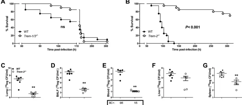
pseudo-malleiand observed them for 14 days (Fig 2A and 2B). There was no significant difference in
survival betweenTrem-1/3-/-and WT mice following a lethalB.pseudomalleichallenge: 95% of
Trem-1/3-/-and WT mice died within 6 days after inoculation (Fig 2A). Strikingly however, Trem-2-/-mice were significantly protected: 70% ofTrem-2-/-survived until the end of the 14-day
observation period while all WT mice died within 6 days (P<0.001;Fig 2B).
Enhanced bacterial clearance in
Trem-2
-/-mice
To substantiate the finding thatTrem-2-/-mice are protected during melioidosis, we
deter-mined bacterial loads in lung and BALF as well as in blood, liver and spleen 72h post-infection.
Relative to WT mice,Trem2-/-mice displayed strongly reduced bacterial loads both at the
Fig 1. Increased TREM-1 and TREM-2 expression in experimental melioidosis.TREM-1 and TREM-2 mRNA expression was determined in wild type (WT) mice prior to infection or at 24 or 72h post-infection with 5 x 10² CFUB.pseudomalleiintranasally. TREM-1 mRNA expression in lung (A) and liver (B) was determined. Likewise, TREM-2 mRNA expression was measured in lung (C) and liver (D) tissue. Data are presented as fold induction compared to the mRNA expression in uninfected mice (all RNA data are normalized to GAPDH). Data are mean±SEM, n = 4–5 mice/group. *P<0.05,**P<0.01, compared to gene-expression at t = 0h (Mann-WhitneyUtest).
primary site of infection (P<0.01 for lung and BALF;Fig 2C and 2D) as well as in distant
organs and the systemic compartment (P<0.01 for blood and spleen;Fig 2E–2G). 72h
post-infection 100% of WT but only 20% ofTrem2-/-mice had become bacteraemic. These findings
indicate that TREM-2 plays a key deleterious role during experimental melioidosis by antago-nizing bacterial clearance leading to increased dissemination of infection.
Trem-2
-/-mice demonstrate reduced lung inflammation
Since TREM-2 has been described as a negative regulator of inflammation [19,20], we next
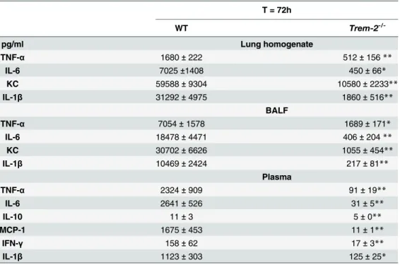
assessed the inflammatory response in the pulmonary compartment. Therefore we studied the extent of inflammation in lung homogenates and BALF. We observed markedly decreased
lev-els of pro-inflammatory cytokines TNF-α, IL-6, IL-1βand the chemokine KC in both lung
homogenates and BALF of TREM-2 deficient mice compared to controls (P<0.01–0.05;
Table 1). To further obtain insight into the involvement of TREM-2 in the inflammatory
response followingB.pseudomalleiinfection, we semi-quantitatively scored lung histology
slides generated fromTrem-2-/-and WT mice. However, all mice displayed severe pulmonary
inflammation and no differences were observed between the mouse strains (Fig 3A–3C).
Neu-trophil recruitment to the lung is an essential part of the inflammatory host response to melioi-dosis. Therefore, we determined the granulocyte influx into the pulmonary compartment by
Ly6G-immunostaining in WT andTrem-2-/-mice 72h post-infection withB.pseudomallei
(Fig 3D–3F). This immunostaining recognizes Gr-1, that is granulocyte-specific,
Fig 2. Survival ofTrem-2-/-mice, but not ofTrem-1/3-/-mice, is enhanced in experimental melioidosis.Survival was observed for every 4-6h, up to a maximum of 14 days after intranasal inoculation with 5 x 10² CFUB.pseudomalleiin wild-type (WT; closed circles) andTrem-1/3-/-mice (open circles;A). Similarly, survival of WT (closed circles) andTrem-2-/-mice (open circles) was determined (B) (n = 20 per group). ThePvalue indicates significance of the difference in survival betweenTrem-2
-/-and WT mice (Kaplan-Meier analysis, followed by a log-rank test). ns = not significant. In addition, WT (closed circles) andTrem-2-/-mice (open circles) were infected with 5 x 10² colony forming units (CFU) ofB.pseudomalleiintranasally (n = 5–6 mice per group)
Corresponding to the diminished bacterial loads and decreased levels of cyto- and chemokines
in lung tissue, a reduced influx of granulocytes in lungs ofTrem-2-/-mice was found (P<0.05,
Fig 3D).
Trem-2 deficiency leads to decreased distant organ injury during
experimental melioidosis
To evaluate the role of TREM-2 in the systemic inflammatory response, we determined plasma
cytokine levels 72h post-infection withB.pseudomallei. Consistent with the lower pulmonary
cytokine levels and bacterial loads, we found that the plasma levels of TNF-α, IL-6, IL-1β,
MCP-1, IL-10, IFN-γand KC were all significantly reduced inTrem-2-/-mice compared to
WTs (P<0.01–0.05,Table 1). Furthermore, we obtained spleen pathology scores and
per-formed routine clinical chemistry tests to evaluate hepatic, renal and systemic injury. In line
with the observed decreased splenic bacterial loads,Trem-2-/-mice showed less inflammation
compared to WT mice 72h after inoculation withB.pseudomallei(P<0.05;Fig 4A). Plasma
AST levels ofTrem-2-/-mice were decreased when compared to controls 72h post-infection,
reflecting decreased hepatocellular injury in these animals (P<0.05;Fig 4B). Consistently, we
Table 1. Cytokine response in lung homogenates, BALF and plasma of WT andTrem-2-/-mice during experimental melioidosis.
T = 72h
WT Trem-2
-/-pg/ml Lung homogenate
TNF-α 1680±222 512±156**
IL-6 7025±1408 450±66*
KC 59588±9304 10580±2233**
IL-1β 31292±4975 1860±516**
BALF
TNF-α 7054±1578 1689±171*
IL-6 18478±4471 406±204**
KC 30702±6626 1055±454**
IL-1β 10469±2424 217±81**
Plasma
TNF-α 2324±909 91±19**
IL-6 2641±526 31±5**
IL-10 11±3 5±0**
MCP-1 1675±453 11±1**
IFN-γ 158±62 17±3**
IL-1β 1123±303 125±25*
Cytokine levels in lung homogenate, broncho-alveolarfluid (BALF) and plasma were measured after intranasal infection with 5 x 10² CFU wild-typeB.pseudomallei. Wild-type (WT) andTrem-2-/-mice were
sacrificed 72 h after infection. Data are represented as means±SEM (n = 5-6/group). TNF-α= Tumor
necrosis factor-α; IL = Interleukin; MCP-1 = Monocyte Chemoattractant Protein-1; KC = Keratinocyte
Chemoattractant; IFN-γ= Interferon-γ *P<0.05
**P<0.01.
observed a trend towards lower ALT, BUN and LDH levels inTrem-2-/-mice compared to
con-trols suggesting less organ damage respectively (Fig 4C–4E).
Lack of TREM-2 leads to a reduced inflammatory response ex vivo, but
does not impact on phagocytosis of
B
.
pseudomallei
by macrophages
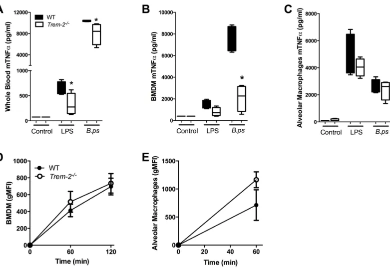
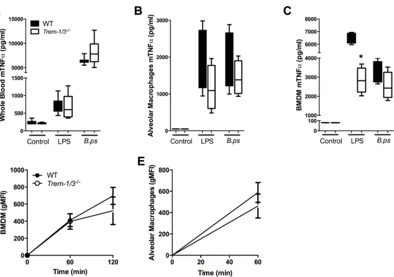
Having established that TREM-2 plays an important deleterious role during experimental melioidosis and is involved in the inflammatory response, we next assessed what cells are responsible for these effects. It is known that blood monocytes, alveolar macrophages (AM)
and BMDM express TREM-2 [25], therefore we harvested these cells and first stimulated them
overnight with the TLR4-ligand LPS andB.pseudomallei. We found a clear trend towards
Fig 3. Reduced neutrophil influx in lungs ofTrem-2-/-mice, without affecting lung pathology.
Lung pathology was determined in wild-type (WT; black bars) andTrem-2-/-mice (white bars) infected with 5 x 10² CFUB.pseudomalleiat 72h post-infection as described in the Methods section (A). Representative lung slides of WT (B) andTrem-2-/-mice (C) (original magnification 10x). Neutrophil influx was defined by Ly6G positivity (expressed as % of total lung surface;D). Representative photographs of Ly6G-immunostaining for granulocytes on lung slides of WT (E) andTrem-2-/-mice (F) (original magnification 10x). Data are expressed as mean±SEM, n = 5–6 mice per group per time point.*P<0.05. (Mann-WhitneyUtest).
lower TNF-αlevels when whole blood, AM or BMDM ofTrem-2-/-mice were stimulated with
LPS (Fig 5A–5C). This effect was even more pronounced after stimulation withB.
pseudomal-lei: the TNF-αresponse of whole blood and BMDM derived from TREM-2 deficient mice was
significantly reduced compared to controls (P<0.05;Fig 5A and 5B). Considering TREM-2’s
known phagocytic properties [24,25] and the observed lower local and systemic bacterial loads
in TREM-2-deficient mice, we determined the phagocytic capacity of AM and BMDM
har-vested from WT andTrem-2-/-mice. Despite a trend towards enhanced phagocytosis of
FITC-labelledB.pseudomalleiby TREM-2 deficient macrophages, no significant differences were
found (Fig 5D and 5E). In line, TREM-2 did not impact on the intracellular killing ofB.
pseudo-malleiby BMDM (S1 Fig).
Limited role of TREM-1/3 in the host defense during experimental
melioidosis
In a final set of experiments we studied the role of TREM-1 in the host defense againstB.
pseu-domalleiusingTrem-1/3-/-mice. In contrast to the data derived fromTrem-2-/-mice, no
differ-ences in bacterial counts in lung or BALF were observed betweenB.pseudomallei-challenged
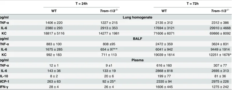
Trem-1/3-/-and WT mice (Fig 6A and 6B). In line, TREM-1 deficiency did not impact on lung pathology and cytokine levels, except for decreased KC levels, which did not influence Fig 4. Reduced distant organ damage inTrem-2-/-mice.
At 72h post-infection with 5 x 10² CFUB.pseudomalleiintranasally splenic injury (A) in WT (black bars) andTrem-2-/-mice (white bars) was quantified as described in the Methods section. Plasma levels of aspartate transaminase (AST;B), alanine transaminase (ALT;C), Lactate dehydrogenase (LDH;D) and blood urea nitrogen (BUN;E) in WT andTrem-2-/-mice were determined. Data are expressed as mean±SEM. n = 5–6 mice per group per time point.*P<0.05 (Mann-WhitneyUtest).
pulmonary neutrophilic content as determined by Ly6-stainings (Fig 6E and 6F,Table 2). However, TREM-1 did influence bacterial dissemination as bacterial loads in blood and liver
were significantly decreased inTrem-1/3-/-mice compared to WTs 72h after infection (P<0.01;
Fig 6C and 6D). We next evaluated TREM-1’s role in systemic inflammation and end organ damage. At 72h post-infection, the levels of key regulatory cytokines in the systemic
compart-ment (TNF-α, IL-6, IL-10, MCP-1 and IFN-γ) did not differ betweenTrem-1/3-/-mice and WT
(Table 2). Induced pathology of the spleen (Fig 6G) was similar inTrem-1/3-/-and WT mice. In correspondence with the lower hepatic bacterial counts at 72h, we found lower levels of the
hepatocellular injury markers AST and ALT levels inTrem-1/3-/-mice compared to WT mice
(Fig 6H and 6I). LDH levels, reflecting general organ injury, were elevated inTrem-1/3-/-mice at 24 h, while they were reduced compared to their WT counterparts at 72h post-infection
(P<0.05;Fig 6J). No difference in plasma BUN levels was observed between mice strains
(Fig 6K).
Fig 5. TREM-2 deficiency reduces cellular responsivenessex vivo.Whole blood(A), bone marrow derived macrophages (BMDM;B) and alveolar macrophages (AM;C) of WT (black bars) andTrem-2-/-mice (white bars) were stimulated with medium,E.coliLPS (100 ng/ml) or heat-inactivatedB.
pseudomallei(107 CFU/ml or MOI of 50). Supernatant was collected after 20 h of stimulation and assayed for TNF-α. In addition, WT andTrem-2-/-BMDM
(D)and AM(E)were incubated at 37°C with FITC labeled heat-inactivatedB.pseudomalleiafter which time-dependent phagocytosis was determined. Data are presented as mean±SEM and are representative of two or three independent experiments. n = 4 or 8 per group.*P<0.05 (Mann-WhitneyU test).
Fig 6. Effect of TREM-1 deficiency on bacterial clearance, pulmonary neutrophil influx and organ damage during experimental melioidosis.WT (closed circles/black bars) andTrem-1/3-/-mice (open circles/ white bars) were intranasally infected with 5 x 10² CFU ofB.pseudomalleiand sacrificed 24 and 72 h post-infection, followed by determination of bacterial loads in lung homogenate(A),BALF(B),blood(C)and liver(D).Neutrophil influx as determined by % Ly6G positive surface of lung slides was calculated for WT andTrem-1/3
-/-mice(E).Lung(F)and spleen(G)pathology was scored as described in the Methods section. Aspartate transaminase (AST;H),alanine transaminase (ALT;I), lactate dehydrogenase (LDH;J)and blood urea nitrogen (BUN;K) were measured as a marker for end organ damage. Data are expressed as mean±SEM. n = 7–8 mice per group.*P<0.05;**P<0.01
(Mann-WhitneyUtest).
doi:10.1371/journal.pntd.0004747.g006
Table 2. Cytokine responses in lung homogenates, BALF and plasma of WT andTrem-1/3-/-mice during experimental melioidosis.
T = 24h T = 72h
WT Trem-1/3-/- WT Trem-1/3
-/-pg/ml Lung homogenate
TNF-α 1406±220 1227±215 2135±312 2312±386
IL-6 2380±293 2913±353 17694±3121 29910±4668
KC 18817±5116 14277±1981 71606±6071 69866±8092
pg/ml BALF
TNF-α 883±100 808±95 2472±359 3624±831
IL-6 1675±285 654±97** 6041±942 9449±1914
KC 992±183 711±113 19039±1614 12251±1678*
pg/ml Plasma
TNF-α 12±1 9±1 616±160 307±77
IL-6 143±36 133±19 2868±818 2695±313
IL-10 8±2 20±6 199±77 81±36
MCP-1 263±63 92±25* 2335±94 2975±226
IFN-γ 28±4 26±4 1606±445 1275±242
Cytokine levels in plasma, lung homogenate and broncho-alveolarfluid (BALF) were measured after intranasal infection with 5 x 10² CFU wild-typeB. pseudomallei. Wild-type (WT) andTrem-1/3-/-mice were sacrificed 24 and 72 h after infection. Data are represented as means±SEM (n = 7 or 8/group per time point). TNF-α= Tumor necrosis factor-α; IL = Interleukin; MCP-1 = Monocyte Chemoattractant Protein-1; KC = Keratinocyte Chemoattractant;
IFN-γ= Interferon-γ *P<0.05
**P<0.01.
TREM-1 deficiency does not impact on ex vivo cytokine responsiveness
and phagocytosis nor intracellular killing of
B
.
pseudomallei
TREM-1 is abundantly expressed on monocytes and macrophages following exposure toB.
pseudomallei[31]. In line with previous findings [11],Trem-1/3-/-BMDM produced less
TNF-αin response to LPS stimulation (P<0.05;Fig 7B). Surprisingly, no differences in cellular
responsiveness were found between AM and whole blood derived from WT andTrem-1/
3-/-mice (Fig 7A–7C). Lastly, we wished to determine whether TREM-1 contributes to
phago-cytosis and/or killing ofB.pseudomallei. No differences in phagocytic and killing capacities
between WT and TREM-1 deficient BMDM were observed (Fig 7D and 7E).
Discussion
TREM-1 and TREM-2 are innate immune receptors that have demonstrated to either amplify or regulate TLR and NLR signaling after recognition of pathogen-associated molecular
Fig 7. No effect of TREM-1 deficiency on the cellular responsiveness and phagocytosis or intracellular killing ofB.pseudomallei.Whole blood (A), bone marrow derived macrophages (BMDM;B) and alveolar macrophages (AM;C)of WT andTrem-1/3-/-mice were stimulated with medium,E.coli LPS(100 ng/ml) or heat-inactivated wild typeB.pseudomallei(107 CFU/ml at a MOI of 50). TNF-αlevels were measured in the supernatant obtained after 20 h of stimulation. BMDM (D) and AM (E) of WT andTrem-1/3
-/-mice were incubated at 37°C with FITC labeled heat-inactivatedB.pseudomalleiafter which time-dependent phagocytosis was determined. Data are expressed as mean±SEM and are representative of two or three independent experiments. n = 4 or 8 (for the whole blood assay) per group.*P<0.05 (Mann-WhitneyUtest).
patterns. Our study is the first to examine the role of both TREM-1 and TREM-2 during exper-imental melioidosis. We observed increased TREM-1 and TREM-2 expression during experi-mental melioidosis, both at the local site of infection and systemically. Subsequently, we found
that TREM-2 impairs the host defense against murineB.pseudomallei-induced sepsis, as
dem-onstrated by an improved survival of infectedTrem-2-/-mice as a direct result of diminished
bacterial dissemination, decreased inflammation and less organ damage. Ourex vivostudies
suggest that the protective effect of TREM-2 deficiency in part results from the diminished capacity of TREM-2-deficient macrophages to elicit a pro-inflammatory response which is an important contributor to organ injury in the event of sepsis. TREM-1 was also found to play a
detrimental role duringB.pseudomalleiinfection, which is in line with our earlier finding that
blocking TREM-1 could improve survival during melioidosis [31]. However when compared
to TREM-2 the role of TREM-1 in the host response againstB.pseudomalleiseems to be
limited.
Previous studies have demonstrated that soluble TREM-1 levels are up-regulated in plasma
of patients with sepsis, pneumonia and melioidosis [31,41,42]. In addition, it is known that
surface TREM-1 expression is increased on monocytes of melioidosis patients [31]. However,
soluble TREM-1 levels in septic patients do not always correlate to the expression of
mem-brane-bound TREM-1 on different myeloid cell types [31,43]. Less is known about the kinetics
of TREM-2 expression during infection. A recent study demonstrated that during sepsis TREM-2 expression on ascites-retrieved cells of patients with abdominal sepsis was increased
[27]. Correspondingly, TREM-2 was up-regulated on AM of mice infected withS.pneumoniae
[25]. In line with these earlier studies, we now show that both TREM-1 and TREM-2 mRNA
expression is elevated in lung and liver tissue of mice infected withB.pseudomallei. Further
research however is warranted to study the cell surface protein expression of TREM-2 on neu-trophils and macrophages during melioidosis.
Thein vivorole of TREM-2 in infectious diseases remains ill defined. In a model studyingP. aeruginosakeratitis TREM-2 deficiency increased corneal bacterial loads [44]. More recently, Chenet al. demonstrated that TREM-2 is required for efficient bacterial clearance in a murine
polymicrobial sepsis model using a TREM-2 blocking recombinant protein [27]. In the same
study it was shown that administration of TREM-2 overexpressing bone marrow derived
mye-loid cells improved survival during polymicrobial sepsis, but not endotoxaemia [27]. In sharp
contrast, Gawish et al. demonstrated a beneficial effect of TREM-2 deficiency during
endotox-aemia [45]. The same group also observed a survival benefit ofTrem-2-/-mice duringS.
pneu-moniaepneumonia [25], while no effect on mortality of TREM-2 deficiency was seen duringE.
colisepsis [45]. To evaluate how TREM-2 deficiency led to increased clearance ofB.
pseudo-mallei, we assessed the functional roles of macrophages that express TREM-2 [19,25].
TREM-2 is known to be involved in direct killing [27,44] and phagocytosis of bacteria by macrophages
[24,25]. Interestingly, we did not find impaired bacterial killing or phagocytosis ofB.
pseudo-malleiby BMDM or AM ofTrem-2-/-mice. Several characteristics of this facultative
intracellu-lar bacterium when compared to other bacteria might in part explain these discrepancies;B.
pseudomalleiis capable of invading both phagocytic and non-phagocytic cells [46] and circum-vents intracellular defense mechanisms efficiently in order to replicate and spread to adjacent cells [47,48].
TREM-2 is traditionally regarded as a negative regulator of thein vitroinflammatory
response in response to TLR-ligands [19,21,45] In contrast, our study now demonstrates that
TREM-2 deficiency leads to a reduced inflammatory response toB.pseudomalleibothex vivo
elements, can explain these inconsistencies: differences in mice strains used (BALB/C versus C57Bl/6), different experimental murine models (e.g. caecal ligation and puncture (CLP)-model versus a intranasal inhalation (CLP)-model for sepsis), differences in TREM-2 blockade (e.g. by
using TREM-2 deficient mice or TREM-2 antibodies) and lastly the difference of anin vitro
approach in contrast to ourex vivocellular challenge model. Interestingly, a recent study
showed augmented inflammation by TREM-2 deficient peritoneal macrophages in response to
LPS [45], while the same group observed the reversed phenotype in alveolar macrophages [25],
underlining possible cell-specific functions of TREM-2. Of importance, neutrophil recruitment
to the lung, an important defense mechanism during melioidosis [32,51], was reduced in
Trem-2-/-mice during experimental melioidosis as determined by Ly6-staining. This may be a potential result of the decreased inflammatory response and production of chemokines
follow-ing infection. In this respect, it is noteworthy, that IL-1β–which we and others have shown to
be involved in excessive deleterious neutrophil influx during experimental melioidosis [37,52]
—was also significantly reduced inTrem-2-/-mice. No differences were observed in the influx
of macrophages (S2 Fig). Excessive inflammation and neutrophil influx and activation can lead
towards multi-organ failure [53], which is almost universally seen in lethal cases of melioidosis.
Distant organ injury was significantly reduced inTrem-2-/-mice, potentially as a result of a
reduced influx of inflammatory cells.Trem-2-/-mice displayed an evidently reduced
inflamma-tory response, which resulted in a strong survival benefit. In addition, it is well known thatB.
pseudomalleican replicate intracellularly [28], and neutrophils may act as its permissive host
cell [52]. We could therefore hypothesize that the anti-inflammatory phenotype and the
reduced bacterial loads seen in TREM-2 deficient mice are a result of decreased intracellular bacterial replication at the infection site, due to reduced neutrophilic influx.
Taken together, during melioidosis, TREM-2 deficiency resulted in a restricted inflamma-tory response, thereby decreasing organ damage and mortality. Future research should focus
on the potential of anti-TREM-2 treatment ofB.pseudomallei-infected mice.
TREM-1 amplifies TLR-responses and therefore might dangerously enhance the
inflamma-tory response to bacterial infection [18]. Controversial results have been found on the role of
TREM-1 during bacterial infection. TREM-1 deficiency has shown to be detrimental during
endotoxaemia [17] and polymicrobial sepsis [12,54], while in contrast, moderate levels of
TREM-1 can improve survival during polymicrobial sepsis, but not endotoxaemia [55].
Block-ade of TREM-1 with a peptide called LP17 could partially protect mice fromB.pseudomallei
induced lethality [31]. In this study however, we observed, using the same infection model, that
survival ofB.pseudomallei-infected TREM-1-deficient mice was similar to WTs. This might be
explained by the fact that these mice were completely TREM-1-deficient and in addition lacked TREM-3, a DAP12-coupled activating receptor on murine macrophages, which supposedly
acts as an activating receptor [56]. In contrast, in humans TREM-3 is a pseudogene [56].
How-ever, since DAP12 is known to both potentiate and attenuate TLR-signaling, it is perhaps not
surprising that the net-effect on bacterial clearance ofB.pseudomalleiis not affected.
TREM-1 has other functions next to TLR-signaling enhancement, such as phagocytosis and
the production of reactive oxygen species [57]. Furthermore, TREM-1 has been recently linked
to trans-epithelial migration of neutrophils after infection withP.aeruginosa[15]. Blocking
TREM-1 completely could therefore interfere with these important antibacterial mechanisms.
We did not find a role for TREM-1 in the killing or phagocytosis ofB.pseudomallei, which is in
line with the fact that TREM-1/3 deficiency in neutrophils neither impacts on bacterial killing,
phagocytosis and chemotaxis ofP.aeruginosa[15]. This suggests that other phagocytic
recep-tors on leukocytes are more important for the efficient eradication ofB.pseudomallei[38,58,
Murine models like the one used here, which make use of relatively young mice exposed to an intranasal bacterial inoculum, do show inter-experiment variation, as reflected by differ-ences in bacterial dissemination and as a result inflammation at the latter time-points before mice will succumb to infection. In addition, caution is needed when extrapolating data from
murine experiments to human disease.”
Taking these precautions into mind, we here demonstrate that murine melioidosis is associ-ated with increased TREM-1 and -2 expression. TREM-2 deficiency is beneficial during experi-mental Gram-negative sepsis caused by a clinical relevant pathogen, resulting in lower bacterial loads, reduced organ damage, decreased inflammation and improved survival. When com-pared to TREM-2, TREM-1 plays a limited detrimental role during experimental melioidosis. These results provide new information on the expression and function of TREM-2 during melioidosis and may demonstrate its potential therapeutic usefulness.
Supporting Information
S1 Appendix. Supplemental materials and methods.
(DOC)
S1 Fig. Intracellular killing ofB.pseudomalleiby BMDM is not impaired by TREM-2
defi-ciency.WT andTrem-2-/-BMDM were incubated at 37°C with liveB.pseudomalleiafter which
time-dependent intracellular killing was determined. Data are presented as mean ± SEM and are representative of two independent experiments. n = 6 per group (Mann-Whitney U test). (TIF)
S2 Fig. Similar macrophage influx in BALF of WT andTrem-2-/-during experimental
melioidosis.Macrophage influx in broncho-alveolar lavage fluid (BALF) was determined 72h
post-infection with 5 x 10² CFUB.pseudomalleiin wild-type (WT; black circles) andTrem-2
-/-mice (white circles). Data are presented as mean ± SEM n = 5–6 mice/group (Mann- Whitney
U test). (TIF)
Acknowledgments
We thank Marieke ten Brink and Joost Daalhuisen for expert technical assistance. We are grateful for the help of Regina de Beer who performed histopathological and immunohisto-chemical stainings, and Onno de Boer for his assistance in analyzing immunohistologically stained slides.
Author Contributions
Conceived and designed the experiments: TAFW WJW AFdV TvdP. Performed the experi-ments: TAFW JML HKdJ. Analyzed the data: TAFW TJH JML HKdJ JJTHR AFdV WJW TvdP. Contributed reagents/materials/analysis tools: TJH MC. Wrote the paper: TAFW TJH WJW TvdP MC.
References
1. Hotchkiss RS, Monneret G, Payen D. Sepsis-induced immunosuppression: from cellular dysfunctions to immunotherapy. Nature reviews Immunology. 2013; 13(12):862–74. doi:10.1038/nri3552PMID:
24232462; PubMed Central PMCID: PMC4077177.
3. Wiersinga WJ. Current insights in sepsis: from pathogenesis to new treatment targets. Current opinion in critical care. 2011; 17(5):480–6. doi:10.1097/MCC.0b013e32834a4aebPMID:21900767.
4. Broz P, Monack DM. Newly described pattern recognition receptors team up against intracellular patho-gens. Nature reviews Immunology. 2013; 13(8):551–65. doi:10.1038/nri3479PMID:23846113.
5. Arts RJ, Joosten LA, van der Meer JW, Netea MG. TREM-1: intracellular signaling pathways and inter-action with pattern recognition receptors. Journal of leukocyte biology. 2013; 93(2):209–15. doi:10.
1189/jlb.0312145PMID:23108097.
6. Klesney-Tait J, Turnbull IR, Colonna M. The TREM receptor family and signal integration. Nat Immunol. 2006; 7(12):1266–73. doi:10.1038/ni1411PMID:17110943.
7. Sharif O, Knapp S. From expression to signaling: roles of TREM-1 and TREM-2 in innate immunity and bacterial infection. Immunobiology. 2008; 213(9–10):701–13. doi:10.1016/j.imbio.2008.07.008PMID:
18926286.
8. Aoki N, Kimura S, Xing Z. Role of DAP12 in innate and adaptive immune responses. Current pharma-ceutical design. 2003; 9(1):7–10. PMID:12570670.
9. Tessarz AS, Cerwenka A. The TREM-1/DAP12 pathway. Immunology letters. 2008; 116(2):111–6. doi:
10.1016/j.imlet.2007.11.021PMID:18192027.
10. Read CB, Kuijper JL, Hjorth SA, Heipel MD, Tang X, Fleetwood AJ, et al. Cutting Edge: Identification of Neutrophil PGLYRP1 as a Ligand for TREM-1. Journal of immunology. 2015. doi:10.4049/jimmunol. 1402303PMID:25595774.
11. Bouchon A, Dietrich J, Colonna M. Cutting edge: inflammatory responses can be triggered by TREM-1, a novel receptor expressed on neutrophils and monocytes. J Immunol. 2000; 164(10):4991–5. PMID:
10799849.
12. Bouchon A, Facchetti F, Weigand MA, Colonna M. TREM-1 amplifies inflammation and is a crucial mediator of septic shock. Nature. 2001; 410(6832):1103–7. PMID:11323674.
13. Lin YT, Tseng KY, Yeh YC, Yang FC, Fung CP, Chen NJ. Triggering Receptor Expressed on Myeloid Cells-1 promotes survival during Klebsiella pneumoniae liver abscess in mice. Infection and immunity. 2014. doi:10.1128/IAI.01347-13PMID:24396044
14. Hommes TJ, Hoogendijk AJ, Dessing MC, Van't Veer C, Florquin S, Colonna M, et al. Triggering recep-tor expressed on myeloid cells-1 (TREM-1) improves host defence in pneumococcal pneumonia. The Journal of pathology. 2014; 233(4):357–67. doi:10.1002/path.4361PMID:24752755.
15. Klesney-Tait J, Keck K, Li X, Gilfillan S, Otero K, Baruah S, et al. Transepithelial migration of neutrophils into the lung requires TREM-1. The Journal of clinical investigation. 2013; 123(1):138–49. doi:10.1172/
JCI64181PMID:23241959; PubMed Central PMCID: PMC3533287.
16. Gibot S, Alauzet C, Massin F, Sennoune N, Faure GC, Bene MC, et al. Modulation of the triggering receptor expressed on myeloid cells-1 pathway during pneumonia in rats. The Journal of infectious dis-eases. 2006; 194(7):975–83. doi:10.1086/506950PMID:16960786.
17. Gibot S, Buonsanti C, Massin F, Romano M, Kolopp-Sarda MN, Benigni F, et al. Modulation of the trig-gering receptor expressed on the myeloid cell type 1 pathway in murine septic shock. Infection and immunity. 2006; 74(5):2823–30. PMID:16622220.
18. Weber B, Schuster S, Zysset D, Rihs S, Dickgreber N, Schurch C, et al. TREM-1 Deficiency Can Atten-uate Disease Severity without Affecting Pathogen Clearance. PLoS Pathog. 2014; 10(1):e1003900. doi:10.1371/journal.ppat.1003900PMID:24453980; PubMed Central PMCID: PMC3894224. 19. Turnbull IR, Gilfillan S, Cella M, Aoshi T, Miller M, Piccio L, et al. Cutting edge: TREM-2 attenuates
mac-rophage activation. Journal of immunology. 2006; 177(6):3520–4. PMID:16951310.
20. Hamerman JA, Jarjoura JR, Humphrey MB, Nakamura MC, Seaman WE, Lanier LL. Cutting edge: inhi-bition of TLR and FcR responses in macrophages by triggering receptor expressed on myeloid cells (TREM)-2 and DAP12. J Immunol. 2006; 177(4):2051–5. PMID:16887962.
21. Ito H, Hamerman JA. TREM-2, triggering receptor expressed on myeloid cell-2, negatively regulates TLR responses in dendritic cells. European journal of immunology. 2012; 42(1):176–85. doi:10.1002/
eji.201141679PMID:21956652; PubMed Central PMCID: PMC3444819.
22. Takahashi K, Rochford CD, Neumann H. Clearance of apoptotic neurons without inflammation by microglial triggering receptor expressed on myeloid cells-2. The Journal of experimental medicine. 2005; 201(4):647–57. doi:10.1084/jem.20041611PMID:15728241; PubMed Central PMCID:
PMC2213053.
23. Daws MR, Sullam PM, Niemi EC, Chen TT, Tchao NK, Seaman WE. Pattern recognition by TREM-2: binding of anionic ligands. Journal of immunology. 2003; 171(2):594–9. PMID:12847223.
2009; 184(2):215–23. doi:10.1083/jcb.200808080PMID:19171755; PubMed Central PMCID:
PMC2654305.
25. Sharif O, Gawish R, Warszawska JM, Martins R, Lakovits K, Hladik A, et al. The triggering receptor expressed on myeloid cells 2 inhibits complement component 1q effector mechanisms and exerts detri-mental effects during pneumococcal pneumonia. PLoS pathogens. 2014; 10(6):e1004167. doi:10. 1371/journal.ppat.1004167PMID:24945405; PubMed Central PMCID: PMC4055749.
26. Zhu M, Li D, Wu Y, Huang X, Wu M. TREM-2 promotes macrophage-mediated eradication of Pseudo-monas aeruginosa via a PI3K/Akt pathway. Scandinavian journal of immunology. 2014. doi:10.1111/ sji.12148PMID:24383713.
27. Chen Q, Zhang K, Jin Y, Zhu T, Cheng B, Shu Q, et al. Triggering receptor expressed on myeloid cells-2 protects against polymicrobial sepsis by enhancing bacterial clearance. American journal of respira-tory and critical care medicine. 2013; 188(2):201–12. doi:10.1164/rccm.201211-1967OCPMID:
23721075.
28. Wiersinga WJ, Currie BJ, Peacock SJ. Melioidosis. The New England journal of medicine. 2012; 367 (11):1035–44. Epub 2012/09/14. doi:10.1056/NEJMra1204699PMID:22970946.
29. Simpson AJ. Melioidosis: a clinical model for gram-negative sepsis. Journal of medical microbiology. 2001; 50(8):657–8. PMID:11478666.
30. Schweizer HP. Mechanisms of antibiotic resistance in Burkholderia pseudomallei: implications for treat-ment of melioidosis. Future microbiology. 2012; 7(12):1389–99. doi:10.2217/fmb.12.116PMID:
23231488; PubMed Central PMCID: PMC3568953.
31. Wiersinga WJ, Veer C, Wieland CW, Gibot S, Hooibrink B, Day NP, et al. Expression profile and func-tion of triggering receptor expressed on myeloid cells-1 during melioidosis. The Journal of infectious diseases. 2007; 196(11):1707–16. doi:10.1086/522141PMID:18008257.
32. Wiersinga WJ, de Vos AF, de Beer R, Wieland CW, Roelofs JJ, Woods DE, et al. Inflammation patterns induced by different Burkholderia species in mice. Cellular microbiology. 2008; 10(1):81–7. doi:10.
1111/j.1462-5822.2007.01016.xPMID:17645551.
33. Weehuizen TA, Wieland CW, van der Windt GJ, Duitman JW, Boon L, Day NP, et al. Expression and function of transforming growth factor beta in melioidosis. Infection and immunity. 2012; 80(5):1853–7.
doi:10.1128/IAI.05534-11PMID:22331429; PubMed Central PMCID: PMC3347439.
34. Wiersinga WJ, Wieland CW, Dessing MC, Chantratita N, Cheng AC, Limmathurotsakul D, et al. Toll-like receptor 2 impairs host defense in gram-negative sepsis caused by Burkholderia pseudomallei (Melioidosis). PLoS medicine. 2007; 4(7):e248. doi:10.1371/journal.pmed.0040248PMID:17676990; PubMed Central PMCID: PMC1950213.
35. Kager LM, Wiersinga WJ, Roelofs JJ, Meijers JC, Zeerleder SS, Esmon CT, et al. Endogenous protein C has a protective role during Gram-negative pneumosepsis (melioidosis). Journal of thrombosis and haemostasis: JTH. 2013; 11(2):282–92. doi:10.1111/jth.12094PMID:23216621.
36. Eske K, Breitbach K, Kohler J, Wongprompitak P, Steinmetz I. Generation of murine bone marrow derived macrophages in a standardised serum-free cell culture system. J Immunol Methods. 2009; 342 (1–2):13–9. doi:10.1016/j.jim.2008.11.011PMID:19133267.
37. Koh GC, Weehuizen TA, Breitbach K, Krause K, de Jong HK, Kager LM, et al. Glyburide reduces bacte-rial dissemination in a mouse model of melioidosis. PLoS neglected tropical diseases. 2013; 7(10): e2500. doi:10.1371/journal.pntd.0002500PMID:24147174; PubMed Central PMCID: PMC3798430. 38. Wiersinga WJ, Kager LM, Hovius JW, van der Windt GJ, de Vos AF, Meijers JC, et al. Urokinase
recep-tor is necessary for bacterial defense against pneumonia-derived septic melioidosis by facilitating phagocytosis. Journal of immunology. 2010; 184(6):3079–86. doi:10.4049/jimmunol.0901008PMID:
20142364.
39. Breitbach K, Sun GW, Kohler J, Eske K, Wongprompitak P, Tan G, et al. Caspase-1 mediates resis-tance in murine melioidosis. Infection and immunity. 2009; 77(4):1589–95. doi:10.1128/IAI.01257-08
PMID:19179418; PubMed Central PMCID: PMC2663179.
40. Currie BJ, Ward L, Cheng AC. The epidemiology and clinical spectrum of melioidosis: 540 cases from the 20 year Darwin prospective study. PLoS neglected tropical diseases. 2010; 4(11):e900. Epub 2010/12/15. doi:10.1371/journal.pntd.0000900PMID:21152057.
41. Gibot S, Kolopp-Sarda MN, Bene MC, Cravoisy A, Levy B, Faure GC, et al. Plasma level of a triggering receptor expressed on myeloid cells-1: its diagnostic accuracy in patients with suspected sepsis. Annals of internal medicine. 2004; 141(1):9–15. PMID:15238365.
43. Oku R, Oda S, Nakada TA, Sadahiro T, Nakamura M, Hirayama Y, et al. Differential pattern of cell-sur-face and soluble TREM-1 between sepsis and SIRS. Cytokine. 61(1):112–7. PMID:23046618. doi:10.
1016/j.cyto.2012.09.003
44. Sun M, Zhu M, Chen K, Nie X, Deng Q, Hazlett LD, et al. TREM-2 promotes host resistance against Pseudomonas aeruginosa infection by suppressing corneal inflammation via a PI3K/Akt signaling path-way. Investigative ophthalmology & visual science. 54(5):3451–62. PMID:23611998.
45. Gawish R, Martins R, Bohm B, Wimberger T, Sharif O, Lakovits K, et al. Triggering receptor expressed on myeloid cells-2 fine-tunes inflammatory responses in murine Gram-negative sepsis. FASEB journal: official publication of the Federation of American Societies for Experimental Biology. 2015; 29(4):1247–
57. doi:10.1096/fj.14-260067PMID:25477281.
46. Jones AL, Beveridge TJ, Woods DE. Intracellular survival of Burkholderia pseudomallei. Infection and immunity. 1996; 64(3):782–90. PMID:8641782; PubMed Central PMCID: PMC173838.
47. Kespichayawattana W, Rattanachetkul S, Wanun T, Utaisincharoen P, Sirisinha S. Burkholderia pseu-domallei induces cell fusion and actin-associated membrane protrusion: a possible mechanism for cell-to-cell spreading. Infection and immunity. 2000; 68(9):5377–84. PMID:10948167; PubMed Central
PMCID: PMC101801.
48. Allwood EM, Devenish RJ, Prescott M, Adler B, Boyce JD. Strategies for Intracellular Survival of Bur-kholderia pseudomallei. Frontiers in microbiology. 2011; 2:170. doi:10.3389/fmicb.2011.00170PMID: 22007185; PubMed Central PMCID: PMC3159172.
49. Sieber MW, Jaenisch N, Brehm M, Guenther M, Linnartz-Gerlach B, Neumann H, et al. Attenuated inflammatory response in triggering receptor expressed on myeloid cells 2 (TREM2) knock-out mice fol-lowing stroke. PloS one. 8(1):e52982. PMID:23301011. doi:10.1371/journal.pone.0052982
50. Correale C, Genua M, Vetrano S, Mazzini E, Martinoli C, Spinelli A, et al. Bacterial sensor triggering receptor expressed on myeloid cells-2 regulates the mucosal inflammatory response. Gastroenterol-ogy. 2013; 144(2):346–56 e3. doi:10.1053/j.gastro.2012.10.040PMID:23108068.
51. Easton A, Haque A, Chu K, Lukaszewski R, Bancroft GJ. A critical role for neutrophils in resistance to experimental infection with Burkholderia pseudomallei. The Journal of infectious diseases. 2007; 195 (1):99–107. doi:10.1086/509810PMID:17152013.
52. Ceballos-Olvera I, Sahoo M, Miller MA, Del Barrio L, Re F. Inflammasome-dependent pyroptosis and IL-18 protect against Burkholderia pseudomallei lung infection while IL-1beta is deleterious. PLoS path-ogens. 2011; 7(12):e1002452. doi:10.1371/journal.ppat.1002452PMID:22241982; PubMed Central PMCID: PMC3248555.
53. Broquet A, Roquilly A, Jacqueline C, Potel G, Caillon J, Asehnoune K. Depletion of natural killer cells increases mice susceptibility in a Pseudomonas aeruginosa pneumonia model. Critical care medicine. 2014; 42(6):e441–50. doi:10.1097/CCM.0000000000000311PMID:24732238.
54. Gibot S, Kolopp-Sarda MN, Bene MC, Bollaert PE, Lozniewski A, Mory F, et al. A soluble form of the triggering receptor expressed on myeloid cells-1 modulates the inflammatory response in murine sep-sis. The Journal of experimental medicine. 2004; 200(11):1419–26. PMID:15557347.
55. Gibot S, Massin F, Marcou M, Taylor V, Stidwill R, Wilson P, et al. TREM-1 promotes survival during septic shock in mice. European journal of immunology. 2007; 37(2):456–66. PMID:17230441.
56. Chung DH, Seaman WE, Daws MR. Characterization of TREM-3, an activating receptor on mouse macrophages: definition of a family of single Ig domain receptors on mouse chromosome 17. European journal of immunology. 2002; 32(1):59–66. doi:
10.1002/1521-4141(200201)32:1<59::AID-IMMU59>3.0.CO;2-UPMID:11754004.
57. Radsak MP, Salih HR, Rammensee HG, Schild H. Triggering receptor expressed on myeloid cells-1 in neutrophil inflammatory responses: differential regulation of activation and survival. J Immunol. 2004; 172(8):4956–63. PMID:15067076.
58. Stevens MP, Galyov EE. Exploitation of host cells by Burkholderia pseudomallei. International journal of medical microbiology: IJMM. 2004; 293(7–8):549–55. doi:10.1078/1438-4221-00292PMID:
15149030.
59. Wiersinga WJ, van der Poll T, White NJ, Day NP, Peacock SJ. Melioidosis: insights into the pathogenic-ity of Burkholderia pseudomallei. Nature reviews Microbiology. 2006; 4(4):272–82. doi:10.1038/