Review
Brucellosis as an Emerging Threat in Developing
Economies: Lessons from Nigeria
Marie J. Ducrotoy1, Wilson J. Bertu2, Reuben A. Ocholi2, Amahyel M. Gusi2, Ward Bryssinckx3, Sue Welburn1, Ignacio Moriyo´n4*
1Division of Pathway Medicine and Centre for Infectious Diseases, School of Biomedical Sciences, College of Medicine and Veterinary Medicine, The University of Edinburgh, Chancellor’s Building, Edinburgh, United Kingdom,2Brucellosis Research Unit, National Veterinary Research Institute, Vom, Plateau State, Nigeria,3Avia-GIS, Risschotlei 33, Zoersel, Belgium,4Instituto de Salud Tropical y Depto. Microbiologı´a y Parasitologı´a, Universidad de Navarra, Edificio de Investigacio´n, Pamplona, Spain
Abstract:Nigeria is the most populous country in Africa, has a large proportion of the world’s poor livestock keepers, and is a hotspot for neglected zoonoses. A review of the 127 accessible publications on brucellosis in Nigeria reveals only scant and fragmented evidence on its spatial and temporal distribution in different epidemio-logical contexts. The few bacterioepidemio-logical studies conduct-ed demonstrate the existence ofBrucella abortusin cattle and sheep, but evidence for B. melitensis in small ruminants is dated and unclear. The bulk of the evidence consists of seroprevalence studies, but test standardiza-tion and validastandardiza-tion are not always adequately described, and misinterpretations exist with regard to sensitivity and/or specificity and ability to identify the infecting Brucella species. Despite this, early studies suggest that although brucellosis was endemic in extensive nomadic systems, seroprevalence was low, and brucellosis was not perceived as a real burden; recent studies, however, may reflect a changing trend. Concerning human brucellosis, no studies have identified theBrucella species and most reports provide only serological evidence of contact with Brucella in the classical risk groups; some suggest brucellosis misdiagnoses as malaria or other febrile conditions. The investigation of a severe outbreak that occurred in the late 1970s describes the emergence of animal and human disease caused by the settling of previously nomadic populations during the Sahelian drought. There appears to be an increasing risk of re-emergence of brucellosis in sub-Saharan Africa, as a result of the co-existence of pastoralist movements and the increase of intensive management resulting from growing urbanization and food demand. Highly contagious zoonoses like brucellosis pose a threat with far-reaching social and political consequences.
Introduction
Brucellosis is considered one of the most common global zoonoses [1]. Caused by the genusBrucella (the most common species beingBrucella abortus,B. melitensis, andB. suis), the main clinical signs in animals are abortion and infertility. Brucellosis is highly contagious and is spread through contact with aborted foetuses, vaginal fluids, placentae, placental fluids, and milk, as well as congenitally and venereally. Animals are the only significant source of human brucellosis, and transmission is via direct contact (e.g., veterinarians, abattoir workers, and livestock keepers) and through consumption of unpasteurised dairy prod-ucts. Human brucellosis is a grave and debilitating disease that may lead to permanent sequelae, requires prolonged and combined antibiotherapy, and is fatal in 1%–5% of untreated
cases [2,3]. Clinical signs are often ignored or incorrectly interpreted, and as a result, human brucellosis is severely underreported [1,4,5]. Eradicated in many developed countries after years of effort, brucellosis remains a major neglected zoonosis of low-income nations [1]. Low rates of transmission are typical of brucellosis in extensive systems, and intensification increases the risk of transmission because of higher stocking densities, increased animal contact, and higher birth index [1,6–8]. Increasing co-location of pastoralist nomadism and transhumance with settled and commercial intensive farms may thus create conditions for brucellosis emergence. These circumstances occur in sub-Saharan Africa because of an exceptionally high rural–urban migration caused by the pull of expectation of a better life, and push of unfavourable environmental conditions on agriculture [9,10].
There is a paucity of science-based evidence on brucellosis in sub-Saharan Africa [1,4,11–13], and an appraisal of historical and contemporary epidemiology (prevalence estimates, affected host species, potential reservoirs and Brucella species) is key to implementing measures for sustainable management of this disease. For a better understanding of these circumstances in the sub-Sahara, we present a review of reports on brucellosis in Nigeria.
Nigeria is the most populous country in Africa (over 170 million in 2012; http://esa.un.org/wpp/ASCII-Data/DISK_ NAVIGATION_ASCII.htm) and has an estimated livestock population of 20.49 million cattle, 23.07 million sheep, 28.07 million goats, 6.54 million pigs (http://www.fao.org/ag/againfo/ resources/en/glw/GLW_dens.html), 18,200–90,000 camels, and 210,000 horses (http://faostat.fao.org/site/573/default.aspx# ancor) [14]. Nigeria, India, Ethiopia, and Bangladesh account for 44% of poor livestock keepers globally, Nigeria ranking second
Citation: Ducrotoy MJ, Bertu WJ, Ocholi RA, Gusi AM, Bryssinckx W, et al. (2014) Brucellosis as an Emerging Threat in Developing Economies: Lessons from Nigeria. PLoS Negl Trop Dis 8(7): e3008. doi:10.1371/journal.pntd.0003008 Editor:John Andrew Crump, University of Otago, New Zealand
PublishedJuly 24, 2014
Copyright:ß2014 Ducrotoy et al. This is an open-access article distributed under the terms of the Creative Commons Attribution License, which permits unrestricted use, distribution, and reproduction in any medium, provided the original author and source are credited.
Funding:This research has received funding from the European Union’s Seventh Framework Program (FP7/2007-2013) under grant agreement nu221948, ICONZ (Integrated control of Neglected Zoonoses). Additional funding from the ‘‘Fundacio´n para la Investigacio´n Me´dica Aplicada (FIMA)’’ and from the University of Navarra is also gratefully acknowledged. The funders had no role in study design, data collection and analysis, decision to publish, or preparation of the manuscript.
Competing Interests:The authors have declared that no competing interests exist.
* E-mail: [email protected]
[8]. Livestock production has always been important in Nigeria, and the rapidly emerging livestock sector now ranks second among the 20 poorest countries [8]. With a large pastoralist population, the livestock industry has been a major focus of government attention since the colonial era (Box 1). Approximately 70% of the population live in rural areas, but there is now considerable rural– urban drift. Increasing demand for animal products has resulted in expansion of animal trade, animal and human movements, and intensification of livestock production systems. The geographic, economic, and social conditions across Nigeria determine the ruminant livestock production systems (Box 2) [15].
The climate varies from semi-arid in the North to tropical in the South. It is estimated that over a third of land that was cultivable 50 years ago is now desert across 11 of Nigeria’s northern states and that over 15 million pastoralists are threatened by decreasing access to water and pasture [16]. About half of the semi-arid and sub-humid zones in northern Nigeria are livestock and mixed crop-livestock dominated. Dairy production is concentrated in the North and the beef industry, mostly in the South. Nomadic herdsmen manage about 90% of ruminants and practice seasonal transhu-mance or year-round nomadism [17,18]. The Northeast has a hot, dry climate from January to June and rain from June to September. Transhumance is practiced to accommodate variations in available vegetation and agricultural practices and to avoid tsetse flies [19]. In the humid areas of the southern, western, and eastern states, mixed crop-livestock systems dominate, and sheep, goats, and pigs are more important. Pastoralism has been evolving in Nigeria, with farmers often combining cattle production with crop cultivation [20]. Herd sizes have been decreasing as pastoralists are becoming
more settled, enabling them to pursue crop farming. Mohammed [21] mentions that a large population of agro-pastoralists settling in the hinterlands of the urban centres in Oyo State were cattle pastoralists displaced from their traditional territories in the North by a variety of agro-ecological and socioeconomic factors. This influx stimulated a new system of livestock production.
The majority (80%) of cattle, mainly Zebu, are concentrated in the savannah zone, with only 10% of the remaining 20% (mostly Bos taurus) in the South [15] in a range of management systems (Box 2). Cattle are usually extensively managed, either under nomadic or seminomadic pastoral systems or, to a lesser extent, under traditional village systems, often in contact with small ruminants belonging to the same household. There is more intimate contact between cattle and sheep as they are co-grazed, while goats are left to scavenge free-range. In nomadic systems, small ruminants are sold and exchanged, serving as a ‘‘current account,’’ whereas cattle are traded for status and serve as a ‘‘savings account’’ [22,23]. Commercial, intensive farms are few and are located on the periphery of major towns in northern and western Nigeria. Cattle reared in extensive systems of the North and the Northeast are transported across Nigeria to the abattoirs of the Southwest to meet the high demand from the economically developed South [24,25]. According to early reports, 20% of cattle are imported, mostly from Chad and Niger [13].
Methods
A database search (PubMed, GoogleScholar, Cabdirect, and African Journals Online) was undertaken using broad terms
Box 1. Some Events of Significance in the History of Brucellosis in Nigeria
Pre-colonial era (before 1900)
N
Livestock production (cattle and small ruminants) dominat-ed by nomadic pastoralism (Fulani) in the savannah region of northern Nigeria. Agricultural land open to grazing post-harvest with mutual benefit of Fulani and farmers (fertilising effect of cow dung).British colonial administration
N
1900–1930.grammes, and mixed farming approaches. EstablishmentTsetse eradication, livestock breeding pro-of Government Veterinary Field and Research Centres (Zaria, 1913; headquarters moved to Vom in 1924; expanded to include vaccine production).N
1930s.breeds (White Fulani, Gudali, and Shuwa). ‘‘Mixed FarmingGovernment sets up stock farms to improve local Policy’’ (use of grasslands and pasture by introducing fodder and selected browse plants) to promote agro-pastoralism and range management and livestock productivity.N
1940s.plants in Vom and Agege to meet expatriate populationEstablishment of dairy herds and milk processingdemand in Jos and Lagos.
Independence (1951) to Civil War (1967–1970)
N
1950s.established in Southwest to improve indigenous cattleLivestock Improvement and Breeding Centres (humpless dwarf Muturu and Keteku) by crossing with N’dama breed (from Guinea, Sierra Leone, and Congo). N’dama becomes the breed of choice in Southwest (white Fulani remain dominant in the North).N
Western Nigeria Development Corporation established topromote importation of non-autochthonous breeds (South Devon cattle, Friesians, Holsteins, Brown Swiss, Jerseys) to upgrade local stock and increase milk production (most multiplication centres established in the Southwest, with some in the East and North).N
Programmes to encourage settlement of nomadic pasto-ralists launched (supplementary feeding programme to secure year-round fodder [1962]; grazing reserves [1965 onwards] to protect grazing lands from expanding crop-farms and to resolve clashes over land-use).N
Early 1960sand Agriculture Organization project) using semi-intensive. Smallholder steer fattening scheme (Food management systems introduced in the Southwest to ensure supply to local slaughterhouses.Post-Civil War to present
N
Early 1970s.lished to regulate all aspects of livestock industry and trade.Nigerian Livestock and Meat Authority estab-Heavy investments in intensive feedlot fattening for beef.N
1980s. Investment in direct livestock production reducesas the government focuses on livestock trade policy and oil industry. Dairy plants set up in Minna, Vom, Kaduna, but inadequate prices cause many to close down.
N
Post-1986.Role (GSAPR) in livestock production initiated in 1986 toGovernment Structural Adjustment Programme reform the Nigerian economy, including the livestock sector. The program dwindles, leading to a dominance of the private sector in livestock production. Research institutes (set up in the 1940s) no longer a priority for funding.(Brucel* or zoonos* plus Nigeria or Africa) and screened for brucellosis and Nigeria. References in the identified articles were also screened, yielding a total of 164 publications, of which 37 were unobtainable (mostly local journals). Of the remaining 127 publications, 16 were excluded because they were duplicates or were not supported by diagnostic tests. The cattle and small ruminant studies rejected are presented in Tables S1 and S2, respectively.
We used this broad inclusion criterion because (i) only one study (limited to seroprevalence in cattle) met strict scientific criteria and (ii) a critical appraisal of grey literature allowed us to identify presence of the disease, limitations in the use of diagnostic tests, epidemiological aspects, and gaps from which lessons can be drawn. Both the first and corresponding author read all references. The studies were largely heterogeneous. To summarize their content, we first grouped data by host (cattle, sheep, goats, camels, pigs, horses and donkeys, chickens, dogs, and humans). The data extracted for cattle, small ruminants, and humans are summarised in Tables 1, 2, 3, and 4; Tables S3, S4, S5, S6, S7, S8, S9, S10, S11, S12, S13, S14, S15. Data for other species are discussed in the text (see ‘‘Brucellosis in other animals’’ below). When several hosts were included in the same study, we listed each in the corresponding Table (the common source can be identified in the references cited in the Tables). For cattle and small ruminants, studies were further separated out into farm studies, abattoir or meat market studies, and milk market studies. The farm studies were then further subdivided according to livestock production system (intensive, extensive, or not specified). Where multiple surveys (e.g., abattoir and farm) were reported in a single study, each survey was listed separately. Data were extracted from each reference on:
N
population origin,N
sampling method (probability or nonprobability sampling),N
sampling approach (brucellosis investigation, randomsam-pling, multistage samsam-pling, systematic samsam-pling, purposive selection, convenience sampling, etc.),
N
diagnostic test used and cut-off (see below),N
bias and/or gaps in sampling method description,N
location of study,N
period of sampling,N
sample size (total number of animals/humans sampled and total number of herds/flocks if information available),N
seroprevalence (individual and herd/flock if available). The intensive farm population (Rows A and C in Tables 1, 2, 3, and 4 and in Tables S3, S5, S10, S12) corresponds to commercial, government or research institutes, and the extensive farm population (Rows B and D in Tables 1, 2, 3, and 4 and in Tables S4, S5, S11, and S12) to Fulani or Indigene (one study only) herds/flocks exclusively. Based on personal field experience in Nigeria, we considered differences in livestock management (for example, nomadic and seminomadic Fulani) across herds of the same category to be of limited significance and merged the values. Studies where the population was not specified were categorised as such (Row E in Tables 1, 2, 3, and 4 and Tables S6 and S13). Some studies conducted surveys in extensively and intensively reared livestock in parallel, and the data for these have been considered separately under Row C and D of Tables 1, 2, 3, and 4 and in Tables S5 and S12. Data from abattoir or meat market studies are summarised in Row F of Tables 1, 2, 3, and 4 (and Tables S7 and S14) and milk market studies in Row G of Table 1 (and Table S8).Box 2. Characteristics of Ruminant Livestock Production Systems in Nigeria.
EXTENSIVE (SUBSISTENCE) North—Pastoral systems (Nomadic or seminomadic)
Exclusive pastoralist
N
Livestock only (range, crop residues)N
Large herdsN
Year-round movements, large range, no perma-nent homesteadTranshumant
N
Livestock more than crop (range)N
Large herdsN
Seasonal migration (quality of grazing and tsetseflies)N
Permanent homestead Agro-pastoralistsN
Livestock more than crop (grazing near environs)N
Medium-size herdsN
Semi-settled, low-range cattle movements South and North—Traditional or village system (seden-tary)Seasonal tethering
N
Crop more than livestock (cut-and-carry)N
Small herdsFattening
N
Crop more than livestock (stall feeding)N
Small herdsScavenging
N
Crop more than livestock (scavenging of foodscraps in village)N
Small herds Compound dairyingN
Crop more than livestock (stall-feeding or grazingclose to homestead)N
Small herdsINTENSIVE AND SEMI-INTENSIVE (COMMERCIAL) All areas
Mixed farming
N
Crop equals livestock (integrated cropping withlivestock rearing)N
Variable size South and NorthPeri-urban and modern husbandry
N
Livestock only (crop residues, agricultural by-products, grazing)N
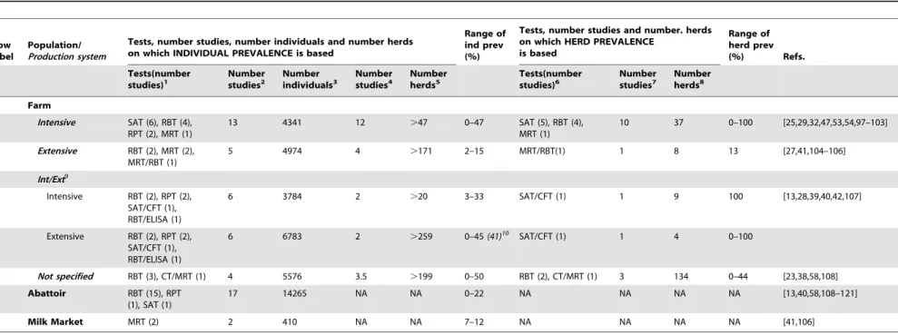
Variable sizeTable 1.Summary of brucellosis serology studies in cattle in Nigeria.
Row label
Population/
Production system
Tests, number studies, number individuals and number herds on which INDIVIDUAL PREVALENCE is based
Range of ind prev (%)
Tests, number studies and number. herds on which HERD PREVALENCE
is based
Range of herd prev
(%) Refs.
Tests(number
studies)1 Numberstudies2 Numberindividuals3 Numberstudies4 Numberherds5 Tests(numberstudies)6 Numberstudies7 Numberherds8
Farm
A Intensive SAT (6), RBT (4), RPT (2), MRT (1)
13 4341 12 .47 0–47 SAT (5), RBT (4),
MRT (1)
10 37 0–100 [25,29,32,47,53,54,97–103]
B Extensive RBT (2), MRT (2), MRT/RBT (1)
5 4974 4 .171 2–15 MRT/RBT(1) 1 8 13 [27,41,104–106]
Int/Ext9
C Intensive RBT (2), RPT (2), SAT/CFT (1), RBT/ELISA (1)
6 3784 2 .20 3–33 SAT/CFT (1) 1 9 100 [13,28,39,40,42,107]
D Extensive RBT (2), RPT (2), SAT/CFT (1), RBT/ELISA (1)
6 6783 2 .259 0–45(41)10
SAT/CFT (1) 1 4 0–100
E Not specified RBT (3), CT/MRT (1) 4 5576 3.5 .199 0–50 RBT (2), CT/MRT (1) 3 134 0–44 [23,38,58,108]
F Abattoir RBT (15), RPT
(1), SAT (1)
17 14265 NA NA 0–22 NA NA NA NA [13,40,58,108–121]
G Milk Market MRT (2) 2 410 NA NA 7–12 NA NA NA NA [41,106]
1Range of diagnostic tests and respective number of studies for each test on which individual prevalence values in table have been based (see text). 2Number of studies on which total number of individuals sampled and individual prevalence values have been based.
3Sum of animal sample size for each study for which individual prevalence data is available.
4Number of studies, out of total number of studies on which individual prevalence is based, which report number of herds sampled.
5Minimum estimate of number of herds sampled for each production system category. Not all studies reported number of herds sampled, hence true value must be superior (
.) to that in table.
6Range of diagnostic tests and respective number of studies on which herd prevalence values in table have been based (see text). 7Number of studies on which total number of herds sampled and herd prevalence values have been based.
8Sum of number of herds sampled for each study for which herd prevalence data is available. 9Studies sampling extensive and intensive flocks in parallel.
10Value of 41% prevalence corresponds prevalence non-adjusted for sensitivity and specificity (apparent prevalence = [true prevalence (0.879
+0.99821)]+120.998]; 0.998 = specificity of RBT*ELISA in test series; 0.879 = sensitivity of test series, see Mai et al. 2012).
doi:10.1371/journal.pntd.0003008.t001
PLOS
Neglected
Tropical
Diseases
|
www.plosntds
.org
4
July
2014
|
Volume
8
|
Issue
7
|
Table 2.Summary of brucellosis serology studies in sheep (S) and goats (G) in Nigeria.
Row label
Population
Production system
Tests, number studies and number individuals on
which INDIVIDUAL PREVALENCE is based Range ofind prev (%)
Tests, number studies and number flocks on
which FLOCK PREVALENCE is based Range of flockprev (%) Refs.
Test (number studies)1
Number studies2
Number Individuals3
Test (number studies)4
Number studies5
Number flocks6
Species S G S G S G S G S G S G S G S G
Farm
A Intensive RBT (4), RPT (1), SAT (1)
RBT (2), RPT (1)
6 3 594 234 0-76 0-33 RBT (4), SAT (1)
RBT (2) 5 2 5 2 100 100 [45,47,53,101,122,123]
B Extensive RBT (1) RBT (2) 1 2 210 643 5 6-29 NA8 NA 0 0 NA NA NA NA [22,124]
Int/Ext7
C Intensive RBT (2), SAT (1) RBT (2) 3 2 734 1053 0-21 5-21 NA NA 0 0 NA NA NA NA [54–56]
D Extensive RBT (2), SAT (1) RBT (2) 3 2 570 557 2-13 6-16 NA NA 0 0 NA NA NA NA
E Not specified RBT (1) SAT (2), RBT (1)
1 3 50 985 2 0-5 NA NA 0 0 NA NA NA NA [44,54,123]
F Abattoir RBT (6), SAT (1) RBT (8), SAT (2)
7 10 1376 6656 0-15 0-17 NA NA NA NA NA NA NA NA [44,50,51,55,57,58,113,117,118,123]
1Range of diagnostic tests and respective number of studies for each test on which individual prevalence values in table have been based (see text). 2Number of studies on which total number of individuals sampled and individual prevalence values have been based.
3Sum of animal sample size for each study for which individual prevalence data is available.
4Range of diagnostic tests and respective number of studies on which flock prevalence values in table have been based (see text). 5Number of studies on which total number of flocks sampled and herd prevalence values have been based.
6Sum of number of herds sampled for each study for which flock prevalence data is available. 7Studies sampling extensive and intensive flocks in parallel.
8Not applicable.
doi:10.1371/journal.pntd.0003008.t002
PLOS
Neglected
Tropical
Diseases
|
www.plosntds
.org
5
July
2014
|
Volume
8
|
Issue
7
|
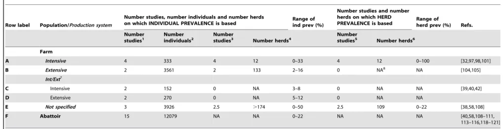
Table 3.Summary of brucellosis RBT studies in cattle in Nigeria.
Row label Population/Production system
Number studies, number individuals and number herds
on which INDIVIDUAL PREVALENCE is based Range ofind prev (%)
Number studies and number herds on which HERD
PREVALENCE is based Range ofherd prev (%) Refs.
Number
studies1 Numberindividuals2 Numberstudies3 Number herds4 Numberstudies5 Number herds6
Farm
A Intensive 4 333 4 12 0–33 4 12 0–100 [32,97,98,101]
B Extensive 2 3561 2 133 2–16 0 NA8 NA [104,105]
Int/Ext7
C Intensive 2 152 0 NA 3–8 0 NA NA [39,40,42]
D Extensive 2 270 0 NA 5–12 0 NA NA
E Not specified 3 3926 2.5 .174 0–50 2.5 109 0–22 [38,58,108]
F Abattoir 15 12079 NA NA 0–22 NA NA NA [40,58,108–111,
113–116,118–121]
1Number of studies using RBT on which individual prevalence values in table have been based (see text). 2Sum of animal sample size for each study for which individual prevalence data is available.
3Number of studies, out of total number of studies, on which individual prevalence is based, which report number of herds sampled.
4Minimum estimate or true number of herds sampled for each production system category. Not all studies reported number of herds sampled, hence true value must be superior (
.) to that in table.
5Number of studies using RBT on which herd prevalence values in table have been based (see text). 6Sum of number of herds sampled for each study for which herd prevalence data is available. 7Studies sampling extensive and intensive flocks in parallel.
8Not applicable.
doi:10.1371/journal.pntd.0003008.t003
PLOS
Neglected
Tropical
Diseases
|
www.plosntds
.org
6
July
2014
|
Volume
8
|
Issue
7
|
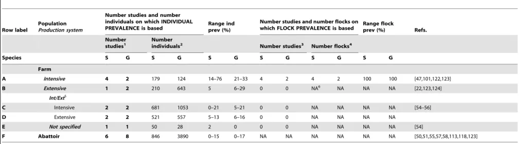
Table 4.Summary of brucellosis RBT studies in sheep (S) and goats (G) in Nigeria.
Row label
Population
Production system
Number studies and number individuals on which INDIVIDUAL
PREVALENCE is based Range indprev (%)
Number studies and number flocks on
which FLOCK PREVALENCE is based Range flockprev (%) Refs.
Number
studies1 Numberindividuals2 Number studies3 Number flocks4
Species S G S G S G S G S G S G
Farm
A Intensive 4 2 179 124 14–76 21–33 4 2 4 2 100 100 [47,101,122,123]
B Extensive 1 2 210 643 5 6–29 0 0 NA6 NA NA NA [22,123,124]
Int/Ext5
C Intensive 2 2 681 1053 0–21 5–21 0 0 NA NA NA NA [54–56]
D Extensive 2 2 521 557 5–13 6–16 0 0 NA NA NA NA
E Not specified 1 1 50 28 2 0 0 0 NA NA NA NA [54]
F Abattoir 6 8 846 3890 0–15 0–17 NA NA NA NA NA NA [50,51,55,57,58,113,118,123]
1Number of studies using RBT on which individual prevalence values in table have been based (see text). 2Sum of animal sample size for each study for which individual prevalence data is available.
3Number of studies using RBT on which herd prevalence values in table have been based (see text). 4Sum of number of herds sampled for each study for which herd prevalence data is available. 5Studies sampling extensive and intensive flocks in parallel.
6Not applicable.
doi:10.1371/journal.pntd.0003008.t004
PLOS
Neglected
Tropical
Diseases
|
www.plosntds
.org
7
July
2014
|
Volume
8
|
Issue
7
|
Most studies screened sera (blood or milk) with more than one serological assay and therefore report a seroprevalence value based on the results of each individual test. The number of cattle and small ruminant studies which have used classical tests such as the rose Bengal test (RBT), card test (CT), serum agglutination test (SAT), rapid plate test (RPT), 2-mercaptoethanol test (2-ME), rivanol test (RIV), coombs test, complement fixation test (CFT), milk ring test (MRT), and more recent diagnostic assays such as the competitive ELISA (C-ELISA), indirect ELISA (I-ELISA), and lateral flow assay (LFA) are summarised in Figure 1. To summarise and compare data we select one test seroprevalence value per study in this preferential order: RBT (or the equivalent Card Test), CFT, RPT, and SAT (all in blood serum). In studies where only milk was screened with MRT, these values are reported. The
rationale for this preferential selection of tests is the superior sensitivity/specificity (in the absence of brucellosis vaccination) of the prioritized tests [26]. Four authors did not report individual test results: Esuruoso [13], who considered samples positive when they were positive for SAT confirmed by CFT for suspicious samples; Alausa [23], who considered samples positive when positive for the card test or MRT or both; Pullan [27], who used MRT screening at herd level and then RBT on individual animals of MRT positive herds; and Mai [28] who confirmed RBT positive or inconclusive samples with C-ELISA. In these cases, we used the positive/negative data provided.
The presentation of average prevalence values calculated from studies using different tests, in different populations, and using different sampling designs is not valid, and so we present only
Figure 1. Number of cattle and small ruminant studies which have used the rose Bengal test (RBT), card test (CT), serum agglutination test (SAT), rapid plate test (RPT), 2-mercaptoethanol test (2-ME), rivanol test (RIV), Coombs test, complement fixation test (CFT), milk ring test (MRT), and more recent diagnostic assays such as the competitive ELISA (C-ELISA), indirect ELISA (I-ELISA), and lateral flow assay (LFA) for serological screening.The data table corresponds to total number of studies that have employed each test for each species. The overall number of studies is greater than the total number of papers retrieved because most papers screened sera with more than one serological assay.
doi:10.1371/journal.pntd.0003008.g001
prevalence ranges. We did not average values across analogous livestock production systems using weighting approaches taking into account test performance or sample size because (i) the lack of standardization of tests (origin of antigens, positive and negative controls, cut-off criteria), (ii) the application of brucellosis vaccination in some of the herds tested in earlier studies, and (iii) nonprobability sampling across studies would have led to misleading estimates of average prevalence. These circumstances limit the interpretation of the range of prevalence values presented in Tables 1 and 2. In an attempt to overcome some of these limitations, we consider the RBT values only in Tables 3 and 4, which yield narrower ranges as they are based on fewer studies and a simpler, more robust test, but the overall pattern when comparing intensive and extensive populations is the same (see below).
Results
Period of sampling and spatial distribution
Historically, two peaks of brucellosis reporting are evident (Figure 2A): the first coincided with establishment of intensive government farms in the 1970s to promote meat production and reduce imports (Box 1); the second with the post-millennium development goals public health agenda, increased interest in neglected zoonotic diseases, and private sector growth. Signifi-cantly, the trough coincides with the oil boom of the 1970s (Box 1). Figure 2B shows studies by animal species and Figure 3, the spatial distribution of animal and human studies.
Cattle brucellosis
To understand brucellosis epidemiology, it is necessary to determine the circulating Brucella species and biovars and, as antibodies are not species specific, bacterial isolation is essential. Since brucellosis was first reported in Nigeria in 1927 [29], only five studies have provided bacteriological data for cattle (Figure 3). In the West, studies in range cattle and in a University herd described the isolation of Brucella strains, probably B. abortus [30]. This species was properly identified in studies in government and private farms and in settled Fulani herds in the Centre and North [31–33]. In total, 58 isolates were classified as B. abortus biovar 1 (54 strains), biovar 2 (1 strain), biovar 3 (2 strains), and biovar 4 (1 strain) (see Table S9). However, re-examination of 20 of the biovar 1 isolates shows characteristics of biovar 3, the dominant biovar in countries proximal to Nigeria [34]. Moreover, VNTR genotyping [35] clusters these 20 strains with biovar 3a rather than 3b, the latter being typically reported in Europe (Ducrotoy, Bertu, Moriyo´n, and Ocholi, unpublished results).B. melitensishas not been reported in cattle, although there is close contact with small ruminants.
The bulk of the evidence is derived from serological studies (Figure 1), but limitations in the application of serological tests make data difficult to interpret. Early studies used RPT or SAT, two tests lacking sensitivity and specificity [26,36,37]. The RBT (or the equivalent Card Test) was applied shortly after its development and has been widely used (Tables 1 and 3; Figure 1). Despite the excellent specificity and sensitivity of RBT [26,36,37], the literature reviewed reflects the misconception that RBT is a test of low specificity which, in the absence of brucellosis vaccination or the false positive serological reaction phenomenon caused by crossreacting bacteria, needs to be confirmed. However, meta-analysis performed using strict criteria [26] shows that RBT specificity is in fact better than that of iELISA and cELISA, two tests used in some works to ‘‘confirm’’ the RBT results. Indeed, the OIE Manual
(http://www.oie.int/en/international-standard-setting/terrestrial-manual/access-online/; Chapter 2.4.3. Bovine Brucellosis) clearly states that these other tests can also sometimes give a positive result because of S19 vaccination or of false-positive serological reactions.
While RBT is a good choice, inadequate standardization results in considerable sensitivity (but not specificity) variation [37]. RBT standardization and origin was inadequately described in 15 out of 46 papers and six investigations used locally prepared antigens. Competitive or indirect ELISA kits were used according to manufacturer instructions but were never validated under local conditions (cut-offs established in brucellosis-free and good hygienic conditions cannot be extrapolated to endemic areas [38]). Across Nigeria, 14,000, 11,000, and 8,000 cattle have been sampled in different studies from abattoirs (animals from both extensive and intensive systems), extensive, and intensive herds, respectively, but the data (Tables 1 and 3; Tables S3, S4, S5, S6, S7, S8, S9; Figures 2A and 3A) illustrate the limitations in time and space of the studies. A total of 1,800 cattle correspond to the North, half this number (1,000) to the West and only small numbers to the East and South. Abattoir studies cannot provide spatial information due to country-wide animal movements (see above). Only five out of the 46 prevalence studies applied probability based sampling methods [28,39–42], and only one describes the method in sufficient detail [28], but even this study is biased, because herds were selected based on proximity to a reliable laboratory and farmer cooperation. Studies of intensive farms have focused mainly on infertility or abortion outbreaks, and few cattle were sampled (Table 1). Most intensive system studies were undertaken in the West before 1986 (Figures 2A and 3), a period of intense interest in the livestock sector (Box 1 and Table 1, Row A). Since 1986, more investigations have been reported in extensive cattle systems (Table 1, Row B) and from abattoirs (Table 1, Row F). Clearly there are few good-quality data on brucellosis in Nigeria, and discussion must bear in mind these limitations.
Extent to which the extensive and intensive cattle management systems are affected by brucellosis
In Nigeria, most cattle are reared extensively in the North and belong to nomadic, seminomadic or transhumant Fulani pasto-ralists. According to early official veterinary records, brucellosis was not regarded as a hazard in these herds [29,43] and most studies conducted independently in the extensive and intensive systems suggest a lower prevalence in the former (Tables 1 and 3, Rows A and B; Table S4). This was the view of early investigators [13,32]. Esuruoso wrote, ‘‘Cattle…in nomadic herds…on the move… are not likely to accumulate infection or spread it from one animal to the other as in settled herds. This factor, and the intense heat of the sun in fairly open country (Sudan Savannah zone) will provide some of the reasons for the low infection rate…in the northern herds… It would appear, therefore, that nomadic herding in Nigeria imposes a natural limit on the rate of brucellosis infection in cattle.’’ This observation is consistent with the low transmission deemed typical of pastoralist systems [7].
The inverse profile can be observed for studies that have looked at intensive and extensive system populations in parallel (Tables 1 and 3, Rows C and D; Table S5). A recent probability sampling study [28] (performed in Adamawa, Kaduna, and Kano, northern Nigeria), reports RBT seroprevalences of 45.1% (nomadic), 22.0% (seminomadic), 23.8% (commercial), and 15.9% (zero-grazing). Using a competitive ELISA kit as the reference, the authors assumed that 42.8% to 24.7% of these RBT results were false positives, but higher prevalence in the extensive than intensive system was also observed with the ELISA. Another recent, but
more limited, work reported higher (but not statistically significant) numbers of RBT positives in extensively than in intensively managed herds (11.6% versus 3.1%, respectively) in Plateau State (North Central Nigeria) [42]. These results suggest that brucellosis prevalence has been on the increase in extensive systems over time [28]. However, in a recent cross-sectional survey using RBT standardised according to OIE criteria, seminomadic Fulani cattle
(n = 2000) showed less than 1% individual seroprevalence in the Kachia Grazing Reserve (Kaduna) (ICONZ, 2013, www. iconzafrica.org). The reasons for the differences between this and earlier work are unclear. Although intensification provides opportunities for better control measures, their implementation cannot be taken for granted because this requires adequate infrastructure and training and, indeed, the risks of transmission
Figure 2. Distribution of studies on brucellosis in Nigeria according to (A) year of publication and (B) host investigated (numbers correspond to cumulative sample size across all studies for each host species).
doi:10.1371/journal.pntd.0003008.g002
Figure 3. Location of brucellosis studies in Nigeria.(A) cattle; (B) sheep and goats; (C) camels and pigs; and (D) humans. doi:10.1371/journal.pntd.0003008.g003
are greatly increased [1,6,7]. None of these recent studies describe control measures in intensively managed herds that could account for the lower prevalence reported. On the other hand, at least in the Kachia Grazing Reserve, Fulani have intuitive disease-reducing management approaches (e.g., rapidly selling or slaugh-tering animals that abort and those with poor fertility or low milk yields), and low reproductive rates reduce transmission [7]. As discussed below, these aspects of brucellosis epidemiology are not trivial, and further studies are necessary to confirm whether there is an increase of brucellosis in extensively managed herds and its distribution across the country. Unfortunately, the gap in information between the early 1980s and late 1990s precludes any possibility of doing this with the data available (Figure 2A).
Extensive nomadic herds as reservoirs of disease
Brucellosis transmission is generally lower in pastoralist systems because of low reproductive rates, animal movements and environmental circumstances [7]. However, brucellosis transmis-sion could increase as a result of the settling of migratory herds and emerge from increased contacts between these herds and unprotected intensive commercial or settled semi-intensive herds. This possibility has seldom been investigated in sub-Saharan Africa. One article provides evidence of this kind of transmission and of its dramatic impact on susceptible populations in the 1970s [23]. In a large brucellosis outbreak in Ibapara, out of ten governments, three private settled, and 12 Fulani herds tested, 11 herds were found to be positive using a combination of the MRT and Card Test. All 11 positive herds belonged to Fulani pastoralists, ‘‘nomadic herdsmen that move only within the district, and within few kilometres from previous settlements.’’ The outbreak coincided with the Sahelian drought that saw a general reduction in the cattle population of Nigeria and prompted an influx and settling of nomadic herds in Ibapara. The outcome was a widespread epidemic of bovine brucellosis with a severe increase in human cases. Fulani herdsmen complained of being unwell and unable to look after their cattle, and 51.5% of herdsmen, 23.5% of abattoir workers, and 3.1% of high school students were serologically positive with the Card Test. Calf losses were reported, resulting in a shortage of meat and protein undernutrition in the local populace.
Brucellosis in small ruminants
Small ruminants represent a major source of meat in Nigeria and are often reared alongside cattle. Their distribution is not known with certainty; Falade et al. [44] cite early sources, according to which 70% of goats were in the North, 20% in the East and 10% in the West, and about 60% of rural households in the northern, 50% in the eastern and 40% in the western states kept goats.15% of sheep and goats were reared under nomadic conditions at the end of the 20th century [22].
Bacteriological evidence for Brucella in small ruminants is scarce (Figure 3; Table S15). An early study claimed the isolation of B. abortus in sheep and goats, but the methodology used in species identification is unclear [45]. B. melitensis biovar 1 (22 strains) andB. abortusbiovar 1 (8 strains) were isolated from goats in western Nigeria [46]. However, the reported biochemical characteristics of theB. melitensisstrains are atypical.B. melitensis was recently described in sheep and goats in northern Nigeria but the ten strains were not definitively typed [24]. A study in Bauchi (central Nigeria) clearly demonstrated B. abortus but not B. melitensisin sheep [33]. Interestingly, sevenB. abortusstrains were isolated from sheep reared in contact with infected cattle [47]. Although B. abortus preferentially infects cattle, it is known to persist in sheep [48] and the significance ofB. abortusinfection in
small ruminants in the mixed breeding systems of sub-Saharan Africa requires further investigation.
There are fewer and more limited serological studies in small ruminants than in cattle (Figure 2B; Tables 2 and 4; Tables S10, S11, S12, S13, S14). Significant misuse of tests were application of MRT (not useful in small ruminants [49]) in four studies and interpretation that animals were infected byB. melitensisbased on a comparison of titres toB. abortusandB. melitensisantigens [50– 52], a discrimination that is not possible by serology and indicates inadequate antigen standardization.
Studies in intensive or semi-intensive systems are not only scarce but also biased because most investigations focused on cattle abortions with simultaneous sampling of small ruminants (com-pare references in Tables 1 and 2 and Tables S3 and S10). In fact, contagion from cattle was often considered the origin of infection. Only one study was performed on intensively or semi-intensively raised small ruminants in the West [44]; the others for this region consisted of abattoir surveys (Tables 2 and 4). Studies in extensive systems were all undertaken in the North (Rows B and D in Tables 2 and 4; Table S11 and S12); hence, the epidemiology in sedentary and nomadic flocks in other regions is unknown. Although values broadly suggest that brucellosis prevalence is higher in intensive than extensive systems for small ruminants (Tables 2 and 4, Rows A, B, C, and D, Tables S10, S11, S12) these trends have to be interpreted with caution.
According to two studies performed in the 1960s, small ruminant brucellosis was not a problem on government farms, but most surveys were undertaken in the cattle-dominated North; hence, no information was available for other regions (Figure 3B) [53,54]. Fifteen years later, one study in northern Nigeria later found significant rates of infection (13.8% and 15.1% averages for sheep and goats, respectively) [55]. This same study reported rates of infection in institutional (i.e., intensive) flocks about four times higher than in local (extensive) flocks for both sheep and goats (Table 2), and attributed the difference to an increased transmis-sion caused by intensification [55]. A recent study [56] found overall prevalence values of 9.3% for sheep and 10.1% for goats, which are comparable to the values found 30 years previously [55], but husbandry-specific values were not obtained.
Ten studies have investigated sheep and goats for brucellosis in trade settings (Table 2, Row F; Table S14), and while values do not reflect the situation at farm level, they confirm the presence of brucellosis in small ruminants in the North. Two abattoirs studies in the West found low prevalence values (0.3%–0.9% and 0% for goat and sheep, respectively) [57,58], but since animals come mostly from other parts of Nigeria, the situation in the West remains unknown.
Brucellosis in other animals
B. abortushas been isolated from horses [33,59], and antibodies have been reported in donkeys [60], dogs [61–63], and fowl [64– 67] in Nigeria (Figure 2B). However, the role of these nonrumi-nant species in disease transmission has never been satisfactorily proven [68] and, as they are unable to act as reservoirs, once brucellosis is eradicated in domestic ruminants, they are consid-ered as spillover hosts or sentinels.
Camels are distributed along the northern borders of Nigeria, and nomadism is common, often across borders. At the turn of the 20th century, estimated numbers of camels in Nigeria varied from 90,000 [14] to 25,000, substantially greater than an estimate of 18,000 in 1978 [69]. BothB. abortusandB. melitensiscan infect camels, butBrucellahas never been isolated from these animals in Nigeria [70–72]. Serological studies are particularly difficult to interpret because brucellosis tests have not been properly
evaluated in these animals [73]. Abattoir studies in northern Nigeria reported 1.3%–14.8% seropositivity using SAT [14,69,74,75] in camels from Nigeria and Chad, Niger, and Cameroon (Figure 3C). In Borno State, two MRT and RBT studies of range camels reported positive animals [70,75]. However, the MRT has been proven useful only in cattle [49], and the RBT is dependent on the effect of acidic pH on ruminant IgG and IgM [76,77]. Since camelids and ruminants differ markedly in immunoglobulin repertoire and structure [78], RBT results should be interpreted with caution. Camels are herded with sheep and goats and, to a lesser extent, cattle [69], and their role in the epidemiology of brucellosis in Nigeria is unclear.
Pigs represent approximately 4.5% of the meat market in Nigeria [79]. An early study claimed isolation of B. suis from animals positive in SAT [80] but a small-scale bacteriological study failed to isolate Brucella [33]. An investigation in government farms during a cattle abortion outbreak [53], a study in intensive and semi-intensive farms in the South [79], and an abattoir study in the West [58] found no or very few RBT positive animals. In contrast, a recent abattoir study in Central Nigeria reported 30% of 281 pigs RBT positive (Figure 3C) [81]. In the absence of bacteriological evidence or protein-based tests, these data have to be interpreted with caution, because pigs are prone to false positive serological reactions with RBT, CFT, and ELISA [82].
Control of animal brucellosis
Brucellosis control was initiated in colonial Nigeria in 1917; vaccination was applied to address widespread bovine abortions in government-owned farms and local production of a liquid S19 vaccine started at this time. A test and slaughter policy was also implemented [83], and its failure was attributed to a lack of rigor in implementation [28]. Production of lyophilised S19 started in 1950 [12], and by 1951, brucellosis eradication and control programmes succeeded in establishing brucellosis-free stock and reducing overall prevalence to less than 5% on government farms [28]. Efforts waned and vaccine production discontinued in 1954 [12] and today there is no government policy for brucellosis control in Nigeria. Nevertheless, local researchers estimated that brucellosis caused approximately 20% financial losses in tradi-tional systems of cattle production in one Nigerian grazing reserve [84] and concluded that, as the nomads settle in these reserves, hygienic measures and brucellosis vaccination are profitable and should be implemented [85]. A recent study identified brucellosis and milk loss as the greatest components of the direct economic losses associated with reproductive disorders in settled herds in Zaria, Nigeria [86].
Human brucellosis
The first cases of human brucellosis confirmed by laboratory tests were reported in Nigeria in 1941 [87] and 1962 [88], and even during this period, underdetection was suspected [89]. A decade later, few laboratories could perform these tests and this, combined with low suspicion, was again thought to lead to underdetection [90]. This review shows that these circumstances have not changed.
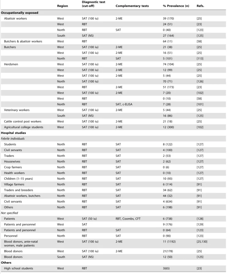
Human seroprevalence data are summarized in Table 5, and Figure 3D shows the geographical location of studies. Although they strongly suggest the importance of the human disease, exact figures cannot be derived from most surveys. The studies based solely on RBT confirm exposure toBrucellaof butchers, abattoir workers, and herdsmen. However, they do not necessarily represent the proportion of true disease, because a positive RBT result can be caused by contact or infection and needs to be
interpreted according to the clinical picture [76]. Several studies complemented RBT with SAT and 2-mercaptoethanol tests, both of which detect only agglutinating antibodies; since these antibodies disappear in long-standing cases, the data only reflect recent infections. Moreover, SAT diagnostic titre varies from 50 to 200 international units (the diagnostic titre most often used in Nigeria was of 100 international units) depending on the origin (urban or rural and endemic or non-endemic areas) and exposure of the patient [76]. Complementary tests that detect non-agglutinating antibodies (competitive ELISA, Coombs, and CFT) were implemented in only two studies, one using competitive ELISA whose diagnostic cut-off for human brucellosis is unknown [76].
There are no reports of Brucellaisolation from human cases, and it is not known to what extent human brucellosis in Nigeria is caused byB. abortus or B. melitensis. Interpretation of human infection caused by B. melitensis or B. abortus on the basis of different titres with B. melitensis and B. abortus antigens is deceptive [91]. Misdiagnosis may be frequent; one abattoir study found that RBT positive individuals often complained of frequent treatments for malaria without showing improvement, while others complained of joint pain and general weakness [58].
Conclusion: Lessons from Nigeria
This review has identified major gaps in epidemiological data, diagnostics, and control, and misconceptions surrounding brucellosis. After 100 years, we know surprisingly little on the disease agent in Nigeria, and good-quality information— essential for evaluation of zoonotic potential and for establish-ment of control measures—is still lacking. Bacteriological studies are necessary to clarify the picture of both animal and human brucellosis. Preliminary evidence suggests thatB. abortusbiovar 3a is dominant or restricted to Africa, but little is known about its virulence and other biological properties. Also, the existence and distribution ofB. melitensisandB. suisneeds to be clarified. Likewise, a judicious choice of serological tests validated under local conditions and an understanding of their value in different contexts is key, as is implementation of clinical protocols and simple affordable tests for routine diagnosis in humans. Most sophisticated serodiagnostic tests were developed in high-income countries many years after brucellosis was eradicated, and these tests are better suited to epidemiological surveillance in well-equipped laboratories. Capacity building is a clear need, and the establishment of a reference laboratory for both human and animal brucellosis in sub-Saharan Africa would be a great asset.
The outbreak investigated by Alausa over 30 years ago [23] may be highly significant, because it shows the dramatic effect of the influx and settling of infected nomadic herds in areas where no control measures are implemented. This can happen in contem-porary Nigeria where rural–urban migration, changing trends in livestock management and increased intensification could re-create the conditions for emergence of disease [6]. Climate change and desertification of the Sahel may also be an important driver for emergence, as it accounts in part for rural–urban migration [9] and is predicted to cause a reduction in the number of crop farmers in favour of livestock keepers [10]. Settling of nomadic Fulani in peri-urban areas and grazing reserves may be advantageous politically and economically, opening market chains for dairy products, offering formalised access to education and healthcare services, and avoiding disputes over land-use and clashes with crop farmers [92]. The emergence of brucellosis could, in these circumstances, have far-reaching social and political implications [84,93,94].
Table 5.Summary of brucellosis studies in humans in Nigeria.
Region
Diagnostic test
(cut-off) Complementary tests % Prevalence (n) Refs.
Occupationally exposed
Abattoir workers West SAT (100 iu) 2-ME 39 (170) [25]
West RBT 24 (51) [23]
North RBT SAT 0 (40) [123]
South SAT (NS) 27 (164) [125]
Butchers & abattoir workers West RBT 64 (11) [58]
Butchers West SAT (100 iu) 2-ME 21 (38) [25]
West SAT (100 iu) 2-ME 16 (51) [25]
North RBT SAT 5 (101) [113]
Herdsmen West SAT (100 iu) 2-ME 74 (104) [25]
West SAT (100 iu) 2-ME 12 (99) [25]
West SAT (100 iu) 2-ME 5 (44) [25]
North SAT (100 iu) 70 (71) [126]
West RBT 2-ME 51 (173) [23]
West SAT (100 iu) 2-ME 7 (20) [102]
West RBT 0 (10) [58]
North RBT SAT, c-ELISA 7 (28) [101]
Veterinary workers West SAT (100 iu) 2-ME 5 (44) [25]
South SAT (NS) 16 (86) [125]
Cattle control post workers West SAT (100 iu) 2-ME 21 (18) [25]
Agricultural college students West SAT (100 iu) 2-ME 12 (300) [102]
Hospital studies
Febrile individuals
Students North RBT SAT 8 (122) [127]
Civil servants North RBT SAT 4 (100) [127]
Traders North RBT SAT 2 (53) [127]
Housewives North RBT SAT 2 (62) [127]
Crop farmers North RBT SAT 0 (6) [127]
Health workers North RBT SAT 0 (10) [127]
Children (1–15 years) North RBT SAT 10 (93) [127]
Village farmers North RBT SAT 6 (114) [91]
Traders and breeders North RBT SAT 34 (62) [91]
Abattoir workers, butchers North RBT SAT 44 (32) [91]
Civil servants North RBT SAT 4 (634) [91]
Others North RBT SAT 6 (198) [91]
Not specified
Patients West SAT (50 iu) RBT, Coombs, CFT 6 (738) [128]
Patients and personnel West SAT 9 (176) [129]
Patients and personnel North RBT SAT 0 (64) [123]
Personnel North RBT SAT 0 (90) [123]
Blood donors, ante-natal women, male patients
West SAT (100 iu) 2-ME 11 (1192) [25,130]
Blood donors West SAT (100 iu) 2-ME 21(178) [25]
Blood donors South SAT (NS) 12 (50) [125]
Others
High school students West RBT 3(65) [23]
doi:10.1371/journal.pntd.0003008.t005
Prophylaxis and control of brucellosis requires contextual adaptation. Most evidence suggests differences in epidemiology between extensive livestock production systems and more intensive systems worldwide [1,7]. This could apply to past situations in Nigeria, but we do not have a clear picture of the present status of the disease. An understanding of the dynamics of brucellosis in nomadic pastoralist systems and at the interface with settled populations is critical. Mass-vaccination approaches may be difficult to implement in extensively managed animals in Nigeria, but it is essential they be applied in the intensive and commercial systems. At a time when cost-effectiveness needs to be demon-strated, brucellosis control measures should be focused on settled populations that are at risk. This appeals to policy-makers, as settled populations are accessible and more amenable to mass-vaccination campaigns than nomadic pastoralist communities. Moreover, since differentiation of infected and vaccinated animals is not critical initially, the most effective vaccines (S19 in cattle and, if necessary, Rev1 in small ruminants [95]) should be used.
Nomadic pastoralism could offer a well-adapted management system for disease mitigation in Nigeria; if the disease exists at low levels, animals exhibit a low overall frequency of abortion and there are few opportunities for disease transmission. One Health and Eco Health approaches to disease reduction and prevention are particularly relevant in pastoralist communities, considering that pastoralism and transhumance is a desirable livelihood strategy in Nigeria [96].
Currently there is no coordinated policy for brucellosis in Nigeria. An assessment of the direct and indirect impact of brucellosis on these communities leading to culturally appropriate and locally adapted control options is overdue. There is a need to undertake a countrywide, evidence-based, and multidisciplinary study of bru-cellosis in the different livestock production systems of Nigeria to
determine the extent, potential impact, and origin of brucellosis and to propose control template strategies of proven efficacy.
Supporting Information
Table S1 Rejected brucellosis serology studies in cattle.
(DOCX)
Table S2 Rejected brucellosis serology studies in sheep and
goats. (DOCX)
Table S3 Brucellosis serology studies in cattle reared under
intensive livestock systems. (DOCX)
Table S4 Brucellosis serology studies in cattle reared under
extensive livestock systems. (DOCX)
Table S5 Brucellosis serology studies in cattle undertaken in
extensive and intensive livestock systems in parallel. (DOCX)
Table S6 Brucellosis serology studies in cattle reared under non-specified livestock systems.
(DOCX)
Table S7 Brucellosis abattoir serology studies in cattle.
(DOCX)
Table S8 Brucellosis milk market milk serology studies in cattle.
(DOCX)
Table S9 Brucellosis bacteriology studies in cattle. (DOCX)
Table S10 Brucellosis serology studies in sheep and goats reared under intensive livestock systems.
(DOCX)
Table S11 Brucellosis serology studies in sheep and goats reared
under extensive livestock systems. (DOCX)
Key Learning Points
N
Despite imperfect evidence, an exhaustive review ofstudies in Nigeria suggests that brucellosis persists at low endemic levels in nomadic pastoralist systems. The settling of nomadic or transhumant pastoralist popula-tions and intensification in livestock management, may favour disease transmission and conditions for brucello-sis outbreaks.N
There is an urgent need to study the dynamics of thedisease at the interface between extensive pastoralist and intensive or settled livestock systems and to implement brucellosis control measures adapted to each of these situations.N
The few attempts to implement a vaccination plus testand slaughter strategy in cattle show that this approach was not sustainable.N
The role of small ruminants and camels in theepidemiology of brucellosis in Nigeria remains unknown because of insufficient bacteriological investigations and, for camels, properly validated serological tests.N
The extent of the public health impact of brucellosis islargely unknown, and bacteriological studies tochar-acterise the Brucella species infecting humans are lacking.
N
There is an imperfect understanding of the animal andhuman disease and of the value of the different diagnostic tests in different epidemiological contexts. One Health courses for veterinarians, medical doctors, and diagnostic laboratory personnel are necessary.Top Five Papers in the Field
1. Ajogi I, Akinwumi JA, Esuruoso GO, Lamorde AG (1998) Settling the nomads in Wase-Zange grazing reserves in the Sudan Savannah zone of Nigeria III. Estimated financial losses due to bovine brucellosis. Nigerian Vet J 19: 86–94.
2. Alausa OK (1979) The investigation and control of a large-scale community outbreak of brucellosis in Nigeria. Public Health 93: 185–193.
3. Esuruoso GO (1974) Bovine brucellosis in Nigeria. Vet Rec 95: 54–58.
4. Grace D, Mutua F, Ochungo P, Kruska R, Jones K (2012) Mapping of poverty and likely zoonoses hotspots. Zoonoses Project 4. Report to the UK Department for International Development. Nairobi, Kenya: ILRI. Avail-able: http://cgspace.cgiar.org/handle/10568/21161. Ac-cessed 27 June 2014.
5. Waters-Bayer AN, Bayer W (1994) Coming to Terms. Interactions between Immigrant Fulani Cattle-Keepers and Indigenous Farmers in Nigeria’s Subhumid Zone. Cahiers d’E´tudes Africaines 34: 213–229. doi:10.3406/ cea.1994.2048
Table S12 Brucellosis serology studies in sheep and goats undertaken in extensive and intensive livestock systems in parallel. (DOCX)
Table S13 Brucellosis serology studies in sheep and goats under non-specified livestock systems.
(DOCX)
Table S14 Brucellosis abattoir serology studies in sheep and goats.
(DOCX)
Table S15 Brucellosis bacteriology studies in sheep and goats.
(DOCX)
Acknowledgments
We are grateful to Professor Ian Maudlin for a critical reading of the manuscript. The constructive comments on data presentation made by an anonymous reviewer are also gratefully acknowledged.
References
1. McDermott J, Grace D, Zinsstag J (2013) Economics of brucellosis impact and control in low-income countries. Rev Sci Tech Off Int Epiz 32: 249–261. 2. Dalrymple-Champneys W (1960) Prognosis.Brucellainfection and undulant
fever in man. Oxford & New York: Oxford University Press. pp. 151–155. 3. Zinsstag J, Schelling E, Solera J, Blasco JM, Moriyo´n I (2011) Brucellosis. In:
Palmer SR, Soulsby L, Torgeson PR, Brown DG, editors. Handbook of Zoonoses. Oxford & New York: Oxford University Press. pp. 54–62. 4. Dean AS, Crump L, Greter H, Schelling E, Zinsstag J (2012) Global burden of
human brucellosis: A systematic review of disease frequency. PLoS Negl Trop Dis 6: e1865. doi:10.1371/journal.pntd.0001865.t004
5. Seimenis A, Morelli D, Mantovani A (2006) Zoonoses in the Mediterranean region. Ann Ist Super Sanita 42: 437–445.
6. Jones BA, Grace D, Kock R, Alonso S, Rushton J, et al. (2013) Zoonosis emergence linked to agricultural intensification and environmental change. Proc Natl Acad Sci USA 110: 8399–8404. doi:10.1073/pnas.1208059110 7. Racloz V, Schelling E, Chitnis N, Roth F (2013) Persistence of brucellosis in
pastoral systems. Rev Sci Tech Off Int Epiz 32: 61–70.
8. ILRI (2012) Mapping of poverty and likely zoonoses hotspots. Nairobi, Kenya: ILRI. 1 p. Available: http://cgspace.cgiar.org/handle/10568/21161. Accessed 27 June 2014.
9. Barrios S, Bertinelli L, Strobl E (2006) Climatic change and rural–urban migration: The case of sub-Saharan Africa. J Urban Econ 60: 357–371. doi:10.1016/j.jue.2006.04.005
10. Jones PG, Thornton PK (2009) Croppers to livestock keepers: livelihood transitions to 2050 in Africa due to climate change. Environ Sci Policy 12: 427– 437. doi:10.1016/j.envsci.2008.08.006
11. McDermott JJ, Arimi SM (2002) Brucellosis in sub-Saharan Africa: epidemiology, control and impact. Vet Microbiol 90: 111–134.
12. Ocholi RA, Kalejaiye JO, Okewole PA (1993) Brucellosis in Nigeria. A review. Trop Vet 11: 15–26.
13. Esuruoso GO (1974) Bovine brucellosis in Nigeria. Vet Rec 95: 54–58. 14. Adamu NN, Ajogi I (1999) Serological investigations of camels (Camelus
dromedarius) slaughtered at Kano municipal abattoir for evidence of brucellosis. Trop Vet 18: 45–48.
15. Aregheore EM (2009) Country Pasture/Forage Resource Profiles. Rome: FAO. Available: http://www.fao.org/ag/agp/AGPC/doc/Counprof/PDF%20files/ Nigeria.pdf. Accessed 19 June 2014.
16. Stewart R (2010) Desertification in the Sahel. Environmental Science in the 21st century - An online textbook. Available: http://oceanworld.tamu.edu/ resources/environment-book/desertificationinsahel.html. Accessed 19 June 2014.
17. Rikin EU (1988) Brucellosis of cattle in Nigeria: proposals for a control program under intensive and extensive husbandry systems. Acta Vet Scand 84 (supplement): 94–97.
18. Suleiman H (1988) Policy issues in agropastoral development in Nigeria. Proceedings of the National Conference on Pastoralism in Nigeria. Nigeria: Ahmadu Bello University Zaria.
19. Majekodunmi AO, Fajinmi A, Dongkum C, Picozzi K, Thrusfield MV, et al. (2013) A longitudinal survey of African animal trypanosomiasis in domestic cattle on the Jos Plateau, Nigeria: prevalence, distribution and risk factors. Parasit Vectors 6: 239. doi:10.1186/1756-3305-6-239
20. Iyayi EA, Okoruwa VO, Babayemi OJ, Busari AA, Peters OF (2003) Livestock production pattern of agropastoralists in peri-urban centres of south-west Nigeria. Nigerian J Anim Prod 30: 87–92.
21. Mohammed TA (1990) A study of peri-urban cattle agro-pastoralism in the derived savanna of Oyo State, southwest Nigeria. Ibadan, Nigeria: ILCA, Humid Zone Programme. 54pp.
22. Brisibe F, Nawathe DR, Bot CJ (1996) Sheep and goat brucellosis in Borno and Yobe states of arid northeastern Nigeria. Small Rumin Res 20: 83–88. 23. Alausa OK (1979) The investigation and control of a large-scale community
outbreak of brucellosis in Nigeria. Public Health 93: 185–193.
24. Bale JO, Nuru S, Addo PB, Adeyinka IA (2003) Bacteriological investigation of sheep and goat milk for brucellosis in government farms in Northern Nigeria. Nigerian J Anim Prod 30: 107–116. Available: http://www.ajol.info/index. php/njap/article/view/3321. Accessed 19 June 2014.
25. Alausa OK, Awoseyi A (1976) Brucellosis: the situation in Western Nigeria. Trop Geogr Med 28: 54–59.
26. Greiner M, Verloo D, De Massis F (2009) Meta-analytical equivalence studies on diagnostic tests for bovine brucellosis allowing assessment of a test against a group of comparative tests. Prev Vet Med 92: 373–381.
27. Pullan NB (1980) Productivity of White Fulani cattle on the Jos Plateau, Nigeria. III. Disease and management factors. Trop Anim Health Prod 12: 77– 84.
28. Mai HM, Irons PC, Kabir J, Thompson PN (2012) A large seroprevalence survey of brucellosis in cattle herds under diverse production systems in northern Nigeria. BMC Vet Res 8: 144. Available: http://www.biomedcentral. com/1746-6148/8/144. Accessed 27 June 2014.
29. Banerjee AK, Bhatty MA (1970) A survey of bovine brucellosis in northern Nigeria (a preliminary communication). Bull Epizoot Dis Afr 18: 333–338. 30. Esuruoso GO (1974) Bovine brucellosis in two southern states of Nigeria; II.
The incidence and implications of infection in range cattle. Bull Epizoot Dis Afr 22: 35–40.
31. Bale OO, Kumi-Diaka J (1981) Serological and bacteriological study of bovine
brucellaefrom livestock investigation and breeding centres in Nigeria. Br Vet J 137: 256–261.
32. Eze EN (1978) Isolation ofBrucellaefrom the Nigerian livestock and the typing of such isolates. Bull Anim Hlth Prof Afr 26: 29–36.
33. Ocholi RA, Kwaga JK, Ajogi I, Bale JO (2004) Phenotypic characterization of
Brucellastrains isolated from livestock in Nigeria. Vet Microbiol 103: 47–53. 34. Sanogo M, Abatih E, Thys E, Fretin D, Berkvens D, et al. (2013) Importance of identification and typing ofBrucellaefrom West African cattle: A review. Vet Microbiol 164: 202–211. doi:10.1016/j.vetmic.2013.02.009
35. Le Fleche P, Jacques I, Grayon M, Al-Dahouk S, Bouchon P, et al. (2006) Evaluation and selection of tandem repeat loci for aBrucellaMLVA typing assay. BMC Microbiol 6: 1–14. doi:10.1186/1471-2180-6-9
36. Davies G (1971) The Rose Bengal test. Vet Rec 88: 447–449.
37. Blasco JM, Garin-Bastuji B, Marı´n C, Gerbier G, Fanlo J, et al. (1994) Efficacy of different rose bengal and complement fixation antigens for the diagnosis of Brucella melitensis infection in sheep and goats. Vet Rec 134: 415–420. 38. Greiner M, Gardner IA (2000) Epidemiologic issues in the validation of
veterinary diagnostic tests. Prev Vet Med 45: 3–22.
39. Junaidu AU, Oboegbulem SI, Salihu MD (2011) Serological survey ofBrucella
antibodies in breeding herds. J Microbiol Biotech Res 1: 60–65.
40. Cadmus SIB, Alabi PI, Adesokan HK, Dale EJ, Stack JA (2013) Serological investigation of bovine brucellosis in three cattle production systems in Yewa Division, south-western Nigeria. J S Afr Vet Assoc 84. doi:10.4102/ jsava.v84i1.217
41. Farouk UM, Ibrahim S, Ajogi I, Bale JO (2013)Brucellaantibodies in milk and risk factors analysis in pastoralist herds in Jigawa State, Nigeria. Eur J Vet Med 2: 29–37.
42. Maurice NA, Wungak SY, Gana BA, Nanven MB, Ngbede EO, et al. (2013) Seroprevalence of bovine brucellosis in northern Plateau State, North Central Nigeria. Asian Pac J Trop Dis 3: 337–340. doi:10.1016/S2222-1808(13)60081-X
43. Anonymous (1958) Annual report on the Department of Veterinary Services of the Northern Region of Nigeria 1955–56. Kaduna: Government Printer. 44. Falade S, Ojo MO, Sellers KC (1974) A serological survey of caprine
brucellosis in Nigeria. Bull Epizoot Dis Afr 22: 335–339.
45. Okoh AE (1980) Abortion in sheep near Kano, Nigeria. Trop Anim Health Prod 12: 11–14.
46. Falade S (1981)Brucellaeisolated from goats. Zentralbl Veterinarmed B 28: 205–209.
47. Ocholi RA, Kwaga JK, Ajogi I, Bale JO (2005) Abortion due toBrucella abortusin sheep in Nigeria. Rev Sci Tech Off Int Epiz 24: 973–979. 48. Luchsinger DW, Anderson RK (1979) Longitudinal studies of naturally
acquiredBrucella abortusinfection in sheep. Am J Vet Res 40: 1307–1312. 49. Alton GG, Jones LM, Angus RD, Verger JM (1988) Techniques for the
brucellosis laboratory. Paris, France: INRA.
50. Junaidu AU, Daneji AI, Salihu MD, Magaji AA, Tambuwal FM, et al. (2010) Seroprevalence of brucellosis in goat in Sokoto, Nigeria. Curr Res J Biol Sci 2: 275–277.
51. Okewole PA, Eze EN, Okoh AE, Oyetunde IL, Odeyemi PS (1988) Small ruminant brucellosis in some parts of Northern Nigeria. Bull Anim Hlth Prof Afr 36: 251–254.