(1) Universidade Federal de São Paulo – UNIFESP, São Paulo, SP, Brasil. (2) Universidade Potiguar – UNP, Natal, RN,
Brasil.
(3) Pontifícia Universidade Católica de Campinas, Campinas, SP, Brasil. (4) Departamento de Pediatria da
Universidade Federal de São Paulo – UNIFESP, São Paulo, SP, Brasil. (5) Departamento de Fonoaudiologia da
Universidade Federal de São Paulo – UNIFESP, São Paulo, SP, Brasil.
Conlict of interest: non-existent
Hearing loss in children exposed to toxoplasmosis
during their gestation
Alterações auditivas em crianças expostas à toxoplasmose
durante a gestação
Carlos Alberto Leite Filho(1) Lorena Carvalho Cavalcanti Lagreca(2) Natália Oliveira de Jesus(1) Cristiana Porchia Corvaro(3) Maria Aparecida Gadiani Ferrarini(4) Ana Isabel Melo Pereira Monteiro(4) Marisa Frasson de Azevedo(5)
Received on: October 02, 2016 Accept on: May 30, 2017
Mailing adress:
Lorena Carvalho Cavalcanti Lagreca Universidade Federal de São Paulo – UNIFESP, São Paulo/SP
Rua José de Oliveira Coelho, 200 – Ap. 64 Vila Andrade – São Paulo - SP CEP: 05727-240
E-mail: [email protected]
ABSTRACT
Purpose: to verify the occurrence and the most frequent type of auditory disorders in children exposed to
toxoplasmosis during pregnancy.
Methods: a retrospective, longitudinal study carried out in a public health institution of São Paulo. Records
of children born between 2010 and 2015 were analyzed and distributed into two groups: study group, composed of 48 children born to mothers with a diagnosis of toxoplasmosis during their pregnancy; and control group, composed of 43 children without congenital infection, who were accompanied due to low birth weight. The children were evaluated two to four times during their irst two years of life, by means of peripheral and central auditory function assessment.
Results: 47 children underwent only two evaluations and only 11 completed all the evaluations up to 24
months. In the control group, 58.1% had normal hearing, 37.2% conductive loss, 4.7% cochlear loss and absence of retrocochlear disorder. In the study group, 56.3% presented normal hearing, 20.8% conductive loss, 2.1% cochlear loss, and 20.8% retrocochlear disorder.
Conclusion: children exposed to toxoplasmosis during pregnancy did not differ from non-exposed
chil-dren in relation to the occurrence of conductive or cochlear hearing loss. However, they showed a higher
occurrence of retrocochlear disorder.
Keywords: Hearing; Toxoplasmosis; Spontaneous Otoacoustic Emissions; Audiometry; Hearing Loss
RESUMO
Objetivo: veriicar a ocorrência e o tipo mais frequente de alteração auditiva em crianças expostas a
toxoplasmose durante a gestação.
Métodos: estudo retrospectivo longitudinal realizado em instituição pública de saúde de São Paulo.
Análise de prontuários de crianças nascidas entre 2010 e 2015 distribuídos em dois grupos: grupo estudo, composto por 48 crianças de mães com diagnóstico de toxoplasmose durante a gestação; e grupo controle, composto por 43 crianças sem infecção congênita que foram acompanhadas por apre-sentar baixo peso ao nascimento. As crianças foram avaliadas de duas a quatro vezes durante os dois primeiros anos de vida, por meio de testes que avaliam a função auditiva periférica e central.
Resultados: 47 crianças izeram apenas duas avaliações e apenas 11 completaram todas as avaliações
até 24 meses. No grupo controle 58,1% apresentaram audição normal, 37,2% perda condutiva, 4,7% perda coclear e ausência de alteração retrococlear, enquanto o grupo estudo apresentou 56,3% de audi-ção normal, 20,8% de perda condutiva, 2,1% de perda coclear e 20,8% de alteraaudi-ção retrococlear.
Conclusão: crianças expostas à toxoplasmose durante a gestação não diferiram das não expostas em
relação à ocorrência de perda auditiva coclear e condutiva. Entretanto, apresentaram maior ocorrência de alteração retrococlear.
INTRODUCTION
Congenital toxoplasmosis is caused by the trans-mission of the protozoan Toxoplasma gondii from the mother to the fetus. The mother is contaminated during the gestational period mainly by the ingestion of water and/or raw meats or inadequately cooked meat contam-inated with sporulated oocysts. The parasite reaches the baby via placenta causing damage of different degrees of severity, which can even be responsible for fetal death. The subclinical (asymptomatic) form of the disease occurs in 70-90% of cases. However, if these are not diagnosed and treated early, 85% may suffer of infections in the eyes during childhood and adoles-cence and 40% can present neurological sequelae in the future. The main characteristics of congenital toxoplasmosis are: neurological and ophthalmological alterations, other symptoms of prematurity, intra-uterine growth retardation, anemia, thrombocytopenia, increased abdominal volume, enlarged lymph nodes, jaundice, sensorineural deafness, among others1-3.
Studies have found a prevalence of congenital toxoplasmosis between 0.6-1.3/1000 live births in different regions of Brazil4-6. It has been suggested that the chance of transmission of toxoplasmosis from mother to fetus ranges from 18.5 to 23%6,7. The risk of
fetal infection is less than 15% in the irst trimester, but
in general, the disease is severe in the newborn. The risk of infection increases from 20 to 50% in the second trimester and from 55 to 80% in the third trimester of pregnancy, but the newborn is asymptomatic or presents a less severe8.
Toxoplasma gondii has been associated with auditory pathway damage since the beginning of the 1950s, with evidence of calcium deposits (similar
to calciications found in the brains of children with
congenital toxoplasmosis) in the spiral ligament and cochlea9. Auditory deicit has been reported in about 20% of cases of congenital toxoplasmosis, especially in children not treated or treated for a very short period10. A study with 174 Brazilian children with toxoplasmosis diagnosed and treated early found a 3.4% cochlear loss, a 4.6% conductive loss, and a 3.4% central alteration11. The occurrence of profound deafness has been almost totally limited to cases with high clinical manifestations12, but another study reports the occur-rence of unilateral or bilateral hearing loss in 26% of the
19 children with subclinical infection13. Other authors have not found an association between parasitosis and hearing loss when children are treated, and doubts persist as to the frequency with which toxoplasmosis
can lead to auditory deicit12,14.
Thus, this study was carried out with the objective of verifying the occurrence and the most frequent type of auditory alteration in children exposed to toxoplas-mosis during gestation.
METHOD
The research was approved by the institution’s research ethics committee under No. 0448/2015 and the written consent was obtained from the head of the outpatient clinic, co-author of the present study.
Retrospective longitudinal study with the analysis of medical records of children born between 2010 and 2015 that were attended at a public institution in the city of São Paulo. From the total of 250 medical records
analyzed, 91 were selected as itting the inclusion and
exclusion criteria. For establishing the study group (SG), the children referred by the Discipline of Pediatric Infectious Disease of the institution were selected for exposure to toxoplasmosis during pregnancy without other indicators of risk for hearing loss15. For the formation of the control group (CG), we selected children who were followed up in the medical sector due to low birth weight, without any risk indicators for hearing loss15. In this way, the SG was composed of 48 children and the CG, for 43 children.
ORL –Otolaryngologist
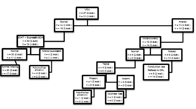
Figure 1. Flowchart of the irst evaluation protocol (at birth)
ORL –Otolaryngologist; VRA - Visual reinforcement audiometry
Figure 2. Flowchart of the protocol of the second and third evaluation (6 to 9 months and 9 to 24 months) with the distribution of the
Acoustic immitance measurements were performed with the Interacoustics brand imitanciometry screener T235 recording the tympanometric curve and the
contralateral acoustic relex search in the frequencies
from 0.5 to 4 kHz. Normal type A tympanometric curve
with acoustic relexes present between 70 and 90 dB
above the auditory threshold was considered in the frequency studied17.
The audiometry with visual reinforcement was performed in children from six months in acoustic booth with supra-aural earphones model TDH39 and Maico brand audiometer MA41, using complex visual reinforcement (toy with movement and light). Thresholds of up to 15 dBHL were considered normal and the mean was of 0.5 to 4 kHz. Bone scan was The TEOAE screening and TEOAE suppression
were performed with a ILO V6 device. For the TEOAE study, the non-linear click type stimulus was used between 75 and 85 dBpeNPS. TEOAEs were considered present when the signal-to-noise ratio was higher than 3 dB in the 1 kHz band and 6 dB in the 2, 3 and 4 kHz bands, with reproducibility higher than 50% and probe stability greater than 70%. The TEOAE suppression was performed using two probes, one in each ear, with alternating presentation of two seconds of linear clicks at 65 dBpeNPS without noise and two seconds of clicks with contralateral noise at 60 dBNPS. At the end of 260 series of clicks, suppression was considered when there was a minimum decrease of 0.2 dB in the general response16.
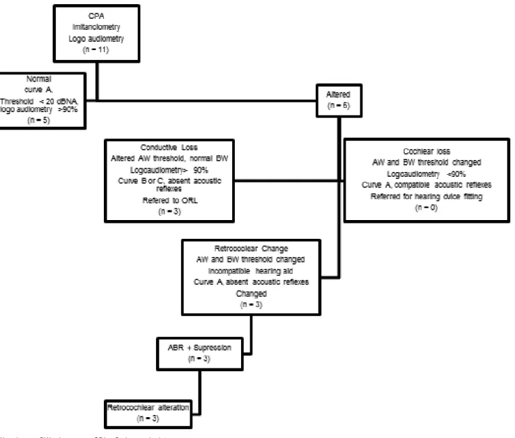
AW – air way; BW – bone way; ORL - Otolaryngologist
• Normal hearing: presence of TEOAE and suppression of TEOAE and BA-EP normal neuro-logical protocol (1st evaluation); Behavioral auditory
thresholds ≤ 15 dBHL, type A tympanometric curve, presence of contralateral acoustic relexes bilate -rally, presence of TEOAE (2nd and 3rd evaluations); Previously mentioned results, plus LRF <20 dBNA and IPRF> 90% (4th evaluation);
• Conductive loss: absence of TEOAE, tympano-metric type B or C, absence of bilaterally
contrala-teral acoustic relexes, BAEP with high electrophy -siological thresholds by AW and normal by BW (1st
evaluation); Behavioral auditory thresholds ≥ 20 dBNA per AW and ≤15 dBNA per VO, type B or C
tympanometric curve, absence of bilaterally
contra-lateral acoustic relexes, absence of TEOAE (2nd
and 3rd evaluations); Previously reported, plus RLI equal to or 10 dB above the mean of 0.5, 1 and 2 kHz and IPRF> 90% (4th evaluation);
• Cochlear loss: absence of TEOAE, type A tympano-metric curve, elevated electrophysiological auditory thresholds by AW and BW in the BAEP with a burst tone of 0.5 to 4 kHz (1st evaluation); Auditory
behavioral thresholds ≥ 20 dBNA per AW and BW,
tympanometric curve type A, absence of TEOAE (2nd and 3rd evaluations); Previously cited results, plus LRF equal to or 10 dB above the mean of 0.5, 1 and 2 kHz and IPRF <90% (4th evaluation);
• Retrocochlear alteration: presence of TEOAE, altered BAEP with absence of waves (auditory neuropathy spectrum) or central alteration (increased interpeaks I-III, III-V or I-V); Absence of suppression of TEOAEs, tympanometric curve type A (1st, 2nd and 3rd evaluations); Results, plus lower than expected RFI in relation to auditory behavioral thresholds (4th evaluation).
Statistical analysis was performed using the IBM SPSS Statistics program (version 23.0) and sought a difference between groups regarding the occurrence of auditory alterations using Pearson’s chi-square test and
Fisher’s exact test. The level of signiicance was set at
5% (p <0.05).
RESULTS
The presence of children in the 2nd (6 to 9 months), 3rd (9 to 24 months) and 4th (24 months) evaluations in each group is presented in Table 1. There was no
statis-tically signiicant difference regarding the presence of
children in the evaluations, indicating that both groups were monitored in a similar way.
performed with a bone vibrator positioned on the mastoid and contralateral masking was used when necessary18.
The CPA was performed in children aged two years or more with the same equipment. The child was condi-tioned to associate the detected sound with a motor act (insert piece into the toy). The investigation of the speech recognition threshold (SRT) was performed
with the identiication of requested igures with a
gradual decrease in the sound pressure level of the stimulus up to 50% of the child’s correct answers were obtained. The percentage of speech recognition index
(PSRI) was performed with the identiication of igures
at 40 dBNS in relation to the average of the thresholds of the frequencies of 0.5, 1 and 2 kHz18.
The BAEP was performed with the Intelligent Hearing Systems Smart-EP equipment with insertion earplugs and surface electrodes positioned at points CZ, FZ, A1 and A2. The click stimulus was presented at 80 dBNA. Waves I, III and V and interpeak intervals I-III, III-V and I-V were recorded. The analysis of the absolute and interpeak latencies was performed using the criterion proposed by Gorga19. Electrophysiological threshold research was performed with a burst tone of 0.5 to 4 kHz by airway (AW) and by bone (BW) in children with suspected cochlear loss 20.
For airway threshold research, at least 2000 stimuli were presented. The sound pressure level of the tone burst stimulus decreased by 20 dB from 80 dBnNA until the V wave was no longer visualized. Then, the intensity was increased 10 by 10 dB until the lowest intensity was obtained, in which the V wave appeared in a smaller amplitude, being considered the electrophysi-ological threshold. A normal threshold of 30 dBnNA was considered for each frequency, corresponding to 20 dBNA20.
In the BAEP record by bone way, a bone vibrator was placed in the mastoids and an alternating stimulus was presented at 50 dBNA, decreasing by 10 in 10 dB. As an electrophysiological threshold, the lowest
intensity at which the V wave was identiied and repli -cated by the examiner was considered. An
electro-physiological threshold ≤ 20 dBnNA was considered
normal20.
From the results obtained in the exams, children
were classiied in relation to hearing as: normal hearing,
The comparisons between the groups in relation to the results of the auditory evaluations obtained from the children who attended two, three and four evaluations are presented in Tables 2, 3 and 4, respectively.
The groups did not differ in relation to the occurrence of conductive and cochlear losses when compared to children with the same number of hearing evaluations in each group. There was a tendency of the GE to present a higher occurrence of retrocochlear alterations in the children who attended two evaluations.
Table 1. Quantity of audiological evaluations per child in each group
Group
Number of evaluations
p-value
2 3 4
n (%) n (%) n (%)
SG 24 (51,1) 18 (54,5) 6 (54,5)
0,956
CG 23 (48,9) 15 (45,5) 5 (45,5)
Total 47 (100) 33 (100) 11 (100)
Pearson’s chi-square test SG: study group CG: control group
Table 2. Occurrence of hearing changes in children present to two evaluations
Result of audiological evaluation
Group
p-value
CG GE
n (%) n (%)
Normal 15 (65,2) 15 (62,5)
-Conductive 6 (26,1) 3 (12,5) 0,464
Cochlear 2 (8,7) 1 (4,2) > 0,999
Retrococlear 0 (0,0) 5 (20,8) 0,057#
Total 23 (100) 24 (100)
Fisher’s exact test. SG: study group CG: control group
# - Value with a tendency to statistical signiicance at 5% (p < 0,05)
Table 3. Occurrence of hearing changes in children present to three evaluations
Result of audiological evaluation
Group
p-value
CG SG
n (%) n (%)
Normal 7 (46,7) 10 (55,6)
-Conductive 8 (53,3) 6 (33,3) 0,479ª
Cochlear 0 (0,0) 0 (0,0) IC
Retrococlear 0 (0,0) 2 (11,1) 0,509b
Total 15 (100) 18 (100)
Pearson’s chi-square test (a) and Fisher’s exact test (b)
The inal audiological diagnosis considering all the
evaluations performed in each child in both groups is presented in table 5. There was a difference between the auditory evaluation distributions of the two groups (p-value = 0.004), with a higher occurrence of retro-cochlear alteration in the SG. From the 10 children
diagnosed with retrocochlear alteration, seven presented no TEOAE suppression, two presented an increase of the I-III interpeak in the BAEP, character-izing a low brain stem change, and one presented alteration in both suppression and BAEP. There were no cases of spectrum of auditory neuropathy.
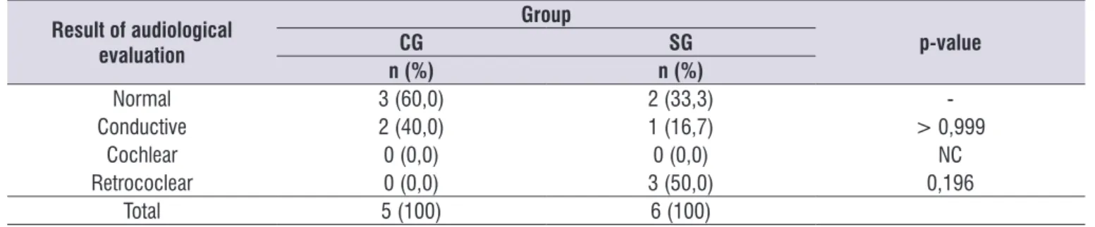
Table 4. Occurrence of hearing changes in children present to four evaluations
Result of audiological evaluation
Group
p-value
CG SG
n (%) n (%)
Normal 3 (60,0) 2 (33,3)
-Conductive 2 (40,0) 1 (16,7) > 0,999
Cochlear 0 (0,0) 0 (0,0) NC
Retrococlear 0 (0,0) 3 (50,0) 0,196
Total 5 (100) 6 (100)
Fisher’s exact test SG: study group CG: control group IC – Incalculable
Table 5. Occurrence of hearing changes in the total sample
Result of audiological evaluation
Group
p-value
CG SG
n (%) n (%)
Normal 25 (58,1) 27 (56,3)
-Conductive 16 (37,2) 10 (20,8) 0,338a
Cochlear 2 (4,7) 1 (2,1) 0,611b
Retrococlear 0 (0,0) 10 (20,8) 0,004*b
Total 43 (100) 48 (100)
Pearson’s chi-square test (a) and Fisher’s exact test (b)
* - Statistically signiicant value at the level of5% (p < 0,05) SG: study group
CG: control group
DISCUSSION
Congenital toxoplasmosis is a potential risk indicator for sensorineural hearing loss15. The infection can be detected in the prenatal period by performing the maternal serology and medicated early. All SG children were followed up at the Pediatric Infectology outpatient clinic and were treated for one year with sulfadiazine, pyrimethamine and folinic acid and were referred for audiological evaluation and follow-up in the sector of Children’s Audiology.
There was a decrease in the sample number inversely proportional to the number of auditory evaluations, indicating the children’s evasion along
the audiological follow-up. This was expected, since evasion is a trend observed in longitudinal studies11.
There were conductive losses in all evaluations,
with no statistically signiicant difference between the groups. In the inal diagnosis, about 1/3 of the children
presented conductive loss, similar to that obtained in the literature21. Conductive loss, although transitory,
causes luctuation in hearing resulting from recurrent
otitis media, and may cause language/learning problems due to inconsistent auditory input during the critical auditory developmental period.
of the group without risk of hearing loss and one child
(2.1%) of the study group, diagnosed in the irst two
evaluations and referred for hearing aid adaptation and speech therapy. The incidence of hearing loss is 1 to 3: 1000 in the general population15. The greater occurrence of cochlear loss in the CG of the present
study was not expected and may be justiied by the
reduced number of the sample or by the fact that 50% of the cochlear hearing losses are of genetic origin22. The occurrence of cochlear loss in the SG (2.1%) is similar to the result of a study with treated children with congenital toxoplasmosis, which obtained 3.8% of cochlear losses11. These results are in contrast to studies conducted in Europe and the United States which, with the same drug therapy used in the present study, observed a reduction of auditory sequelae with early detection and treatment of congenital toxoplasmosis14,23.
The main inding of the present study was the high
occurrence of retrocochlear alterations in children exposed to toxoplasmosis during pregnancy (20.8%), differing statistically from the control group (0%).
The presence of retrocochlear alterations makes the child with congenital toxoplasmosis, even when treated, at risk for alterations in auditory processing and language. In fact, 29.6% of language alterations were found in children with congenital toxoplasmosis treated early and followed up to three years of age11. Thus, the child with congenital toxoplasmosis, even when treated early, needs to be accompanied by a multidisciplinary team throughout its development, in order to detect early changes in auditory processing and language, which may negatively affect school learning.
The retrocochlear alteration in congenital
toxoplas-mosis could be related to the postnatal inlammatory
response of the internal acoustic meatus to the presence of T. gondii. Such an immunological reaction would damage the vestibulocochlear nerve, which is found in the internal acoustic meatus, and would justify the greater occurrence of retrocochlear alterations in the SG24.
The occurrence of retrocochlear auditory disorders in 20.8% of the SG children in the present study is similar to that found in the literature, between 21.1 and 35.5%4,11,25. However, it differed from other studies
in which the prevalence found was signiicantly lower
or even zero14,23,26,27. This difference is due to the fact that many studies only perform the evaluation of the peripheral auditory system, without including BAEP and suppression of TEOAE. In the present study, the medial
olivocochlear efferent system presented alteration in eight of the 10 children with retrocochlear alterations. Such a system acts on the speech comprehension in the noise, in the selective attention that implies in future alterations of the auditory processing, which would justify to include the evaluation of this structure by means of the suppression of the TEOAE.
In addition, the literature points out the method-ological differences between the studies, mainly in relation to the selection of the audiological evaluation tests, diagnosis and early treatment; and periodicity of the treatment performed as the cause of the discrep-ancies between the results of the studies28. The main differences also occur in relation to audiological monitoring: many studies do not follow the cases periodically. A study conducted in Brazil followed 174 children with toxoplasmosis for up to 36 months, obtaining results similar to those described in the present study11.
The occurrence of retrocochlear alterations in children exposed to toxoplasmosis during pregnancy indicates the need to evaluate the peripheral and central auditory system in this population as well as monitoring the development of hearing and language skills.
A limitation of this study due to its clinical and retrospective nature was the lack of application of all
procedures in all children, considering the service low
chart of the service in which the study was performed (Figures 1 to 3). Another limitation of this study was the
dificulty of keeping track of all children up to two years
of age due to circumvention.
CONCLUSION
Children exposed to toxoplasmosis during gestation did not differ from those not exposed in relation to cochlear and conductive hearing loss. However, they had a higher occurrence of retrocochlear alterations.
REFERENCES
1. Sáfadi MAP, Farhat CK. Toxoplasmose. In: Farhat CK, Carvalho LHFR, Succi RCM. Infectologia pediátrica. 3a ed. São Paulo: Atheneu; 2008. p. 953-64.
2. Liesenfeld O. Toxoplasmose. In: Goldmann L, Ausiello D. Cecil Medicina. 23a ed. Rio de Janeiro: Elsevier; 2009. p. 2767-73.
MB, Campos SO, Hilário MOE (eds.). Pediatria Diagnóstico e Tratamento. 1a ed. Barueri: Manole; 2013. p. 209-19.
4. Andrade GMQ, Resende LM, Goulart EMA,
Siqueira AL, Vitor RWA, Januario JN. Deiciência
auditiva na toxoplasmose congênita detectada pela triagem neonatal. Rev Bras Otorrinolaringol. 2008;74(1):21-8.
5. Vasconcelos-Santos DV, Azevedo DOM, Campos
WR, Oréice F, Queiroz-Andrade GM, Carellos
EVM et al. Congenital Toxoplasmosis Brazilian Group CTBG/UFMG. Congenital toxoplasmosis in southeastern Brazil: results of early ophthalmologic examination of a large cohort of neonates. Ophthalmology. 2009;116(11):2199-205.
6. Varella IS, Canti ICT, Santos BR, Coppini AZ, Argondizzo LC, Tonin C et al. Prevalence of acute toxoplasmosis infection among 41,112 pregnant women and the mother-to-child transmission rate in a public hospital in South Brazil. Mem Inst Oswaldo Cruz. 2009;104(2):383-8.
7. Jenum PA, Stray-Pedersen B, Melby KK, Kapperud G, Whitelaw A, Eskild A et al. Incidence of Toxoplasma gondii infection in 35,940 pregnant women in Norway and pregnancy outcome for infected women. J Clin Microbiol. 1998;36(10):2900-6.
8. Artigao FB, Martín FC, Corripio IF, Mellgren AG, Guasch CF, Miranda MCF et al. Guía de la Sociedad Española de Infectología Pediátrica para el diagnóstico y tratamiento de la toxoplasmosis congénita. Anales de Pediatría. 2013;79(2):116. e1-16.
9. Wright I. Congenital toxoplasmosis and deafness. An investigation. Pract Otorhinolaryngol (Basel). 1971;33(6):377-87.
10. Cecatto SB, Garcia RID, Costa KS, Abdo TRT, Rezende CEB, Rapoport PB. Análise das principais
etiologias de deiciência auditiva em Escola
Especial “Anne Sullivan”. Rev Bras Otorrinolaringol. 2003;69(2):235-40.
11. Resende LM, Andrade GMQ, Azevedo MF,
Perissinoto J, Vieira BC.Congenital Toxoplasmosis Brazilian Group CTBG/UFMG. Congenital toxoplasmosis: auditory and language outcomes in early diagnosed and treated children. Sci Med. 2010;20(1):13-9.
12. Remington JS, McLeod R, Thulliez P, Desmonts G. Toxoplasmosis. In: Remington JS, Klein JO, Wilson CB, Baker CJ (eds.). Infectious diseases of the fetus
and newborn infant. 7th ed. Philadelphia: Elsevier Saunders; 2011. p.918-1041.
13. Wilson CB, Desmonts G, Couvreur J,
Remington JS. Lymphocyte transformation in the diagnosis of congenital toxoplasma infection. N Engl J Med. 1980;302(14):785-8.
14. McLeod R, Boyer K, Karrison T, Kasza K, Swisher C, Roizen N et al. Toxoplasmosis Study Group. Outcome of treatment for congenital toxoplasmosis, 1981-2004: the National Collaborative Chicago-Based, Congenital Toxoplasmosis Study. Clin Infect Dis. 2006;42(10):1383-94.
15. Joint Committee on Infant Hearing. Year 2007 Position Statement: principles and guidelines for early hearing detection and intervention programs. Pediatrics. 2007;120(4):898-921.
16. Jesus NO, Angrisani RG, Maruta EC, Azevedo MF. Efeito de supressão nas emissões otoacústicas em lactentes termo e pré-termo. CoDAS. 2016;28(4):331-7.
17. Jerger J. Clinical experience with impedance
audiometry. Arch Otolaryngol. 1970;92(4):311-24. 18. Northern JL, Downs MP. Audição na infância. 5a
ed. Rio de Janeiro: Guanabara Koogan; 2005. 19. Gorga M, Kaminski J, Beauchaine K, Jesteadt
W, Neely S. Auditory brainstem responses from children three months to three years of age: normal patterns of response II. J Speech Hear Res. 1989;32(2):281-8.
20. Stapells DR, Gravel JS, Martin BA. Thresholds for auditory brain stem responses to tones in notched noise from infants and young children with normal hearing or sensorineural hearing loss. Ear Hear. 1995;16(4):361-71.
21. Ho V, Daly KA, Hunter LL, Davey C. Otoacoustic emissions and tympanometry screening among 0-5 year olds. Laryngoscope. 2002;112(3):513-9. 22. Petit C. Genes Responsible for human hereditary
deafness: symphony of a thousand. Nat Genetics. 1996;14(4):385-91
23. Austeng ME, Eskila A, Jacobsen M, Jenum PA, Whitelaw A, Engdahl B. Maternal infection with
Toxoplasma gondii in pregnancy and the risk
of hearing loss in the offspring. Int J Audiol. 2010;49(1):65-8.
24. Salviz M, Monctoya JG, Nadol JB, Santos F. Otopathology in congenital toxoplasmosis. Otol Neurotol. 2013;34(6):1165-9.
gondii in children: a case-control study. Clin Otolaryngol. 2008;33(3):269-73.
26. McGee T, Wolters C, Stein L, Kraus N, Johnson D, Boyer K et al. Absence of sensorineural hearing loss in treated infants and children with congenital toxoplasmosis. Otolaryngol Head and Neck Surg. 1992;106(1):75-80.
27. Muhaimeed HA. Prevalence of sensorineural
hearing loss due to toxoplasmosis in Saudi children: a hospital based study. Int J Pediatr Otolaryngol. 1996;34(1-2):1-8.