BRAINSTEM AUDITORY EVOKED POTENTIAL RESPONSE
IN THE PROGNOSIS OF SUPERFICIAL COMA
Potencial evocado auditivo de tronco encefálico
no prognóstico do coma supericial
Libia Camargo Ribeiro Leite (1), Maira de Victor Francisco (2) , Sinésio Grace Duarte (3),
Cristiane Fregonesi Dutra Garcia (4) , Samanta Natália Bizinoto (5)
(1) Degree in Speech Pathology from the University of Franca
– UNIFRAN, SP, Brazil; Graduate Student of Rehabilita-tion Science at the Medical School of the University of São Paulo (FMUSP).
(2) Speech pathologist for the Municipal Government of
Guaira, SP; Degree in Speech Pathology from the Univer-sity of Franca – UNIFRAN, SP, Brazil.
(3) Physician; Chief of Neurosurgery Services of Santa Casa
de Franca and São Joaquim / Regional Hospitals; Coordi-nator / teacher at the UNIFRAN Medical School, Franca, SP, Brazil;
(4) Speech pathologist; PhD – Associate Professor of Speech
Pathology at the Medical School of the Federal University of Rio de Janeiro – UFRJ, Rio de Janeiro, RJ, Brazil.
(5) Speech pathologist at the Santa Casa de Misericórdia de
Franca Foundation- FSCMF, Franca, SP, Brazil; Post Gra-duation in Hospital Speech Pathology at Faculty of Medi-cine of São José do Rio Preto, SP, Brazil; Post-graduation in dysphagia and dysphonia at CEFAC.
Conlict of interest: non-existent
INTRODUCTION
The coma is a state in which consciousness is reduced to the point the individual cannot be awakened even with strong sensory stimuli that would otherwise do it. Therefore, coma can be
deined as the state in which the individual is not
aware of himself and his environment, with absence
or extreme decrease in consciousness level. This
state of unconsciousness is variable in intensity
and is directly related to the extent or structure of
the central nervous system affected. It is caused by lesion or dysfunction of the ascending reticular
activating system (ARAS), of the cerebral cortex
diffusely or both. Countless pathologies can lead the individual to coma, such as metabolic disorders, cranial traumas, infections of the nervous system
ABSTRACT
The coma is the persistent reduction consciousness level, not responsive to stimuli, due to low brain activity. To check the consciousness level, a feature often used is the Glasgow Coma Scale. Another method that was been showing up is the Brainstem Evoked Response Audiometry, that evaluates the activity of the ascending auditory pathways from the midbrain to the peripheral route. The test is simple, immune to depressant drugs and electrically charged environments, being the most
suitable potential for monitoring the coma. The aim of his study was to examine the characterization
of Brainstem Evoked Response Audiometry in mild coma (Glasgow 7-8) and its contribution. We conducted a prospective cross-sectional study in two patients in coma (Glasgow 7), state secondary to head trauma. The test results showed the presence of electrical activity on the entire route in both cases studied, with differences changes relative to the decrease in interpeak latency, morphology, and replication of the waves. These differences were contemplated whit the evolution the each case:
case 1 was favorable, but the second died. These results conirm the indings in the literature that
describes the Brainstem Evoked Response Audiometry normal are associated with good outcome,
while abnormal results are poor prognosis lags.
(meningitis), intracranial hypertension and intoxi -cation, among others1-3.
The level of consciousness can be assessed by
neurological examination and by some quantiied
parameters in the Glasgow Coma Scale (GCS), namely: eye response, motor response, and verbal response, which may be obtained by various
stimuli, ranging from spontaneous activity to verbal stimuli and even to painful stimuli (Table 1). The advantages of this scale are the ease of evaluation and record and its universal use, which favors the standardization of language referring to levels of coma2-5.
Parameters Response Observed Score
Eye opening
Spontaneous 4
In response to speech 3
In response to painful stimulation 2
None 1
Better Verbal
Response 5
Confused 4
Inappropriate words 3
Incomprehensible sounds 2
None 1
Better Motor Response
Follows commands 6
Localizes and removes stimulus 5
Localizes stimulus 4
Flexes in response 3
Extends in response 2
None 1
Table 1 – Glasgow Coma Scale
Source: Koizumi e Araújo (2005, p. 142) 5.
The score varies from three to ifteen, and
these values are representative of severe coma (lower values) state of full alertness (higher values). Intermediate intervals indicate the levels of coma, i.e., values between three and four, deep coma;
between ive and six, moderate coma; between
seven and eight, light coma5.
Besides the neurological examination, the
use of an objective resource in the evaluation of the brainstem, where the ARAS is located, can
provide signiicant information about the function
of this structure. The Evoked Auditory Brainstem Response (ABR) can provide such data, as it consists in recording the response to auditory stimuli that generate a chart in the form of waves. This
test presents seven waves that appear in the irst
ten milliseconds (ms) after the presentation of the auditory stimulus. Each wave has different places of origin: wave I – distal portion of the brainstem
them, the waves I, III and V are those that offer the most important parameters for the interpretation of ABR 6-8. This information indicates the electrophysi-ological activity of the auditory system; monitors the synapses of the auditory pathways starting from the auditory nerve, passes through the entire segment of the brain stem responsible for most of the body’s vital functions, to the inferior colliculus in the midbrain level. The design of this potential can be analyzed by morphology, replication, latency and amplitude of waves 9-11.
The ABR exam is the most suitable potential
PRESENTATION OF CASES
Initially, the project was referred to the Research Ethics Committee of the Santa Casa de Misericórdia in the city of Franca on June 1, 2011, which was approved under protocol number 103/201. For the cases included in this study, the relatives or guardians of patients were contacted and informed about the objectives of the research, the
proce-dures to be performed, its risks and beneits, and
thus the authorization for inclusion in the study was obtained by signing the Term of Consent, pursuant to Resolution 196/96.
We conducted a cross-sectional prospective study with comatose patients in the ICU of the Santa Casa de Misericórdia of Franca, between 13 and 30 September 2011.
We considered the following criteria for inclusion
and exclusion of patients in the study:
Inclusion Criteria:
• Clinical diagnosis of coma with score 3-8, according to the ECG;
• No alterations in the acoustic meatus;
• Authorization of participation from family or guardian.
Exclusion criteria:
• Infectious diseases of the central nervous system;
• Demyelinating diseases, brainstem stroke, tumors located near the auditory nerve or along the auditory tract;
• Reported hearing loss or other pathologies that could compromise the presence of ABR waves;
• Artifact and background noise that exceeded
10% of the presented stimuli;
• The information about the patient was obtained from medical records and family.
For the procedure, we used the following materials:
• Equipment Biologic version 5.70, Model 317, two channels, coupled to a conventional computer, installed in its proper place;
• HEINE mini 2000 otoscope and specula of different sizes;
• Surface electrodes and material for their attachment;
• Personal protective equipment (gloves, masks, aprons);
• Material for recording data.
Following these criteria, two patients were selected and tested, being both male.
Case 1
Individual of twenty years and two months of
age, in a coma for six days, secondary to traumatic
brain injury (TBI) due to motorcycle accident. His GCS score was 7 from admission until the day of
the exam.
Case 2
Individual of thirty-eight and nine months of age, hospitalized for twenty days due to traumatic brain injury followed by coma after suffering beatings. On admission he presented Glasgow 6 and at the time of assessment, G7.
Initially, the patient was stabilized, taken to the
examination room within the ICU in a bed stretcher,
and he was properly monitored. The following steps were performed:
• Patient´s identiication and personal data (age,
gender, etiology of coma, time evolution of the state of coma);
• Patients were normothermic (36-37 °C).
• Inspection of the external acoustic meatus
(EAM);
• Preparation of the attachment sites for the electrodes (cleaning and placement of conductive paste);
• Attaching the electrodes: active electrode (positive) positioned on the high forehead, reference electrode (negative) positioned in the earlobe tested and ground electrode (neutral) placed in the contralateral earlobe;
• Connection to the equipment;
• Recording of ABR waves for analysis at the intensity of 90 dB HL (decibel hearing level);
• Removal of electrodes from the patient and taking him back to his bedside in the ICU;
All procedures were performed in the ICU.
In the analysis of the exam, we considered the
aspects related to waveform morphology, i.e., the presence of waves I, III and V, the replication of the waves in order to eliminate the variability and subjectivity of interpretations; the absolute latency of responses of each wave ( I, III, V) and the relative latency between the interpeak intervals (I-III, III-V,
I-V), following the criteria described by Hall15 (Table 3), according to the reference commonly used in clinical practice; the ipsilateral relation of waves I-V, in which the amplitude of wave V should be greater than wave I, and the interaural
comparison of waves V, with maximum difference
of 0.3 ms, based on the proposal of Figueiredo and Castro Júnior (2003)16.
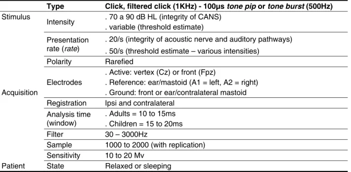
Type Click, filtered click (1KHz) - 100µs tone pip or tone burst (500Hz)
Stimulus
Intensity . 70 a 90 dB HL (integrity of CANS) . variable (threshold estimate)
Presentation rate (rate)
. 20/s (integrity of acoustic nerve and auditory pathways) . 50/s (threshold estimate – various intensities)
Polarity Rarefied
Electrodes
. Active: vertex (Cz) or front (Fpz)
. Reference: ear/mastoid (A1 = left, A2 = right) Acquisition . Ground: front or ear/contralateral mastoid
Registration Ipsi and contralateral Analysis time
(window)
. Adults = 10 to 15ms . Children = 15 to 20ms
Filter 30 – 3000Hz
Sample 1000 to 2000 (with replication) Sensitivity 10 to 20 Mv
Patient State Relaxed or sleeping Table 2 – Parameters for ABR catchment
Source: Katz, J (2008) 6
Waves Correspondent (likely) Latency-adults (ms)
I Distal portion to the brainstem of auditory nerve 1,5 a 1,9 II Proximal portion to the brainstem of the auditory nerve 2,5 a 3,0
III Choclear nucleus 2,5 a 4,1
IV Superior olivary complex 4,3 a 5,2
V Lateral lemniscus 5,0 a 5,9
VI Inferior Colliculus
VII Medial geniculate body
INTERPEAKS I – III 2,14
III – V 1,89
I – V 4,02
Table 3 – Values for latency of waves I, III, V and their intervals I-III, III-V, I-V
RESULTS
The ABR results demonstrated the presence of electrical activity on the entire route studied in both cases, with indication of different alterations in regards to the reduction in interpeak latencies, morphology, and replication of the waves. Such differences were contemplated with the evolution of each case.
Case 1
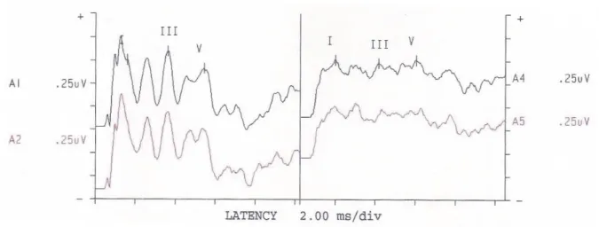
• Adequate morphology for registration A1 (left ear) and altered to registration A4 (right ear);
• Adequate replication for both registrations, being A2 for the left ear and A5 for the right ear.
• Absolute latency of waves I, III, V with appro-priate values for both ears;
• Interpeak latency in intervals I-III, III-V, I-V with appropriate values for both ears;
• Wave I amplitude greater than wave V for both registrations, which may indicate impairment in the auditory processing13;
• Interaural comparison in waves V with value of 0.20 ms, which does not indicate retrocochlear impairment.
Figure 1 – Registration of ABR – Case 1
The exam results indicated preserved activity of electrical stimuli conduction in the extension of
the auditory pathway evaluated, i.e., the peripheral segment and brainstem.
The design of the exam indicated the preser -vation of the function of the auditory pathways in the brainstem signaling integrity of ARAS and,
conse-quently, a better prognosis, conirmed by clinical
reassessment after twenty days when the patient was on the ward and in the process of discharge, with GCS score of 14.
Case 2
• Alteration in morphology, and the A6 registration (right ear) was better than A3 (left ear);
• Alteration in replication for both registrations, being A4 for the left ear and A8 for the right ear;
• Absolute latency of waves I, III, V, with adequate values for both records;
• Interpeak latency intervals I-III, III-V, I- V, with altered values for both registrations;
• Amplitude of wave I greater than wave V for both registrations;
• Interaural comparison of waves V with a value of 0.16 ms, which does not indicate retrocochlear impairment.
The exam results indicated preserved activity of
electrical stimuli conduction; however, the activity
was impaired, as described above. These indings suggest a worse prognosis, which was conirmed by
DISCUSSION
ABR stands out as a competent method in
measuring the electrophysiological proile of
brainstem. This happens because of the ascending auditory pathways, which occupy the entire segment of this structure in the central nervous system, which is responsible for the functions of the human organism, from the simplest ones, such as primitive
relexes until the integrated relexes, such as those
responsible for the heart rate, breathing and blood pressure control 7,11,12.
The deterioration of the function of the nervous
system, most often, is rostrocaudal, this is, irstly it starts in the cortex, then through the subcortical regions and inally in the brainstem 7,11,12.
The assessment of the neurophysiological integrity of the brainstem by the ABR occurs through the synchrony of the neural element, which can be observed by wave overlapping, appropriate morphology, wave latency and interpeak intervals in normal individuals 10,12,17.
The literature contains reports of ABR as an
eficient method for monitoring the states of coma,
contributing to the clinical evaluation and prognosis, and also to the diagnosis of brain death. This is
due to the fact that it is an exam which is objective,
non-invasive, highly reliable, trustworthy, immune to depressant medications of the central nervous system and with a good reproducibility, even in environments such as the ICUs, which are electri-cally charged 10,11,13-15.
In this paper, we found a few studies that have
conirmed the effectiveness of the ABR on the
System. Other authors conirmed the same propo -sition in their studies 15,18-21.
The cases described in this study had the etiology of head injury coma due to motorcycle accidents and beatings. In the literature, we found that the main causes that lead an individual to coma
are the exogenous intoxication, the most common
being alcohol abuse, traumatic brain injury (TBI) and cardiovascular diseases 22.
Traumatic brain injury, the cause of the coma,
is deined in this present study as an aggression
to the brain caused by temporary or permanent
external physical force, which may cause a state
of decreased or altered consciousness in the individual 23. Among the leading causes of TBI are motor vehicle accidents (50%), falls (21%), hold-ups and assaults (12%), sports and recreation (10%) 24,25. Some authors have also stated that the proportion of motor vehicle accidents is 90% for motorcycles and 9% for other vehicles 26.
Studies have shown that patients with preserved morphology in ABR research (presence of waves I, III and V), with adequate registry replication, with absolute latencies and interpeak intervals within the normal standard of normality evolved better in most cases and were discharged. Such data may highlight the role of ABR as a resource in monitoring and determining prognosis of coma 11-13.
Case 1 showed no alterations in the analysis
of the ABR exam. We observed only a change of
morphology in the right ear, the side on which the patient fell during the accident. The patient who suffered traumatic brain injury may show alterations resulting from impairment of the auditory pathway at
4. Rabello GD. In: Stávale M. Bases da Terapia Intensiva Neurologica. 2ª ed. São Paulo: Santos; 2011. p. 281
5. Koizumi MS, Araújo GL. Escala de Coma de Glasgow – Subestimação em pacientes com respostas verbais impedidas. Acta Paul. Enferm. 2005;18(2):136-42.
6. Katz, J. Tratado de audiologia clínica. 4ª ed. São Paulo: Manole; 1999.
7. Musiek FE, Rintelman WF. Perspectivas atuais em avaliação auditiva. São Paulo: Manole; 2001. REFERENCES
1. Goldman L, Ausiello D. CECIL, Tratado de Medicina Interna. 22ª ed. Rio de Janeiro: Elsevier; 2005.
2. Bacheschi LA, Nitrini R. A neurologia que todo médico deve saber. 2ª ed. São Paulo: Atheneu; 2008.
3. Greenberg MS. Handboob of Neurosurgery. 7ª ed. Tampa, Florida: Thieme; 2010. p. 279.
In the second case, which showed poor morphology, however, with the presence of waves I, III and V, with inadequate replication (for there was no overlapping in the design), interpeak intervals latency with altered values, showed abnormality in the auditory pathways at the level of brainstem,
as well as impairment in this system. Such indings
are signals of worse prognosis, which occurred with the patient in the study. In the literature, in similar situations, the patients also died because of brain impairment 11,13.
CONCLUSION
In the present study, it was possible to conirm the importance of using ABR as an auxiliary method
in the evaluation of the comatose patient and the
relationship between the examination indings and the prognosis. This was exempliied by the cases
described, contemplated by their results, in which the presence of normal ABR was associated with good evolution of the clinical case, whereas altera-tions in the ABR signaled poor prognosis.
In case 1, it was ascertained that the ABR waveform morphology, worse on the right, when
compared to the left side, was justiied by the
affected side in TBI; however, the possibility of verifying the integrity of auditory functions indicated a good prognosis, culminating in the discharge of the patient.
In case 2, the ABR test results indicated impaired
activity, conirmed by the worse prognosis, and the
patient evolved to death.
RESUMO
O coma é a redução persistente do nível de consciência, arresponsivo a estímulos, devido à baixa ati
-vidade cerebral. Para veriicar o nível de consciência, um recurso frequentemente utilizado é a Escala
de Coma de Glasgow. Outro método que se destaca é o Potencial Evocado Auditivo de Tronco Encefálico, o qual avalia a atividade elétrica das vias auditivas ascendentes, desde o trajeto
perifé-rico até o mesencéfalo. O exame é simples, imune a medicamentos depressores e ambientes ele -tricamente carregados, sendo o mais adequado dos potenciais para a monitoração dos estados de
coma. O presente estudo teve por objetivo veriicar as características do Potencial Evocado Auditivo
de Tronco Encefálico no estado de coma leve (Glasgow 7 – 8) e suas respectivas contribuições. Foi realizado um estudo prospectivo transversal em dois pacientes em coma (Glasgow 7), estado
secundário a traumatismo cranioencefálico. Os resultados do exame evidenciaram presença de ativi
-dade elétrica em toda extensão da via estudada, em ambos os casos, com indicações de diferentes
alterações, quanto à redução na latência entre os intervalos, morfologia e replicação das ondas. Tais diferenças foram contempladas com a evolução de cada caso: caso 1 evoluiu a alta hospitalar e caso
2 evoluiu a óbito. Os resultados conirmaram os achados da literatura, que descreve que a presença
do Potencial Evocado Auditivo de Tronco Encefálico normal está associada à boa evolução do caso
clínico, enquanto alterações no exame podem sinalizar para um mau prognóstico.
8. Møller AR, Janetta PJ. Compound action potentials recorded intracranially from the auditory
nerve in man. Exp. Neurol. 1981;74:862-74.
9. Jardim M, Person OC, RapoportPB. Potencial
evocado auditivo de tronco encefálico como auxílio
diagnóstico de morte encefálica. Pró-Fono R. Atual. Cient. 2008;20(2):123-8.
10. Sousa LCA, Piza MRT, Ferez M, Rodrigues LS, Ruiz DB, Schmidt VB. O BERA como instrumento de avaliação funcional do tronco cerebral em cirurgias com hipotermia profunda e parada circulatória total. Rev. Bras. Otorrinolaringol. 2003;69(5):664-70. 11. Luccas FJC, Lopes JA, Caivano ABS, Lourenço FMR, Silva MMR, Silva MLI. in Stavale M. Bases da Terapia Intensiva Neurologica; 2ª ed. Santos; 2011. p. 431.
12. Souza Jr AA , Cuehuen JAN, Fukuda Y , Almeida JM , Gonçalves YP , Lara R. Audiometria de Tronco Encefálico (ABR) e estadiamento clínico (Glasgow) no diagnóstico de morte encefálica em candidatos à doação de órgãos. Rev. Bras. Ter. Intensiva. 2004;16(2):82-7.
13. Sousa LCA, Piza MRT, Alvarenga KF, Cóser PL.
Eletroisiologia da audição e emissões otoacústicas.
1ª ed. São Paulo: Novo Conceito Saúde; 2008. p. 49-82, 349-53.
14. Marseillan RF, Oliveira JAA, Vecchio FD. Audiometria de respostas elétricas do tronco cerebral humano. Rev. Bras. de Otorrinolaringol. 1997;43:229-38.
15. Hall III JW. Handbook of auditory evoked responses. Boston: Allyn and Bacon; 1992.
16. Figueiredo MS, Castro Jr NP de. Potenciais evocados auditivos precoces. In: Campos CAH de, Costa HOO. Tratado de otorrinolaringologia. São Paulo: Roca; 2003. p. 522-9.
17. Sousa LCA, Rodrigues LS, Pizza MRT, Ferreira DR, Ruiz DB. Achado ocasional de doenças neurológicas durante a pesquisa da surdez infantil através do BERA. Rev. Bras. de Otorrinolaringol. 2007;73(3):424-8.
18. Uziel A, Benezech J. Auditory brain-stem responses in comatose patients:
relationship with brain-stem relexes and
levels of coma. Eletroencephalogr. Clin. neurophisiol.1978;45:515-24.
19. Facco E, Munari M, Liviero MC, Caputo P, Martini A, Toffoletto F et al. Serial recordings of auditory brainstem responses in severe head injury: relationship between test timing and prognostic power. Intensive Care Med. 1988;14:422-8.
20. MjoenS, Nordby HK, TorvikA. Auditory evoked brainstem responses (ABR) incoma due to severe head trauma. Acta Otolaryngol. 1983;95:131-8. 21. Tsubokawa T, Nishimoto H, Yamoto T, Kitamura M, Katayama Y, MoriyasuN. Assessment of brainstem damage by the auditory brainstem response in acute severe head injury. Journal of Neurology, Neurosurgery and Psyquiatry. 1980;43:1005-11.
22. Andrade AF de, Carvalho RC, Amorim RLO
de, Paiva WS, Figueiredo EG, Teixeira MJ. Coma
e outros estados de consciência. Rev Med. 2007;86(3):123-31.
23. Morgado FL, ROSSI LA. Correlação entre a escala de coma de Glasgow e os achados
de imagem de tomograia computadorizada em
pacientes vítimas de traumatismo cranioencefálico. Radiol Bras. 2011;44(1):35-41.
24. Oliveira SG, Wibelinger LM, Luca RD. Traumatismo Cranioencefálico: uma revisão
bibliográica. [homepage na internet]. Site Fisioweb Wgate; 27 set. 2005 [ Acesso em: 04 out. 2011]; [cerca de 15p.] Disponível em: http://www.wgate.
com.br/conteudo/medicinaesaude/fisioterapia/ neuro/traumatismo_tce.htm
25. Carlotti Jr CR, Boullosa JLR, Dias LAA, Oliveira RS, Colli BO. Traumatismos cranioencefálicos. Medicina.1995;28(4):765-76.
26. Koizumi MS. Padrão das lesões nas vítimas de acidentes de motocicleta. Rev. Saúde Pública. 1992;26:306-15.
27. Marangoni AT, Santos RBF, Suriano IC, Ortiz
KZ, Gil D. Avaliação eletroisiológica da audição
em indivíduos após traumatismo cranioencefálico.
Rev. CEFAC [periódico na Internet] 2013; [acesso em: 25 de março de 2013];15(1):58-68]. Disponível
em:
http://www.scielo.br/scielo.php?pid=S1516-18462011005000138&script=sci_arttext
Received on: November 21, 2012 Accepted on: April 15, 2013
Mailing address:
Libia Camargo Ribeiro Leite