Breathing pattern in weaning patients: comparison
of two inspired oxygen fractions
A influência de duas frações inspiradas de oxigênio no padrão
respiratório de pacientes sob desmame ventilatório
INTRODUCTION
Supplemental inspired oxygen fraction (FiO2) is a mechanical
ventila-tion parameter often used to optimize tissue oxygenaventila-tion. However, an
in-adequate adjustment of FiO2 may lead to hypoxia or hyperoxia and,
conse-quently, to noxious effects.(1-3) Several cellular alterations and an increased
anaerobic metabolism are some of the consequences of tissue hypoxia.(4)
When an individual is in a situation of acute hypoxemia, there may be an increased stimulus of peripheral chemoreceptors and therefore increase of respiratory drive, which is defined as the lowest central stimulus able to generate a motor response in the inspiratory muscles.(5) In patients
under mechanical ventilation, respiratory drive is directly related to the patient and mechanical ventilator interaction.(6) The respiratory pattern is
directly influenced by the drive and is regarded as a set of factors related
Gisele do Carmo Leite Machado Diniz1, Walter Araújo Zin2,
Fernando Antônio Botoni3, Aldemar
Vilela de Castro4, Maria da Glória
Rodrigues-Machado5
1. Master, Professor of the Physiotherapy Department, Pontifícia Universidade Católica de Minas Gerais - PUC-Minas - Betim (MG), Minas Gerais, Brazil. 2. PhD, Professor of the Biophysics Institute Carlos Chagas Filho,
Universidade Federal do Rio de Janeiro - UFRJ - Rio de Janeiro (RJ), Brazil. 3. PhD, Professor of the Department of Internal Medicine, Faculdade de Medicina - Universidade Federal de Minas Gerais - UFMG - Belo Horizonte (MG), Minas Gerais, Brazil.
4. PhD of the Ophtalmology Clinic at Hospital Governador Israel Pinheiro - HGIP - Belo Horizonte (MG), Minas Gerais, Brazil.
5. Master, Professor from Faculdade de Ciências Médicas de Minas Gerais - Belo Horizonte (MG), Minas Gerais, Brazil.
ABSTRACT
Background and objectives: An in-spired oxygen fraction (FiO2) of 40% is often used for weaning patients, but lower FiO2 values are also recommended, if arteri-al oxygen pressure (PaO2)/ FiO2 ≥150–200 mmHg. his study aimed to compare spiratory variables and vital data values re-corded during use of suicient FiO2 (ideal) to maintain peripheral oxygen saturation at 92% with values recorded during use of FiO2 established at 40% (baseline) in wean-ing patients.
Methods:Prospective cross-over study. Respiratory variables (respiratory frequency, tidal volume, occlusion pressure, inspira-tory time/total time ratio) and vital data (blood pressure and heart rate) were collect-ed sequentially at 30 and 60 minutes with baseline FiO2, followed by ideal FiO2. hese were compared to a generalized linear mod-el for repeated measurements. Comparisons between baseline and ideal FiO2 values, and
arterial blood gases were evaluated by the Student’s t or Wilcoxon tests.
Results: In 30 adult patients the me-dian of ideal FiO2 was 25% (IQ25%-75% 23-28). his was signiicantly lower than baseline FiO2 (40%) (p< 0.001). No sig-niicant diference was found in the PaO2/ FiO2 ratio between baseline FiO2 (269±53) and ideal FiO2 (268±47). Tidal volume was signiicantly lower during use of ideal FiO2 (p=0.003) and blood pressure was signii-cantly higher during use of baseline FiO2 (p=0.041), but there was no clinical signii-cance. he remaining variables were not af-fected by reduction in FiO2. he ideal FiO2 did not inluence remaining variables.
Conclusions: hese results suggest that FiO2 levels suicient to ensure a SpO2≥92% did not alter breathing patterns or trigger clinical changes in weaning patients.
Keywords: Respiration; Respiratory mechanics; Mechanical ventilation; Oxy-gen inhalation therapy; Ventilator weaning
he article was developed based upon a Masters in Health Sciences by the Institute for Social Welfare of Minas Gerais - IPSEMG - Belo Horizonte (MG), Brazil.
Submitted December 4, 2008 Accepted September 4, 2009
Author for correspondence:
Gisele do Carmo Leite Machado Diniz Centro Clínico de Fisioterapia da PUC Minas - Betim
to respiratory frequency and depth, such as flow, min-ute volume, inspiration and expiration times as well as associated variables such as the inspiratory time/total time ratio (Ti/Ttot).(7)
Toxic effects of oxygen are not well established in humans, but when administered in high doses or for a prolonged period, oxygen can cause pulmonary and systemic injuries.(8,9) In case of hyperoxia, the
princi-pal mechanism involved in these injuries is oxidative stress.(10,11) This can lead to degenerative processes of
organic biomolecules, with subsequent cell failure and death.(12) Concerning pulmonary inflammation,
acti-vation and recruitment, of neutrophils and alveolar macrophages may occur , resulting in hyaline mem-brane formation, edema, hyperplasia and proliferation of type II alveolar epithelial cells, type I epithelial cells destruction, interstitial fibrosis and vascular pulmo-nary remodelling.(13)
To avoid harmful effects of hypoxia or hyperoxia
on the organism, a FiO2 higher than that of the
sur-rounding air is recommended as adjuvant therapy when arterial oxygen pressure (PaO2) is below 60 mmHg or SaO2 ≤ 90%.(14) For weaning adult patients,
criteria for assessment of mechanical ventilation dis-continuation are adequate oxygenation (eg, PaO2/ FiO2 ratio ≥ 150 to 200; requiring positive
end-ex-piratory pressure [PEEP] ≤ 5 to 8 cmH2O; FiO2 ≤ 40
to 50%).(15) Many patients undergoi prolonged
me-chanical ventilation(16,17) and consequently prolonged
use of oxygen. These patients should receive a FiO2 sufficient to meet their needs without changes in their breathing patterns and vital data. As such, , the aim of this study was to compare respiratory variables and vital data values recorded during use of FiO2 at suf-ficient levels to maintain peripheral oxygen saturation
(SpO2) ≥ 92% with variables recorded during use of a
baseline FiO2 at 40% in stable patients being weaned
from mechanical ventilation. The secondary objective was to determine the effect of exposure time on these variables of each FiO2 level. The hypothesis was that such patients would exhibit no significant alterations in their respiratory variables and vital data values, be-cause a SpO2 level considered safe enough to avoid hypoxia in stable patients would be ensured.
METHODS
Study Patients
This was a prospective cross-over study that took place between April and December 2006, in one
in-tensive care unit (ICU). The sample comprised 30 weaning patients, over 18 years of age, who had been on mechanical ventilation for more than 48 hours, due to different causes of respiratory failure. At the time of the study, all patients were on weaning from mechanical ventilation and on a 40% baseline FiO2. Criteria used for considering patients as undergoing weaning from mechanical ventilation were those de-scribed in literature.(15)
Exclusion criteria were hemodynamic instability, severe cardiomyopathy or recent acute coronary syn-drome. Patients with hemoglobin levels below 8 g/
dL; those without adequate monitoring of SpO2; with
significant hydroelectrolytic, acid-base and metabolic disorders, neuromuscular diseases or need for sedation were also excluded. This study was approved by the Ethics Committee of the Governador Israel Pinheiro Hospital. Terms of informed consent were signed ei-ther by the patients or legal guardians.
Study Protocol
Respiratory pattern and drive variables were as-sessed from the mechanical ventilator monitor. The mechanical ventilator used was the Servoi® (Maquet
Critical Care AB, Solna, Sweden). This model has an
automatic calibration of FiO2 and continuously
moni-tors pressure in the first 100 milliseconds of an oc-cluded inspiration (P0.1) (used to estimate respiratory drive)(18) and the Ti/Ttot ratio (reflecting contraction
duration of the inspiratory muscles).(7) Respiratory
fre-quency (f), tidal volume (VT) and Ti/Ttot ratio were recorded from a single measurement. P0.1 was obtained from the average of three consecutive measurements. Patients on pressure support ventilation (PSV) were studied. Professionals who had no knowledge about this study defined all ventilatory parameters in accor-dance with each patient‘s clinical conditions.
Heart rate (HR), mean arterial pressure (MAP) and
SpO2 were monitored using Dixtal heart and oximetric
monitors (DX 2010®, Dixtal Biomédica, São Paulo) and
values were recorded throughout all phases of the study. he sampled arterial blood was analyzed periodically using a calibrated ABL 520 gasometer (Radiometer®,
Copenhagen, Denmark). Complementary data was ob-tained from each patient at the time of collection.
denominated baseline FiO2 and was carried out with
the patient on 40% FiO2. he second was denominated
ideal FiO2 because it used an acceptable SpO2 for stable patients,(14) in which SpO
2 was adjusted to a level
suf-icient to maintain it at 92% for Caucasians and 95% for Black individuals.(19) Ideal FiO
2 was determined
af-ter completion of the irst phase; the FiO2 was adjusted to 25% for all patients and observed for ten minutes. According to Cakar et al.,(20) this is suicient time for a
balance of the PaO2 and SpO2 following FiO2 alterations in stable patients. After 10 minutes, FiO2 was readjust-ed only if the desirreadjust-ed SpO2 had not yet been achieved and the same stabilization period was maintained until the ideal FiO2 was obtained. Once this FiO2 was deter-mined, the second phase of the study began.
Data collection of respiratory variables and vital data were made every 30 and 60 minutes after onset of each phase to determine a possible inluence of time. Arteri-al blood sample for blood gas anArteri-alysis and lactate mea-surement was collected only 30 minutes after onset of each phase, to minimize the discomfort of radial artery punction. Pressure support and positive end-expiratory pressure (PEEP) remained unchanged throughout the study period. In accordance with the routine protocol of the service, FiO2 of each patient was readjusted to 40% after completion of the second stage.
Statistical analysis
Data were analyzed using the SPSS 11.5 (SPSS Inc. Chicago, Illinois) and Prism 3 (GraphPad Software, San Diego) software programs. The information col-lected was presented either in absolute values, median (IQ25%-75%) or as the mean ± SD.
Respiratory variables and vital data were analyzed using the generalized linear model for repeated mea-surements with the Wilk’s Lambda test, which in-vestigated two effects: FiO2 (baseline and ideal) and time (30 and 60 minutes). According to the test for normality, the paired Student’s t test or the Wilcoxon
test were used to compare PaO2, arterial carbon
diox-ide pressure (PaCO2), pH, arterial oxygen saturation (SaO2), lactate, SpO2, ideal FiO2 in relation to baseline FiO2, and the PaO2/ FiO2ratio between the two study phases (use of different FiO2 values). A two-sided p-value < 0.05 was considered significant.
RESULTS
All the 30 patients initially recruited completed the protocol. Of these 21 were male, mean age was
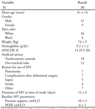
61±14 years and main reasons for mechanical venti-lation were: pneumonia, complications after abdomi-nal surgery, sepsis and stroke. Their demographic and clinical characteristics are shown in table 1. The
median of ideal FiO2 was 25% (IQ25%-75% 23-28).
This result was significantly lower than baseline FiO2
(40%) (p<0.001). PaO2, SaO2 and SpO2 were signifi-cantly lower at 30 minutes during use of ideal FiO2 (p<0.001, p<0.001, p<0.001, respectively), whereas no significant difference was observed regarding PaCO2
(p=0.21) (Table 2).Lactate (1.42±0.56 and 1.41±0.52
mmol/L) and PaO2/ FiO2 (268±47 and 269±53) values
obtained from ideal FiO2 and baseline ideal, respec-tively, were not significantly different between the two phases. Among all patients studied, four (13%) had a PaO2 < 60 mmHg during use of ideal FiO2. Table 3 shows the PaO2, SaO2, SpO2, f and HR values during the two study phases.
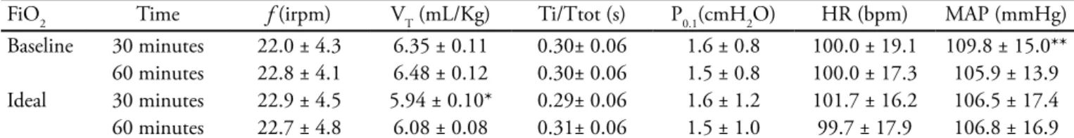
Respiratory variables and vital data assessed throughout the study time using different FiO2 values are shown in table 4. Tidal volume (VT) was signifi-cantly lower at 30 minutes during use of ideal FiO2
Table 1- Patient clinical features
Variable Result
N 30
Mean age (years) Gender
Male Female Skin color
White Black Weight (Kg) Hemoglobin (g/dL) APACHE II Artiicial airway
Tracheostomy cannula Oro-tracheal tube Reason for use of MV
Pneumonia
Complications after abdominal surgery Sepsis
Stroke Other
Duration of MV at time of study (days) Baseline MV parameters
Pressure support, cmH2O PEEP, cmH2O
61 ± 14
21 9
26 4 74 ± 9 9.2 ± 1.2 14 (9,5-20)
18 12
7 7 5 4 7 12 ± 6
in comparison to that at 30 and 60 minutes after on-set of baseline FiO2, and no significant difference was observed in intragroup analyses. When considering
interaction between the two FiO2 levels used and the
two patient exposure times for each FiO2, a significant difference was observed only in the MAP variable. It was significantly higher during the first 30 minutes
of the baseline FiO2 phase then at other times of
ex-posure. The remaining variables demonstrated no sig-nificant alterations with regard to different FiO2 levels or exposure times.
Table 2 – Gas exchange parameters observed during the use of baseline FiO2 and ideal FiO2
Baseline FiO2 Ideal FiO2 P value FiO2 (%) 40.0 24.9 ± 2.5
25 (21-30)
<0. 001* PaO2 (mmHg) 107.4 ± 21.2 65.6 ± 7.8 <0.001* PaCO2 (mmHg) 36.2 ± 6.8 34.7 ± 6.4 0.21 SaO2 (%) 97.8 ± 1.2 92.5 ± 2.4 <0.001* SpO2 (%) 97.7 ± 0.9
98 (97-98)
92.7 ± 1.34 92 (92-93)
<0.001*
FiO2 - inspired oxygen fraction; PaO2 - Arterial Oxygen Pressure; PaCO2 - Arterial Carbon Dioxide Pressure; SaO2 - Arterial Oxygen Saturation and SpO2 - Peripheral Oxygen Saturation. Results are pre-sented in median (IQ 25%-75%) or mean ± SD. *p < 0.05 compared with baseline FiO2
DISCUSSION
The main finding of the present study was that re-duction of 40% FiO2 to a level sufficient to assure SpO2 ≥ 92% did not alter the breathing pattern and/or trigger clinical changes in stable patients undergoing weaning from mechanical ventilation. However, there was a significant difference between baseline FiO2 and tideal FiO2 values for each patient. The clinical impli-cation of these findings is that it is possible to reduce FiO2 without affecting the PaO2 / FiO2, ensuring gas exchange.
No significant alterations in variables related to the respiratory pattern and drive were observed in this study, except for a reduction in VT 30 minutes after onset of the ideal FiO2 phase. However, this find-ing does not seem to have clinical significance, as the VT value considered ideal during weaning from me-chanical ventilation ranges from 4 to 6 mL/kg of ideal
weight. One reason for reduction in VT is the
inhibi-tion of chemoreceptors through reducinhibi-tion of PaCO2.
In our study, no significant variation was observed in PaCO2 levels during the different FiO2 regimens used. This may be due to the fact that patient respiratory patterns did not change. Values of average lactate and Ti/Ttot ratio remained within ranges considered
nor-Table 3 – Comparison of gas exchange parameters and vital data of the four patients with hypoxemia during the use of ideal FiO2
PaO2 (mmHg) SaO2 (%) SpO2 (%) f (irpm) HR (bpm)
Patient Baseline FiO2
Ideal FiO2
Baseline FiO2
Ideal iO2
Baseline FiO2
Ideal FiO2
Baseline FiO2
Ideal FiO2
Baseline FiO2
Ideal FiO2
1 86.9 52.8 97.1 88.2 97 92 24 26 80 84
2 85.3 57.4 96.7 89 97 92 24 25 91 93
3 82.1 52.5 96.4 87.7 96 92 18 23 85 92
4 88.4 56.1 97.2 88.8 97 92 24 25 88 96
FiO2 – inspired oxygen fraction; PaO2 - arterial oxygen pressure; SaO2 - arterial oxygen saturation; SpO2 - peripheral oxygen saturation; f - respiratory frequency; HR – heart rate
Table 4 - Respiratory variables and vital data observed during the use of baseline and ideal inspired oxygen fraction
FiO2 Time f (irpm) VT (mL/Kg) Ti/Ttot (s) P0.1(cmH2O) HR (bpm) MAP (mmHg) Baseline 30 minutes 22.0 ± 4.3 6.35 ± 0.11 0.30± 0.06 1.6 ± 0.8 100.0 ± 19.1 109.8 ± 15.0** 60 minutes 22.8 ± 4.1 6.48 ± 0.12 0.30± 0.06 1.5 ± 0.8 100.0 ± 17.3 105.9 ± 13.9 Ideal 30 minutes 22.9 ± 4.5 5.94 ± 0.10* 0.29± 0.06 1.6 ± 1.2 101.7 ± 16.2 106.5 ± 17.4 60 minutes 22.7 ± 4.8 6.08 ± 0.08 0.31± 0.06 1.5 ± 1.0 99.7 ± 17.9 106.8 ± 16.9
mal with both FiO2 levels and no significant differ-ences between them was found.
Unlike our results, Volta et al.(21) found that
respi-ratory pattern and drive were modulated by variations
in FiO2. The authors observed that reduction of FiO2
was associated to a significant increase of VT, f, P0.1 and dyspnea. However, differences of the ventilatory parameters used may explain the divergence from our results. Volta et al. compared predetermined FiO2
lev-els (21 and 30%) to the 40% FiO2 and found a
signifi-cant difference only when FiO2 was decreased from 40
to 30%. The pressure support level used for similar VT
in the two studies was also greater in the population of the Volta et al. study. This suggests that our patients were most likely at a greater mechanical advantage, which may have reflected positively on our results. In patients under adequate pressure support level, there is less overload on respiratory muscles, which trans-lates to lower P0.1 values.(22) Differences in methods of
measuring time applied to the variables may also have favored divergences between results. Pesenti et al.(23)
found an increased hypoxic respiratory drive after 20 minutes, even when SpO2 was maintained at values considered adequate (90 to 95%). However, differenc-es between the populations studied are also evident, especially because our patients were not under the ef-fect of sedatives. The diagnostic heterogeneity of our population provides a greater clinical applicability of results. Furthermore, our patients were studied for a longer time than those of the studies cited, which may also have influenced findings.
Sensitivity to increase or decrease of oxygen level occurs through specialized chemoreceptor cells that regulate respiratory and cardiovascular response. This takes place acutely through activation of pre-existing proteins and chronically by regulation of genetic tran-scription.(24) However, hypoxic stimulation in the
ca-rotid body, only occurs where there is an important
reduction in arterial oxygen content or when PaO2 is
lower than 60 mmHg. This stimulates neurosecretion by the glomic cells and causes the sensation of dysp-nea.(25) Although dyspnea was not objectively assessed
in this study, there was no report of respiratory dis-comfort by patients and, upon inspection, no acces-sory muscle action was observed.
No significant HR alterations were observed in this
study. Thomson et al.(26) found an increased HR when
assessing the effect of hypoxemia on the cardiovas-cular function of healthy volunteers. However, these individuals were exposed to 80 percent SpO2, which
is different from that in our population. Regarding MAP, a significantly higher value was observed in the first 30 minutes of the study. This may have occurred because patients were alert and possibly anxious in re-lation to the initial procedures of the study.
Absence of clinical and breathing pattern changes in our results may be explained by two main reasons. The first is that, at the time of the study, our patients no longer exhibited signs or symptoms of acute re-spiratory failure as demonstrated by a close to nor-mal PaO2/ FiO2 . Despite this clinical condition, all patients were using a 40% baseline FiO2. Moreover,
average PaO2 was higher than 65 mmHg in the
popu-lation studied. Four patients had a PaO2 of less than 60 mmHg, which probably occurred due to the 2 to 4% variability presented by most pulse oximeters. Ad-justment of FiO2 to an “ideal” value in this study was
based on SpO2, however, blood gas analyses were used
only for control of blood gas data. For that, purpose FiO2 was set at a value that would ensure a SpO2 of 92%. Oximeter variability may have caused an im-proper adjustment of FiO2 in these patients and there-fore a PaO2 of less than 60 mmHg. Respiratory fre-quency and heart rate increased in these four patients, probably in response to hypoxemia. The ideal FiO2 should be higher than the adjusted, therefore risk of hypoxemia in these patients could be avoided if the SpO2 cut-off point were raised to 94%.(27,28)
There are some limitations in our study: (1) lack of a control group, (2) non-randomization of the pa-tients studied and (3) non-assessment of the clinical
outcome of these patients. According to Benchetrit,(7)
choice of control individuals is difficult in studies that involve ventilatory alterations due to the considerable variability regarding the diverse components of the re-spiratory pattern. In order to minimize this bias, each individual served as his own control. Assessment of the clinical outcome of weaning patients submitted to different FiO2 levels could provide relevant informa-tion on oxygen use in this populainforma-tion. The total time of each patient on mechanical ventilation after data collection was not assessed, since this was not an ob-jective of the present study.
CONCLUSION
This study suggests that FiO2 levels sufficient to
ensure a SpO2 ≥ 92% did not alter breathing patterns
RESUMO
Introdução e objetivos: Frações inspiradas de oxigênio (FiO2) ≤ 40% são recomendadas durante o desmame ventilatório se pressão arterial de oxigênio (PaO2)/FiO2 ≥150–200 mmHg, O objetivo desse estudo foi comparar as variáveis respiratórias e os dados vitais coletados durante a utilização de uma FiO2 suiciente para manter a saturação periférica de oxigênio em 92% (ideal) com aquelas coletadas durante uma FiO2 rotineiramente ajustada em 40% (basal) em pacientes sob desmame ventilatório.
Métodos: Estudo prospectivo cruzado. As variáveis freqü-ência respiratória, volume corrente, pressão de oclusão, relação tempo inspiratório/tempo total, pressão arterial e freqüência cardíaca foram coletados, seqüencialmente, aos 30 e 60 minutos sob FiO2 basal (40%) e, em seguida sob FiO2 ideal. Essas foram comparadas pelo modelo linear generalizado para medidas repe-tidas. Para comparar os valores basal e ideal da FiO2 e da PaO2
foram utilizados os testes t Student ou Wilcoxon.
Resultados: Em 30 pacientes adultos a mediana da FiO2 ideal foi 25% (IQ25%-75% 23-28), signiicativamente menor que a basal (40%) (p< 0,001). A relação PaO2/FiO2 não apre-sentou diferença signiicativa entre a FiO2 basal (269±53) e a FiO2 ideal (268±47). O volume corrente foi signiicativamente menor durante a utilização da FiO2 ideal (p=0,003) e a pressão arterial foi signiicativamente maior durante a utilização da FiO2 ideal (p=0,041), mas sem signiicância clínica. A FiO2 ideal não inluenciou as demais variáveis.
Conclusão: Esses resultados sugerem que níveis de FiO2 su-icientes para manter uma SpO2≥92% não alteraram o padrão respiratório ou provocaram alterações clínicas em pacientes sob desmame ventilatório.
Descritores: Respiração; Mecânica respiratória; Ventilação mecânica; Oxigenoterapia; Desmame do respirador
REFERENCES
1. Crapo JD. Morphologic changes in pulmonary oxygen to-xicity. Annu Rev Physiol. 1986;48:721-31.
2. Barazzone C, Horowitz S, Donati YR, Rodriguez I, Piguet PF. Oxygen toxicity in mouse lung: pathways to cell death. Am J Respir Cell Mol Biol. 1998;19(4):573-81.
3. Barazzone C, White CW. Mechanisms of cell injury and death in hyperoxia: role of cytokines and Bcl-2 family pro-teins. Am J Respir Cell Mol Biol. 2000;22(5):517-9. 4. Scheuler KM. Tissue oxigenation and capacity to
de-liver O2do the two go together? Transfus Apher Sci. 2004;31(1):45-54.
5. Treacher DF, Leach RM. Oxygen transport-1. Basic prin-ciples. BMJ. 1998;317(7168):1302-6.
6. Dick CR, Sassoon C. Patient-ventilator interactions. Clin Chest Med. 1996;17(3):423-38.
7. Benchetrit G. Breathing pattern in humans: diversity and individuality. Respir Physiol. 2000;122(2-3):123-9. 8. Bryan CL, Jenkinson SG. Oxygen toxicity. Clin Chest
Med. 1988;9(1):141-52. Review.
9. Durbin CG Jr, Wallace KK. Oxygen toxicity in the criti-cally ill patient. Respir Care. 1993;38:739-53.
10. Quinn DA, Moufarrej RK, Volokhov A, Hales CA. Inte-ractions of lung stretch, hyperoxia, and MIP-2 produc-tion in ventilator-induced lung injury. J Appl Physiol. 2002;93(2):517-25.
11. Sinclair SE, Altemeier WA, Matute-Bello G, Chi EY. Augmented lung injury due to interaction between hyperoxia and mechanical ventilation. Crit Care Med. 2004;32(12):2496-501.
12. Weinberger B, Laskin DL, Heck DE, Laskin JD. Oxygen toxicity in premature infants. Toxicol Appl Pharmacol.
2002;181(1):60-7.
13. Jackson RM. Molecular, pharmacologic, and clinical as-pects of oxygen-induced lung injury. Clin Chest Med. 1990;11(1):73-86.
14. Kallstrom TJ; American Association for Respiratory Care (AARC). AARC Clinical Practice Guideline: oxygen the-rapy for adults in the acute care facility--2002 revision & update. Respir Care. 2002;47(6):717-20.
15. MacIntyre NR, Cook DJ, Ely EW Jr, Epstein SK, Fink JB, Hefner JE, Hess D, Hubmayer RD, Scheinhorn DJ; American College of Chest Physicians; American Associa-tion for Respiratory Care; American College of Critical Care Medicine. Evidence-based guidelines for weaning and discontinuing ventilatory support: a collective task force facilitated by the American College of Chest Physi-cians; the American Association for Respiratory Care; and the American College of Critical Care Medicine. Chest. 2001;120(6 Suppl):375S–95S.
16. MacIntyre NR, Epstein SK, Carson S, Scheinhorn D, Christopher K, Muldoon S; National Association for Medical Direction of Respiratory Care. Management of patients requiring prolonged mechanical ventilation: report of a NAMDRC consensus conference. Chest. 2005;128(6):3937-54.
17. Steban A, Alía I, Ibañez J, Benito S, Tobin MJ. Modes of mechanical ventilation and weaning. A national survey of Spanish hospitals. he Spanish Lung Failure Collaborative Group. Chest. 1994;106(4):1188-93.
18. Whitelaw WA, Derenne JP. Airway occlusion pressure. J Appl Physiol. 1993;74(4):1475-83. Review.
20. Cakar N, Tuörul M, Demirarslan A, Nahun A, Adams A, Akýncý O, et al. Time required for partial pressure of ar-terial oxygen equilibration during mechanical ventilation after a step change in fractional inspired oxygen concentra-tion. Intensive Care Med. 2001;27(4):655-9.
21. Volta CA, Alvisi V, Bertacchini S, Marangoni E, Ragaz-zi R, Verri M, Alvisi R. Acute efects of hyperoxemia on dyspnoea and respiratory variables during pressure support ventilation. Intensive Care Med. 2006;32(2):223-9. 22. Perrigault PF, Pouzeratte YH, Jaber S, Capdevila XJ, Hayot
M, Boccara G, et al. Changes in occlusion pressure (P0.1) and breathing pattern during pressure support ventilation. horax. 1999;54(2):119-23.
23. Pesenti A, Rossi N, Calori A, Foti G, Rossi GP. Efects of short-term oxygenation changes on acute lung injury patients undergoing pressure support ventilation. Chest. 1993;103(4):1185-9.
24. Michiels C. Physiological and pathological responses to hypoxia. Am J Pathol. 2004;164(6):1875-82.
25. Weir EK, López-Barneo J, Buckler KJ, Archer SL. Acute oxygen-sensing mechanisms. N Engl J Med. 2005;353(19):2042-55. Review.
26. homson AJ, Drummond GB, Waring WS, Webb DJ, Maxwell SR. Efects of short-term isocapnic hyperoxia and hypoxia on cardiovascular function. J Appl Physiol. 2006;101(3):809-16.
27. Perkins GD, McAuley DF, Giles S, Routledge H, Gao F. Do changes in pulse oximeter oxygen saturation predict equivalent changes in arterial oxygen saturation? Crit Care. 2003;7(4):R67.