Ação do anestésico tópico diluído e da mitomicina sobre a
sintomatologia e re-epitelização corneana no pós-operatório
da ceratectomia fotorrefrativa
Ana Carolina Freitas Morais Fortes
1, Edmundo José Velasco Martinelli
2, Luís Gustavo Ribeiro
3,
Jesse Haroldo de Nigro Corpa
4, Fernando Antonio Galhardo Tarcha
4, José Ricardo Carvalho Lima Rehder
51 Fellow at the Refractive Surgery Unit, ABC Medical School (FMABC), Santo André/SP, Brazil. 2 Head of the Refractive Surgery Unit, ABC Medical School (FMABC), Santo André/SP, Brazil. 3 Assistant at the Refractive Surgery Unit, ABC Medical School (FMABC), Santo André/SP, Brazil. 4 Collaborator at the Refractive Surgery Unit, ABC Medical School (FMABC), Santo André/SP, Brazil. 5 Professor of Ophthalmology, ABC Medical School (FMABC), Santo André/SP, Brazil.
The authors declare no conflict of interest.
Received for publication: 1/11/2012 - Accepted for publication: 22/4/2013
The action of topic diluted anesthetic and
mitomycin on the symptomatology and
corneal re-epithelialization at postoperative
photorefractive keratectomy
A
BSTRACTObjective: To evaluate the effects of diluted topic anesthetic proximetacaine on the symptoms and corneal re-epithelialization in postoperative photorefractive keratectomy (PRK). Methods: A prospective, comparative and double-blind Study. Patients were divided into two groups: With (n=32) and Without (n=24) the use of Mitomycin C at 0.02% (indicated for eyes with 4 or more diopters). Each patient received two bottles of eye drops one had artificial tear carmellose sodium 0.5%, and other had the same along with 0.125% proximetacaine. The choice of which eye would receive one or another was random. Each patient was asked, at the 1st, 2nd and 3rd postoperative days, about the type of presenting symptom on each eye and its intensity (assessed by Pain Visual Analogue Scale). The corneal re-epithelialization of each eye was evaluated at the first and fifth postoperative days. Results: The scores were significantly lower in the eyes in which anesthetic was used for both groups. The decrease in symptomatology was significantly superior in the eyes that received concomitantly the diluted anesthetic and the mitomycin when compared to the eyes that received only the anesthetic. There were only three cases of corneal re-epithelialization retardation in mitomycin group. Conclusion: The diluted proximetacaine, at the concentration and dosage proposed in this study, is safe and non-toxic to the corneal epithelium, providing significant relief for PRK postoperative symptoms. When combined, the diluted symptoms.
I
NTRODUCTIONP
hotorefractive keratectomy (PRK) is widely used to correct refractive errors using excimer laser. The technique involves removing the epithelial layer of the cornea in order to apply the laser on the stromal surface, correcting the refractive error by photoablation. Removal of the epithelium and part of the stroma exposes free nerve endings on the cornea, causing significant postoperative pain. Post-PRK pain is usually relieved with hydrophilic bandage contact lenses, non-steroidal anti-inflammatory agents (NSAIDs), and oral analgesics(1,2).Studies in the literature report on the use of topical anaesthetic agents (“comfort drops”) to control postoperative pain in patients submitted to PRK. It has been shown that 1.0% tetracaine effectively controls pain after PRK, but it is only safe if used strictly during the first 48 hours(3). Tetracaine reduces corneal sensitivity, which is an
important protection mechanism, and it is toxic to the cornea when used repeatedly and for a long period of time(4).
The anaesthetic proxymetacaine causes less corneal toxicity than tetracaine, but chronic and repeated use has also been associated with severe corneal complications(5-7).
Lower concentrations of proximetacaine have been used in an attempt to reduce or eliminate its corneal toxicity. Studies have concluded that its use during the first week after PRK is safe in rabbit(8) and human(9) eyes.
Several studies(10) indicate that proxymetacaine and other
local anaesthetics may reduce the permeability of the nerve cell membrane to sodium ions. This prevents the fundamental change which is needed to produce an action potential.
The present study is relevant for several reasons: (1) PRK often produces considerable postoperative pain; one study(3)
has shown that 1.0% tetracaine is effective in controlling pain but it reduces corneal sensitivity and is toxic when used repeatedly and for a long period of time(4), while proxymetacaine produces
less corneal toxicity than tetracaine; (2) the use of lower concentrations of proxymetacaine has reduced or eliminated the potential for corneal toxicity(8); (3) while tetracaine is
considered safe if instilled every 30 minutes during the first 48 hours(3), proxymetacaine is considered safe for use throughout
the first postoperative week(9); (4) this study investigates an
ori-ginal experimental question; and (5) this study may yield results relevant to general practice, public health, and ophthalmology.
O
BJECTIVEThis study aimed to assess the effects of topical 0.125% proxymetacaine on post-PRK symptoms and whether it delays the regeneration of corneal epithelial defects.
M
ETHODSSample
The study included 56 patients of both sexes, aged 20-35 years, seen at the Refractive Surgery Unit of the Eye Clinic of ABC Medical School, Department of Ophthalmology. The inclusion criterion was a difference lower than 1.50 dioptre (spherical equivalent) between the two eyes. Exclusion criteria were: intraoperative complications in any of the operated eyes, interrupting the use of medication in one or both eyes before the end of the study period, failure to correctly adhere the prescription of eye drops during the study, and the patient’s wish to drop out of the study. All patients underwent simultaneous bilateral PRK between July and August 2012.
Patients were divided into two groups: Group I included 32 patients whose refractive error was equal or greater than 4 D (spherical equivalent) and who received 0.02% mitomycin C intraoperatively for 20 seconds; Group II included 24 patients whose refractive error was under 4 D and who were operated without mitomycin.
All patients participated in the study voluntarily, and the study followed the ethical standards for research involving human subjects set out in resolution 196/96 of the Brazilian National Health Council (1996). The study protocol was approved by the Ethics Committee for Research Involving Human Subjects of ABC Medical School (01207212.3.0000.0082). All patients gave their Free and Informed Consent.
Procedure
On the day of surgery, patients received two bottles of eye drops with the labels covered by white tape reading RE or LE, for the right eye and left eye, respectively. One of the bottles contained a placebo consisting of artificial tear eye drops (Refresh
R
ESUMOObjetivo: Avaliar a ação do anestésico tópico proximetacaína diluído, sobre a sintomatologia e re-epitelização corneana no pós-operatório de ceratectomia fotorrefrativa (PRK). Métodos: Estudo prospectivo, comparativo e duplo cego. Os pacientes foram divididos em dois grupos: Com (n=32) e Sem (n=24) o uso de mitomicina C 0,02% no intraoperatório (indicada para ametropia igual ou acima que 4 dioptrias). Cada paciente recebeu dois frascos de colírio: um com a lágrima artificial carmelose sódica 0,5% como placebo e o outro com a mesma acrescida do anestésico proximetacaína a 0,125%. A escolha do olho a receber um ou outro colírio foi aleatória. Cada paciente foi questionado, no 1o, 2o e 3o pós-operatórios, sobre o tipo de sintoma apresentado em cada olho e sua
intensidade (medida por meio da Escala Analógica Visual de Dor). O estado da re-epitelização corneana de cada olho foi avaliado no 1o e 5o dias pós-operatórios. Resultados: Os escores da intensidade da sintomatologia foram significativamente menores nos
olhos que utilizaram anestésico, tanto para os pacientes do grupo que recebeu mitomicina como para os pacientes do grupo que não. A diminuição da sintomatologia foi significativamente maior nos olhos que receberam concomitantemente o anestésico e a mitomicina. Ocorreu retardo da re-epitelização corneana em apenas 3 casos do grupo que utilizou mitomicina. Conclusão: O anestésico tópico diluído na concentração e posologia propostas neste estudo mostrou-se seguro e não tóxico ao epitélio cornenano, proporcionando alívio significante da sintomatologia pós-operatória do PRK. Quando associados, o anestésico tópico diluído e a mitomicina apresen-taram uma ação sinérgica nesse alívio.
Figure 1: Visual Analogue Scale adapted to measure the intensity of postoperative symptoms.
Figure 2: Sheet where patients recorded the intensity of symptoms on postoperative days 2 and 3.
Chart 1
Percentages of postoperative symptoms in groups I (red) and II (blue)
Tears™, 0.5% carmellose sodium, Allergan Produtos Farmaceuticos Ltda.), while the other contained the same artifi-cial tear eye drops plus the anaesthetic agent (Anestalcon™, 0.5% proxymetacaine, Alcon Labs. do Brasil Ltda.) diluted to 0.125%. The bottles were given to the patients by an assistant who had previously randomly determined which bottle would be used in each patient’s eye and registered that information in a table to which neither the physician nor the patient had access. The eye drops had been manipulated by the surgeons a few hours before the procedures. The dilution of proxymetacaine in artificial tears to 0.125% was made by adding 1 ml of proxymetacaine to 3 ml of artificial tears. This procedure was done under strict aseptic conditions. Patients were instructed to instil one drop in each eye 3 times a day for 3 consecutive days. All 56 patients underwent PRK in both eyes in a surgical theatre, in the following steps: Topical anaesthesia and antisepsis of the periocular region; instillation of balanced frozen saline for 20 seconds(11); 180-degree marking of the site for removal of the
epithelium; mechanical removal of the corneal epithelium with a blunt (mechanical) spatula in the central 10 mm. The procedure was initiated in the corneal periphery 1 mm from the limbus and proceeded towards the centre, followed by photoablation with excimer laser Nidek EC 5000.
Immediately after photoablation, MMC 0.2 mg/ml (0.02%) was applied for 20 seconds on treated area. The surface was then washed with 20 ml of balanced saline, followed by instillation of balanced frozen saline for 20 seconds. A bandage contact lens was applied until complete epithelialisation.
Postoperatively, in addition to the study eye drops, patients were prescribed Vigamox™ (moxifloxacin hydrochloride, Alcon Labs. do Brasil Ltda.) antibiotic eye drops every 8 hours for 6 days (starting the day before surgery); Acular LS (0.5% ketorolac tromethamine, Allergan Produtos Farmaceuticos Ltda.) non-steroidal anti-inflammatory eye drops every 6 hours for 4 days; Florate™ (fluorometholone acetate, Alcon Labs. do Brasil Ltda.) corticosteroid eye drops every 8 hours for 3 weeks; and Arcoxia™ 90mg (etoricoxib, Merck and Dohme) anti-inflammatory and analgesic tablets every 12 hours for 3 days. Immediately after surgery, all patients received hydrophilic bandage contact lenses which were maintained during the corneal re-epithelialisation period and removed after full epithelial regeneration.
The day after surgery, patients were examined to assess corneal re-epithelialisation through biomicroscopy and were asked about their symptoms in each eye.
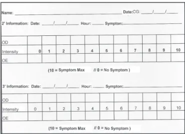
The intensity of symptoms was assessed using the Visual Analogue Scale (VAS), which was adapted for this study due to its simple, objective, and sensitive method for assessing symptom severity. The scale consists of a 10-cm-long line in which one end corresponds to zero or “no symptoms” and the opposite end corresponds to 10 or “maximum symptom intensity”. The intensity of symptoms is obtained by measuring the distance between the zero point and the location marked by the patient (Figure 1). Symptom intensity was recorded by the patients themselves at the end of postoperative days 2 and 3, one hour after the last instillation of eye drops, on a sheet of paper containing the graded tables (Figure 2).
On postoperative day 5, patients returned for an assessment of corneal healing and brought the tables containing the assessment of pain intensity which they had completed at home.
R
ESULTSThe results of the quantitative analysis of postoperative symptoms in each eye were evaluated using SPSS (Statistical Package for Social Sciences) version 17.0. The results for group I (which received 0.02% mitomycin C intraoperatively) and group II (no mitomycin) were analysed separately.
The symptoms reported by patients were: pain, burning, photophobia, gritty feeling, tearing, scratching, discomfort, and twinges. The percentages of postoperative symptoms (Chart 1) were calculated separately for groups I and II. The greatest differences between groups were for tearing, which was more frequent in group II, and gritty feeling, which was more frequent in group I.
Pain Burning
PhotophobiaGritty feeling Tearing
ScratchingDiscomfortTwinges
No Mitomycin
· Group I:
The results showed that, for patients who received mitomycin intraoperatively, the use of topical 0.125% proxymetacaine affected postoperative symptoms. The eyes that received artificial tears plus proxymetacaine had VAS scores 1.7, 2.5 and 4.0 times lower on postoperative days 1, 2 and 3, respectively, than those that used artificial tears only. That is, the eyes treated with proxymetacaine presented less severe postoperative symptoms than those treated with artificial tears only (Table 1). The Wilcoxon signed-rank test showed that these differences were statistically significant (pd”0.05).
· Group II:
The results showed that, for the eyes that did not receive mitomycin, the use of topical 0.125% proxymetacaine also affected postoperative symptoms. The eyes of patients in Group II that used proxymetacaine had VAS scores 1.7, 2.1 and 1.8 times lower than those that used artificial tears only on postoperative days 1, 2 and 3, respectively. That is, the eyes treated with proxymetacaine presented less severe postoperative symptoms than those treated with artificial tears only (Table 2). The Wilcoxon signed-rank test showed that these differences were statistically significant (pd”0.05).
Table 1
Comparison of the effect of proxymetacaine on the scores of symptom severity on postoperative days 1, 2 and 3,
as measured by the Visual Analogue Scale in the eyes of patients in Group I.
N M SD Min Max Med p-Value
Day1, artificial tears
+proxymetacaine 32 2,91 2,62 0,00 10,00 3,00
≤ 0,05
Day1, artificial tears
only 32 5,22 2,89 0,00 10,00 5,00
Day2, artificial tears
+proxymetacaine 32 2,47 2,11 0,00 8,00 2,00
≤ 0,05
Day2, artificial tears
only 32 5,38 2,59 0,00 10,00 5,00
Day3, artificial tears
+proxymetacaine 32 1,75 2,02 0,00 8,00 1,00
≤ 0,05
Day3, artificial tears
only 32 4,00 2,31 0,00 9,00 4,00
N = number of eyes; M = Mean; SD = standard deviation; Min = Minimum value; Max = Maximum value; Med = Median; (p) = Significant difference (pd”0.05) for the effect of proxymetacaine over symptom intensity scores on postoperative days 1, 2 and 3.
N M SD Min Max Med p-Value
Day1, artificial tears
+proxymetacaine 24 3,71 2,77 0,00 10,00 3,50
≤0,05
Day1, artificial tears
only 24 6,29 2,14 1,00 10,00 6,00
Day2, artificial tears
+proxymetacaine 24 4,00 2,72 0,00 9,00 3,50
≤
0,05
Day2, artificial tears
only 24 7,08 2,15 2,00 10,00 7,50
Day3, artificial tears
+proxymetacaine 24 3,29 2,05 0,00 7,00 3,00
≤0,05
Day3, artificial tears
only 24 6,00 2,55 1,00 10,00 5,50
N M SD Min Max Med p-Value
Day1, artificial tears
+proxymetacaine 32 2,91 2,62 0,00 10,00 3,00
Day2, artificial tears
+proxymetacaine 32 2,47 2,11 0,00 8,00 2,00 ≤ 0,05
Day3, artificial tears
+proxymetacaine 32 1,75 2,02 0,00 8,00 1,00
Table 2
Comparison of the effect of proxymetacaine on the scores of symptom severity on postoperative days 1, 2 and 3,
as measured by the Visual Analogue Scale in the eyes of patients in Group II.
N = number of eyes; M = Mean; SD = standard deviation; Min = Minimum value; Max = Maximum value; Med = Median; (p) = Significant difference (pd”0.05) for the effect of proxymetacaine over symptom intensity scores on postoperative days 1, 2 and 3.
Table 3
Comparison of symptom severity scores on postoperative days 1, 2 and 3, as measured by the Visual Analogue Scale in the eyes of patients in Group I who received proxymetacaine.
N = number of eyes; M = Mean; SD = standard deviation; Min = Minimum value; Max = Maximum value; Med = Median; (p) = Significant difference (pd”0.05) for the effect of proxymetacaine over symptom intensity scores on postoperative days 1, 2 and 3.
Table 4
Comparison of symptom severity scores on postoperative days 1, 2 and 3, as measured by the Visual Analogue
Scale in the eyes of patients in Group I who did not receive proxymetacaine.
N M SD Min Max Med p-Value
Day1, artificial tears
only 32 5,22 2,89 0,00 10,00 5,00
Day2, artificial tears
only 32 5,38 2,59 0,00 10,00 5,00 ≤0,05
Day3, artificial tears
only 32 4,00 2,31 0,00 9,00 4,00
N M SD Min Max Med p-Value
Day1, artificial tears
+proxymetacaine 24 3,71 2,77 0,00 10,00 3,50
Day2, artificial tears
+proxymetacaine 24 4,00 2,72 0,00 9,00 3,50 ≥ 0,05
Day3, artificial tears
+proxymetacaine 24 3,29 2,05 0,00 7,00 3,00
Table 5
Comparison of symptom severity scores on postoperative days 1, 2 and 3, as measured by the Visual Analogue Scale in the eyes of patients in Group II who received proxymetacaine.
N = number of eyes; M = Mean; SD = standard deviation; Min = Minimum value; Max = Maximum value; Med = Median; (p) = Significant difference (pd”0.05) for the effect of proxymetacaine over symptom intensity scores on postoperative days 1, 2 and 3.
N M SD Min Max Med p-Value
Day1, artificial tears
only 24 6,29 2,14 1,00 10,00 6,00
Day2, artificial tears
only 24 7,08 2,15 2,00 10,00 7,50 ≥ 0,05
Day3, artificial tears
only 24 6,00 2,55 1,00 10,00 5,50
Table 6
Comparison of the effect of proxymetacaine on the scores of symptom severity on postoperative days 1, 2 and 3,
as measured by the Visual Analogue Scale in the eyes of patients in Group II.
N = number of eyes; M = Mean; SD = standard deviation; Min = Minimum value; Max = Maximum value; Med = Median; (p) = Significant difference (pd”0.05) for the effect of proxymetacaine over symptom intensity scores on postoperative days 1, 2 and 3.
The Friedman test was applied to the variables of interest to identify possible differences between the three observation points (postoperative days 1, 2, and 3). The results showed that: 1. For the eyes in Group I that received proxymetacaine, symptom scores showed a statistically-significant decrease over time (pd”0.05). The scores for these eyes were 1.5 times lower on postoperative day 2 than on day 1, and 2 times lower on day 3 than on day 2. That is, the intensity of symptoms in the eyes that received mitomycin and proxymetacaine was progressively lower on every postoperative day (Table 3).
2. For the eyes of patients in Group I that did not receive proxymetacaine, VAS scores were similar on postoperative days 1 and 2, but on day 3 they were 1.25 times lower, and this reduction was statistically significant (pd”0.05). That is, the intensity of symptoms was similar on postoperative days 1 and 2 but improved significantly on day 3 (Table 4).
3. For the eyes of Group II that received proxymetacaine, VAS scores were similar on postoperative days 1 and 2. On day 3 they were 1.2 times lower, but this reduction was not statistically significant (pe”0.05). That is, the intensity of symptoms was similar on postoperative days 1 and 2, with a small, not significant improvement on day 3 (Table 5).
4. For the eyes of patients in Group II that did not receive proxymetacaine, VAS scores were slightly different on postoperative days 1, 2 and 3, but the difference was not statistically significant (pe”0.05). The intensity of symptoms on day 2 was 1.25 times higher than on day 1, and on day 3 it was 1.4 times lower than on day 2. That is, there was a slight, not significant increase in pain severity on postoperative day 2, with a slight, not significant improvement on day 3 (Table 6).
· Overview
The behaviour of median VAS scores in Groups I and II during the 3 postoperative days (Chart 2) shows a greater reduction in symptom severity scores among eyes that received both mitomycin and proxymetacaine (blue), followed by those that used proxymetacaine only (red). Scores also decreased in
the eyes that used mitomycin only (green) compared to those that received neither mitomycin nor proxymetacaine (purple).
Regarding the behaviour of postoperative corneal epithelialisation, delayed re-epithelisation was observed in three patients in the mitomycin group. In two of these patients, there was an epithelialisation defect with stacking of epithelial cells in the eyes that received proxymetacaine. The third patient had an ulcer in the eye that did not receive proxymetacaine.
Chart 2
D
ISCUSSIONThis study sought to address two problems that are relevant for patients undergoing PRK: intense postoperative pain and delayed corneal re-epithelialisation. The usual methods for pain relief, such as hydrophilic bandage contact lenses, NSAIDs, and oral analgesics(1,2), have not proven sufficiently effective in
controlling the postoperative symptoms of PRK nor in preventing delayed corneal re-epithelialisation.
Verma et al.(3) showed that topical anaesthetic agents can
be efficient in controlling postoperative pain in patients undergoing PRK. They used 1.0% tetracaine instilled every 30 minutes during the first 48 hours. This dose was considered safe — the authors observed a small number of cases of delayed corneal re-epithelialisation and/or complications, but only in the group that received mitomycin.
However, post-PRK pain generally persists for the first four postoperative days, and tetracaine is toxic to the cornea when used repeatedly and for a long period of time(4). The drug
reduces corneal sensitivity, which is an important protective mechanism.
Proxymetacaine is less toxic to the cornea than tetracaine, leading researchers to investigate whether its use as a topical anaesthetic agent is safe and effective in reducing post-PRK symptoms and delayed corneal re-epithelialisation.
The use of lower concentrations of proxymetacaine has reduced or eliminated the potential for corneal toxicity(8,12).
However, indiscriminate use leads to corneal changes that can manifest in varying degrees, from simple de-epithelisation to difficult-to-treat corneal ulcers(1,13).
In a study by Maurice & Singh(8) on rabbit eyes, instillation
of 0.3% proxymetacaine for one week did not cause ulceration of the corneal epithelium. Shahinian et al.(9), in a study on humans,
showed that topical 0.05% proxymetacaine induced corneal analgesia but not anaesthesia and did not cause corneal epithelial toxicity; this dose was therefore considered safe for use in the first week post-PRK.
In our study, 0.125% proximetacaine produced a significant improvement in the postoperative symptoms of PRK without interfering with corneal re-epithelialisation. Of all patients included in our study, there were only three cases of delayed corneal regeneration, all of them in the group that received mitomycin: two in eyes that received proxymetacaine and one in the placebo group. The number of cases of delayed epithelialisation was therefore not statistically significant, suggesting that the proxymetacaine solution used in this study is safe in terms of epithelial toxicity, supporting the results of Maurice & Singh(8) and Shahinian et al(9).
In our study, the VAS scores found in almost all patients were consistent with previous studies, which report that postoperative pain generally persists for the first 4 days after PRK(4). VAS scores in the group that received mitomycin without
proxymetacaine, in the group that received neither mitomycin nor proxymetacaine, and even in the group that received proxymetacaine without mitomycin showed that symptom intensity remained essentially unchanged from postoperative days 1 to 3, with minor variations (Chart 2). The only curve that showed a clear decrease, with scores close to zero after 72 hours,
was from the group that received both proxymetacaine and mitomycin.
Mitomycin C is an antimetabolite that acts at the cellular level by blocking DNA and RNA replication and protein synthesis; it is used in various areas in ophthalmology and has recently been employed as a modulator of the corneal healing response in refractive surgery using excimer laser(14). Our results
showed that mitomycin C caused a decrease in the intensity of symptoms even when used alone, with a significant decrease when associated with proxymetacaine, showing that these drugs act synergically.
The limitations of this study were its small sample size, the fact that all patients were young and healthy, and the fact that all patients underwent refractive surgery to correct myopia.
C
ONCLUSIONLow concentrations of proxymetacaine can be considered as an alternative in the management of post-PRK symptoms, considerably reducing pain scores compared to patients that did not receive proxymetacaine. Proxymetacaine was shown to be safe at the concentration and dosage examined in this study; it did not produce corneal toxicity and there were few cases of delayed corneal re-epithelialisation, which appear to be associated with the use of mitomycin. In combination with mitomycin, proxymetacaine significantly reduced symptoms on postoperative days 1 to 3, with pain scores close to zero after 72 hours.
R
EFERENCES1. Cherry PM, Tutton MK, Adhikary H, Banerjee D, Garston B, Hayward JM, et al. The treatment of pain following photorefractive keratectomy. J Refract Corneal Surg. 1994;10(2 Auppl): S222-5.
2. Tutton MK, Cherry PM, Raj PS, Fsadni MG. Efficacy and safety of topical diclofenac in reducing ocular pain after excimer photorefractive keratectomy. J Cataract Refract Surg. 1996;22(5):536-41.
3. Verma S, Corbett MC, Marshall J. A prospective, randomized, double-masked trial to evaluate the role of topical anesthetics in controlling pain after photorefractive keratectomy. Ophthalmol-ogy. 1995;102(12):1918-24.
4. Peyman GA, Rahimy MH, Fernandes ML. Effects of morfine on corneal sensitivity and epithelial wound healing: implications for topical ophthalmic analgesia. Br J Ophthalmol. 1994;78(2):138-41. Comment in Br J Ophthalmol. 1995;79(7):710.
5. Rosenwasser GO, Holland S, Pflugfelder SC, Lugo M, Heidemann DG, Culbertson WW, et al. Topical anesthetic abuse. Ophthalmol-ogy. 1990;97(8):967-72.
6. Marcondes AM. Anestésicos tópicos. In: Vita Sobrinho JB. Farmacologia & terapêutica ocular. Rio de Janeiro: Cultura Medica; 1999. p. 29-34.
7. Epstein DL, Paton D. Keratitis from the misuse of corneal anes-thetics. N Engl J Med. 1968;279(8):396-9.
8. Maurice DM, Singh T. The absence of corneal toxicity with low-level topical anesthesia. Am J Ophthalmol. 1985;99(6):691-6. 9. Shahinian L Jr, Jain S, Jager RD, Lin DT, Sanislo SS, Miller JF.
10. Arshinoff SA, Mills MD, Haber S. Pharmacotherapy of photorefractive keratectomy. J Cataract Refract Surg. 1996; 22(8):1037-44. 11. Durrie DS. BBS “popsicle” method cools eye’s surface. ASCRS
“EyeWorld” Magazine, Refract Surgery Section, Sep 2003:18. 12. Bisla K, Tanelian DL. Concentration-dependent effects of
lidocaine on corneal epithelial wound healing. Invest Ophthalmol Vis Sci. 1992;33(11):3029-33.
13. Cherry PM. The treatment of pain following excimer laser photorefractive keratectomy: additive effect of local anesthetic drops, topical diclofenac, and bandage soft contact. Ophthalmic Surg Lasers. 1996;27(5 suppl):S477-80.
Corresponding author:
Ana Carolina Freitas Morais Fortes
Rua Capote Valente, nº 500 – apto 3016 – Pinheiros CEP 05409-000 – São Paulo (SP), Brasil
Tel: (11) 3069-4000 – Celular: (11) 99306-5778 E-mail: [email protected]