Maximal bite force in young adults
with temporomandibular disorders and
bruxism
Força de mordida máxima em adultos jovens
com disfunção temporomandibular e bruxismo
Abstract: Parafunctional habits, such as bruxism, are contributory factors for temporo-mandibular disorders (TMD). The aim of this study was to evaluate the maximal bite force (MBF) in the presence of TMD and bruxism (TMDB) in young adults. Twelve women (mean age 21.5 years) and 7 men (mean age 22.4 years), composed the TMDB group. Ten healthy women and 9 men (mean age 21.4 and 22.4 years, respectively) formed the control group. TMD symptoms were evaluated by a structured questionnaire and clinical signs/symptoms were evaluated during clinical examination. A visual analogical scale (VAS) was applied for stress assessment. MBF was measured with a gnatodynamometer. The subjects were asked to bite 2 times with maximal effort, during 5 seconds, with a rest interval of about one minute. The highest values were considered. The data were analyzed with Shapiro-Wilks W-test, descriptive statistics, paired or unpaired t tests or Mann-Whitney tests when indicated, and Fisher’s exact test (p < 0.05). TMDB women presented lower values of MBF as compared to those presented by TMDB men and by the control group. MBF for TMDB men was similar to that of the control group. The proportion of TMDB women with muscle pain and facial/teeth/head pain upon waking up was signiicantly higher than that of men. Control women presented signiicantly lower stress scores than the others. It was concluded that MBF was reduced in TMDB women, as they presented more signs and symptoms. Men presented higher MBF values than women, but TMD and bruxism did not signiicantly de-crease MBF. Stress was not an inluencing factor for TMD and bruxism in men.
Descriptors: Temporomandibular joint disorders; Bruxism; Bite force.
Resumo: Hábitos parafuncionais, como o bruxismo, podem contribuir para a disfunção temporomandibular (DTM). O objetivo deste trabalho foi avaliar a força de mordida má-xima (FMM) na presença de DTM e bruxismo (DTMB) em adultos jovens. Doze mulheres (idade média de 21,5 anos) e sete homens (idade média 22,4 anos) compuseram o grupo DTMB. O grupo controle foi formado por 10 mulheres e 9 homens saudáveis, com ida-des médias de 21,4 e 22,4 anos, respectivamente. Os sintomas de DTM foram avaliados com um questionário estruturado, e os sinais/sintomas clínicos foram avaliados no exame clínico. Para avaliar estresse, utilizou-se a escala analógica visual (VAS). A FMM foi men-surada com gnatodinamômetro, e o participante foi orientado a morder com o máximo esforço durante 5 segundos, duas vezes, com intervalo de aproximadamente 1 minuto, considerando-se os valores máximos. Os dados foram analisados pelo teste de Shapiro-Wilks, estatística descritiva, teste t pareado e independente, Mann-Whitney e exato de Fi-sher (p < 0,05). As mulheres do grupo DTMB apresentaram FMM menor que os homens do grupo DTMB e do grupo controle. A FMM entre os homens do grupo DTMB foi simi-lar à do grupo controle. A proporção de mulheres no Grupo DTMB com dor muscusimi-lar e em face/dentes/cabeça ao acordar foi signiicativamente maior do que a proporção de ho-mens. As mulheres do grupo controle apresentaram escores do estresse signiicativamente menores. Concluiu-se que a FMM foi reduzida em mulheres com DTM e bruxismo devido ao maior número de sinais e sintomas. Os homens apresentaram maior FMM do que as mulheres, mas a presença de DTM e bruxismo não diminuiu signiicativamente a FMM. O estresse não inluenciou a presença de DTM e bruxismo nos homens.
Descritores: Transtornos da articulação temporomandibular; Bruxismo; Força de mordida.
Raquel Aparecida Pizolato(a) Maria Beatriz Duarte Gavião(b) Giédre Berretin-Felix(c)
Ana Claudia Martins Sampaio(d) Alceu Sergio Trindade Junior(e)
(a) Graduate Student, Department of Physiological Sciences; (b)Professor,
Department of Pediatric Dentistry – School of Dentistry of Piracicaba, State University of Campinas.
(c) Assistant Professor, Department of Speech Pathology and Audiology; (e)Professor,
Department of Biological Sciences – School of Dentistry of Bauru, University of São Paulo.
(d) Physiologist, Foundation for the Study and Treatment of Craniofacial Anomalies, Physiology Laboratory, University of São Paulo, Bauru, SP, Brazil.
Corresponding author: Raquel Aparecida Pizolato
Faculdade de Odontologia de Piracicaba da Universidade Estadual de Campinas (UNICAMP) - Departamento de Ciências Fisiológicas - Área de Fisiologia Oral Av. Limeira, 901
Piracicaba - SP - Brazil CEP: 13414-903
E-mail: [email protected]
Introduction
The temporomandibular disorder (TMD) is con-sidered a heterogeneous group of psychophysiological disturbances involving facial pain and/or masticatory dysfunction as common characteristics.24 Although
its etiology has been considered multifactorial,13 the
relative importance of individual factors is still con-troversial. Psychosocial variables may play an impor-tant role in adaptation to pain and eventual recovery. TMD patients exhibit a variety of psychological and behavioral characteristics, including increased soma-tization, stress, anxiety and depression.20
Parafunctional habits are considered contributory factors for TMD manifestation. The main parafunc-tional habit involved is bruxism, which is classiied as parafunction because it does not have a functional objective, such as mastication, phonation, or degluti-tion.27 Bruxism is an involuntary masticatory muscle
activity that is characterized by clenching and/or grinding of the teeth.3 Tooth clenching occurs in
most episodes of diurnal bruxism, while in nocturnal bruxism, both clenching and grinding are observed.16
Bruxism has a prevalence of about 10% in the gen-eral adult population, and is usually regarded as one of the possible causative factors, among others, of temporomandibular pain, tooth wear in the form of attrition, and loss of dental implants.18 Two groups
of proposed etiological factors can be distinguished: peripheral (morphological) and central (pathophysi-ological and psych(pathophysi-ological). At present, peripheral morphological factors, e.g. occlusal discrepancies, are considered to play a minor role, if any, whilst central factors, such as disorders in the dopaminer-gic system and stress, have been suggested as more important in this disorder. Smoking and alcohol consumption have also been linked to bruxism, and studies suggest that age, gender and genetic factors may inluence its prevalence.2,16 In short, bruxism is
mainly centrally – not peripherally – mediated.18
In bruxers, the distribution of muscle forces to the teeth and to the temporomandibular joints (TMJ) may result in tooth wear and orofacial pain, as well as hyperactivity and hypertrophy of the mas-ticatory muscles, especially the masseter.7
Neverthe-less, some authors question the role of bruxism as a causal agent of tooth wear,4 while others suggest
that increased tooth wear is related to bite force14
and parafunctional habits.19
The maximal bite force is the effort exerted be-tween the maxillary and mandibular teeth when the mandible is elevated by the masticatory muscles.2
The relationship between high levels of bite force and the presence of bruxism is a controversial is-sue in the related literature. It has been suggested that subjects with bruxism have overtrained masti-catory muscles, leading to hypertrophy and higher bite force,21 but no higher levels of bite force
dur-ing episodes of sleep bruxism26 were found. One
study showed that young dentate adult bruxers and nonbruxers did not present different voluntary maximum bite force values.8 However, patients with
TMD have been reported to have lower values than healthy subjects.2,6
The aim of this study was to evaluate the volun-tary maximal bite force (MBF) in the presence of bruxism and TMD in a sample of young adults.
Material and Methods
Sample
TMD and bruxism symptoms
All subjects answered a structured questionnaire, adapted from Fonseca11 (1992), to assess the
symp-toms with regard to pain in the jaws when function-ing (e.g. chewfunction-ing), unusually frequent headaches (more than once a week and with unknown etiology), dificulty in opening the mouth wide, sounds from the TMJ, and facial/tooth/head pain upon waking up. Questions about oral parafunction comprised tooth-clenching, tooth-grinding at night, and oral habits such as biting nails, lip, cheek, and/or foreign objects. The variable bruxism was constructed by combining daytime tooth-clenching and/or tooth-grinding at night. Each question could be answered with “yes” or “no”. A visual analogical scale (VAS) for stress ranging from 0 to 10 was applied individually.
Clinical signs of TMD
The signs of TMD were assessed by one cali-brated examiner in accordance with the crite-ria proposed by Dworkin, Le Resche10 (1992). A
standardized clinical examination evaluated TMJ sounds and pain during mandibular movements, and TMJ and muscle pain upon palpation. The TMJ was palpated laterally and posteriorly via the audi-tory meatus. The muscles palpated were the origin and insertion of the temporal muscle, of the medial pterygoid muscle, of the supericial portion of the masseter muscle, and of the sternocleidomastoideus muscle. All muscles were palpated bilaterally, with a standard pressure of about 1,000 g.
Maximal bite force (MBF) measurement
To measure MBF, a gnatodynamometer with two strain gages and having 10 mm in height and 10 mm in diameter was used (KFG-1-D16-11 Kyowa Elec-tronic Instruments CO., LTD., Tokyo, Japan). The subjects received detailed experimental instructions and tested biting the equipment several times before the actual recordings were made to build conidence in the test procedure. Next, each subject was asked to bite the device 2 times with maximal effort, during 5 seconds, with a rest interval between trials of about one minute. The bite force was recorded in Kgf and converted to Newton. The highest values between two trials were considered as the subject’s MBF.
Statistical analysis
The Shapiro-Wilks W-test assessed the normal-ity of the distributions. The data were analyzed by descriptive statistics and comparisons between sides, groups and genders were performed using paired or unpaired t tests or Mann-Whitney tests, when indicat-ed, considering the respective powers (α = 0.05). The proportions of signs and symptoms of TMD between genders in the TMDB group were evaluated by Fish-er’s exact test. Statistical tests were two-tailed, and a p
value < 0.05 was considered statistically signiicant.
Results
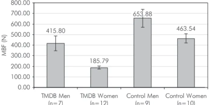
There were no MBF differences between sides so the corresponding values were averaged. They are presented in Graph 1 with their standard error of the mean (SEM) values. The respective p values are in Table 1, showing that the TMDB women had signiicantly lower values than the others. Men had greater MBF values than women in both groups; nevertheless, men in the TMDB group had MBF values similar to those of both genders in the con-trol group. The test powers produced while compar-ing TMDB women to the sub-groups was 1.0; while comparing genders in the control group, 0.5; and while comparing TMDB men to control men, 0.6.
The mean stress scores, SEM values and p values inter-groups are presented in Graph 2 and Table 1, respectively. Women in the control group presented signiicantly less stress than did both genders in the TMDB group.
The clinical signs observed in the TMDB group and the proportions for genders, as well as the prev-alence of subjective symptoms, are shown in Tables 2 and 3.
Discussion
Graph 1 - Mean values of MBF (N) for the TMDB and con-trol groups. MBF: maximal bite force. TMDB: temporoman-dibular disorders and bruxism.
185.79 415.80
653.88
463.54
0.00 100.00 200.00 300.00 400.00 500.00 600.00 700.00 800.00
TMDB Men (n=7)
TMDB Women (n=12)
Control Men (n=9)
Control Women (n=10)
M
B
F
(N
)
of both groups there was no statistical difference in MBF (Table 1), in spite of patients with TMD hav-ing been reported to have lower maximum bite force values than those of healthy subjects.2,13 This
ind-ing could be attributed to men generally reportind-ing greater pain thresholds and tolerance.9 It may thus
be that the presence of TMD had not inluenced muscle strength in the studied sample. Conversely, the number of men in the control group could be considered as an inluencing factor and a limitation
of this study, since the test powers were moderate upon comparing this sub-group with control women and TMDB men.
The proportion of women in the TMDB group with pain in some muscles (supericial masseter, ster-nocleidomastoideus, and medial pterygoid) was sig-niicantly higher than that of men in the same group (Table 2); and so was the proportion of women with facial/teeth/head pain upon waking up, which was presented by all women in the TMDB group
Table 1 -p values between subgroup comparisons of MBF and VAS scores.
Groups TMDB ♂ versus TMDB ♀
TMDB ♂ versus
Control ♂
TMDB ♂ versus
Control ♀
TMDB ♀ versus
Control ♂
TMDB ♀ versus
Control ♀
Control ♂ versus
Control ♀
MBF p 0.011 † 0.059 ‡ 0.591 ‡ < 0.001 † < 0.001 † 0.060 ‡
VAS p (†) 0.767 0.169 0.010 0.065 0.003 0.307
† = Mann-Whitney test. ‡ = Unpaired t-test. MBF: maximal bite force. VAS: visual analogical scale. TMDB: temporomandibular disorders and bruxism. Graph 2 - Mean values of VAS for stress in the TMDB and control groups. VAS: visual analogical scale. TMDB: tem-poromandibular disorders and bruxism.
0 1 2 3 4 5 6 7 8 9 10
TMDB Men (n=7)
TMDB Women (n=12)
Control Men (n=9)
Control Women (n=10)
V
A
S
sco
res
6.92 6.71
3.56
1.90
Table 2 - Proportion of clinical signs according to gender in the TMDB group.
Pain
Men (n = 7)
Women (n = 12) p
n % n %
Deep Masseter 6 86 12 100 ns
Superficial Masseter 2 29 11 92 0.001*
Deep Temporalis 1 14 7 58 ns
Sternocleidomastoideus 3 43 11 92 0.038*
Medial Pterygoid 1 14 7 58 0.002*
Temporomandibular joint 3 43 9 75 ns
*Statistically significant (Exact Fisher’s test). ns: not significant. TMDB: temporomandibular disorders and bruxism.
Table 3 - Proportion of subjective symptoms according to gender in the TMDB group.
Symptoms
Men (n = 7)
Women (n = 12) p
n % n %
Headache 6 86 7 58 ns
Teeth grinding or teeth
clenching 7 100 12 100 ns
Facial/teeth/head pain
upon waking up 4* 57 12* 100 0.029
Stress 7 100 12 100 ns
(Table 3). Decreased bite force was correlated with TMD in women, primarily those with muscle tender-ness.6 Maximum bite force can be reduced by pain
in jaw-closing muscles or in the TMJ,17 thus the
pres-ent results corroborate those of other authors.6,17 On
the other hand, patients with bruxism showed exces-sively large biting forces for each given submaximal load,21 while nocturnal bite force during bruxism can
exceed the amplitude of maximum voluntary bite force during the daytime in patients without TMD.23
Conversely, Cosme et al.8 (2005) did not ind
differ-ence in maximal bite force between young dentate adults with or without bruxism. Thus, in view of the indings of the present study, it is possible to consider that the presence of pain is associated with marked functional impairment that may be a result of relex adaptation and long-term hypoactivity of the jaw muscles, as also previously stated,13 thus decreasing
MBF in women despite the presence of bruxism. In principle, the strength of the mandibular el-evator muscles in terms of maximum bite force var-ies in much the same way for age and gender.5,12 The
gender-related difference found in bite force may be a result of anatomic differences. Men’s masseter muscles have type II ibers with larger diameter and sectional area than those of women, suggesting that hormonal differences might contribute to the com-position of the muscle ibers.12
Nevertheless, in the present study, MBF for men in the TMDB group was similar to that of men and women in the control group. In contrast, women in the TMDB group had signiicantly lower bite forces than those in the control group (Graph 1), suggest-ing that they were more affected. These outcomes could corroborate the evidence of gender differences in pain perception, as women have been reported to show more clinical pain, lower pain threshold and tolerance levels than men and are more vulnerable to the development and maintenance of musculoskeletal pain conditions.9 There is a hypothesis that the
high-er prevalence of chronic orofacial pain in women is a result of gender differences in generic pain mecha-nisms and of as-yet unidentiied factors, unique to the craniofacial system.9 The gender and age distribution
of TMD suggested a possible link between its patho-genesis and the female hormonal axis.28 In addition,
the hypothesis that the overwhelming majority of pa-tients treated for TMD are women could suggest an inluence of the sexual hormone role in TMD. The se-verity of symptoms is also related to the patient’s age. Pain onset tends to occur after puberty and peaks in the reproductive years22, with the highest prevalence
occurring in women aged 20-40 years.15
Stress is known to be an initiating, predispos-ing and perpetuatpredispos-ing factor for physical impair-ment, psychological symptoms and sleep disorders,1
whereas bruxism has been considered to be closely associated with TMD.25 The outcomes of the
self-reported stress (Graph 2, Table 1) showed that only women in the control group presented signiicant-ly lower stress scores than those of the other sub-jects, agreeing with the indings of Lobbezoo et al.18
(2006) according to which the exact contribution of stress to bruxism remains a subject of debate, as the role of psychological factors differs between individuals and is probably smaller than previously assumed in its etiology. When bruxism is present due to emotional stress, the individual can develop constant tooth clenching, leading to alterations in the normal physiological process of the mastica-tory muscles. The capability of the stomatognathic system to adapt physiologically after alterations de-pends on the individual’s system. For some, it is pos-sible to overcome stress without pathologic manifes-tations, which could have occurred with the men of the control group. Therefore, factors like perceived stress should be taken into account when treating bruxism-related temporomandibular pain.1
Conclusions
In the present study, the maximal bite force was reduced in women with TMD and bruxism, as they presented more signs and symptoms. Men present-ed higher bite force values than those of women in both groups, but the presence of TMD and bruxism did not signiicantly decrease men’s bite force. Stress was not an inluencing factor on TMD and bruxism among men.
Acknowledgements
References
1. Ahlberg J, Könönen M, Nissinen M, Rantala M, Sarna S, Lindholm H et al. Self reported stress among multiprofessional media personnel. Occup Med. 2003;53(6):403-5.
2. Ahlberg JP, Kovero OA, Hurmerinta KA, Zepa I, Nissinen MJ, Kononen MH. Maximal bite force and its association with signs and symptoms of TMD, occlusion, and body mass index in a cohort of young adults. Cranio. 2003;21(4):248-52. 3. American Sleep Disorders Association. ICSD – International
Classification of Sleep Disorders, revised: Diagnostic and Cod-ing Manual. Rochester: American Sleep Disorders Associa-tion; 1997.
4. Baba K, Haketa T, Clark GT, Ohyama T. Does tooth wear status predict ongoing sleep bruxism in 30-year-old Japanese subjects? Int J Prosthodont. 2004;17(1):39-44.
5. Bakke M, Holm B, Jensen BL, Michler L, Moller E. Unilateral, isometric bite force in 8-68-year-old women and men related to occlusal factors. Scand J Dent Res. 1990;98(2):149-58. 6. Bonjardim LR, Gaviao MB, Pereira LJ, Castelo PM. Bite force
determination in adolescents with and without temporoman-dibular dysfunction. J Oral Rehabil. 2005;32(8):577-83. 7. Clark GT, Beemsterboer PL, Rugh JD. Nocturnal masseter
muscle activity and the symptoms of masticatory dysfunction. J Oral Rehabil. 1981;8(3):279-86.
8. Cosme DC, Baldisserotto SM, Canabarro S de A, Shinkai RS. Bruxism and voluntary maximal bite force in young dentate adults. Int J Prosthodont. 2005;18(4):328-32.
9. Dao TT, LeResche L. Gender differences in pain. J Orofac Pain. 2000;14(3):169-84.
10. Dworkin SF, LeResche L. Research diagnostic criteria for temporomandibular disorders: review, criteria, examina-tions and specificaexamina-tions, critique. J Craniomandib Disord. 1992;6(4):301-55.
11. Fonseca DM. Disfunção craniomandibular (DCM): diagnóstico pela anamnese [Dissertação de Mestrado]. Bauru: Faculdade de Odontologia de Bauru da USP; 1992.
12. Garner LD, Kotwal NS. Correlation study of incisive bit-ing forces with age, sex, and anterior occlusion. J Dent Res. 1973;52(4):698-702.
13. Hansdottir R, Bakke M. Joint tenderness, jaw opening, chew-ing velocity, and bite force in patients with temporomandibu-lar joint pain and matched healthy control subjects. J Orofac Pain. 2004;18(2):108-13.
14. Johansson A. A cross-cultural study of occlusal tooth wear. Swed Dent J. 1992;86 Suppl:1-59.
15. Kuttila M, Niemi PM, Kuttila S, Alanen P, Le Bell Y. TMD treatment need in relation to age, gender, stress, and diagnostic subgroup. J Orofac Pain. 1998;12(1):67-74.
16. Lavigne GJ, Kato T, Kolta A, Sessle BJ. Neurobiological mechanisms involved in sleep bruxism. Crit Rev Oral Biol Med. 2003;14(1):30-46.
17. Liu ZJ, Yamagata K, Kasahara Y, Ito G. Electromyographic examination of jaw muscles in relation to symptoms and oc-clusion of patients with temporomandibular joint disorders. J Oral Rehabil. 1999;26(1):33-47.
18. Lobbezoo F, Van Der Zaag J, Naeije M. Bruxism: its multiple causes and its effects on dental implants – an updated review. J Oral Rehabil. 2006;33(4):293-300.
19. Manfredini D, Cantini E, Romagnoli M, Bosco M. Prevalence of bruxism in patients with different research diagnostic crite-ria for temporomandibular disorders (RDC/TMD) diagnoses. Cranio. 2003;21(4):279-85.
20. Manfredini D, Landi N, Fantoni F, Segu M, Bosco M. Anxiety symptoms in clinically diagnosed bruxers. J Oral Rehabil. 2005; 32(8):584-8.
21. Mäntyvaara J, Sjöholm T, Kirjavainen T, Waltimo A, Iivonen M, Kemppainen P et al. Altered Control of submaximal bite force during bruxism in humans. Eur J Appl Physiol Occup Physiol. 1999;79(4):325-30.
22. Meisler JG. Chronic pain conditions in women. J Women’s Health. 1999;8(3):313-20.
23. Nishigawa K, Bando E, Nakano M. Quantitative study of bite force during sleep associated bruxism. J Oral Rehabil. 2001;28(5):485-91.
24. Phillips JM, Gatchel RJ, Wesley AL, Ellis E 3rd. Clinical im-plications of sex in acute temporomandibular disorders. J Am Dent Assoc. 2001;132(1):49-57.
25. Pierce CJ, Chrisman K, Bennett ME, Close JM. Stress, an-ticipatory stress, and psychologic measures related to sleep bruxism. J Orofac Pain. 1995;9(1):51-6.
26. Pigno MA, Hatch JP, Rodrigues-Garcia RC, Sakai S, Rugh JD. Severity, distribution, and correlates of occlusal tooth wear in a sample of Mexican-American and European-American adults. Int J Prosthodont. 2001;14(1):65-70.
27. Rugh JD, Ohrbach R. Occlusal parafunction. In: Mohl N, ZarbGA, Carlsson GE, Rugh JD. A Textbook of occlusion. Chicago: Quintessence; 1988. p. 249-61.