www.rbo.org.br/
issn/$–see front matter © 2013 Sociedade Brasileira de Ortopedia e Traumatologia. Published by Elsevier Editora Ltda. All rights reserved. doi: 10.1016/j.rboe.2012.08.001
*Corresponding author at: Clinica de Ortopedia e Traumatologia. Rua 13 de maio, 940. Catanduva, SP, Brazil. CEP 15800-000. Phone/Fax: (17) 3522-3104.
E-mail: [email protected] A RT I C L E I N F O
Article history:
Received on March 18, 2012 Approved on August 20, 2012
Keywords: Pelvis Arthroplasty Hip/radiography
a b s t r a c t
Objective: The aim was to evaluate the effectiveness of traditional preoperative planning with the use of templating. Method: Forty-three anteroposterior X-rays were analyzed by three experienced surgeons (A, B, C) and compared. Cohen’s Kappa concordance test and weighted Kappa indexes using quadratic weighting were used for statistical analysis with a confidence interval of 95%. Results: The preoperative evaluations were divided into the analysis of the sizes of the acetabular cup, stem and plug of the distal femoral canal. Surgeon A obtained a moderate agreement in relation to the acetabular component and substantial agreements in relation to the stem and plug. Surgeon B had moderate agreement in relation to both the acetabulum and the stem and substantial agreement in relation to the plug. Surgeon C obtained moderate agreement in relation to the analysis of the acetabulum and the plug and substantial agreement for the stem. The intraobserver agreement test demonstrated a prevalence of slight agreement in relation to the acetabulum and substantial agreement in relation to the stem and to the plug. Conclusion: Templating used in preoperative planning proved effective; however, there was a prevalence of slight and moderate agreement in relation to the size of the acetabular component.
© 2013 Sociedade Brasileira de Ortopedia e Traumatologia. Published by Elsevier Editora Ltda. All rights reserved.
Original Article
Efficacy of the use of templating in total hip arthroplasty
Fábio Stuchi Devito,
1Alceu Gomes Chueire,
2Cristiane Bonvicine
3,*1PhD in Orthopedics, Faculdade de Medicina de São José do Rio Preto, São José do Rio Preto, SP, Brazil.
2Full Professor of the Department of Orthopedics and Traumatology, Faculdade de Medicina de São José do Rio Preto, São José do Rio Preto,
SP, Brazil.
3MSc in Health Sciences and Lecturer at Universidade Paulista (UNIP), São José do Rio Preto, SP, Brazil.
Introduction
Total hip arthroplasty is a surgical procedure with a high success rate. It provides pain relief and improvement of joint mobility.1 Long-term success with this type of procedure is related to the position and orientation of the acetabular and femoral components, which influence the wear and durability of the prosthesis.2,3
The parameters used in positioning and orienting the acetabular and femoral components, such as the prosthesis stability, restoration of lower-limb length, joint surface wear and osteolysis formation process, are vitally important with regard to the longevity of this joint and the production of femoral impact that would lead to prosthesis dislocation.2,4-6
The positioning and orientation of the acetabular and femoral components should only be determined during the operation. However, some anatomical parameters are difficult to identify during surgery and may be easier to analyze by means of preoperative radiographs, i.e. through good preoperative planning.6
Many revisions are caused by lack of primary stability of the prosthesis, errors in acetabular and femoral positioning, insufficient cement layer, muscle imbalance, design, bone quality and biological response to debris.7 Many of these factors can be considered to be not dependent on the surgeon’s choices, but good planning ought to diminish the risks of prosthesis failure.8
Traditional preoperative planning consists of analysis of radiographs of the pelvis in AP and lateral views, on which a template will be overlain, consisting of a prosthesis design for use in different sizes.6,9 In this manner, the appropriate prosthesis size and correct position for prosthesis placement can be ascertained, along with determining the presence of osteophytes, bone deformities or acetabular dysplasia, the need for bone corrections such as use of bone grafts in acetabular and femoral bone defects, the presence of leg length discrepancy and the availability of the correct prosthesis in the operating theater, so as diminish the duration of the operation and also the complication rate.6,9,10 Therefore, the preoperative planning recommended by Charnley in 197911 has the aims of more adequately restoring the anatomy of the hip, reestablishing the center of rotation of the hips and the femoral “offset”, and seeking to balance the forces that act on the biomechanics of the hips and to equalize the legs.9
Poor planning has a direct influence on the long-term final result from hip arthroplasty, since it leads to poor positioning of the prosthesis, thus causing loosening or early wear.8,12
Sample and method
To analyze the efficacy of using templating, 43 radiographs of the pelvis in AP and lateral views were analyzed by three experienced surgeons who were accustomed to doing this type of preoperative planning, in the hip diseases outpatient
clinic of the Department of Orthopedics and Traumatology, Hospital de Base, Faculdade de Medicina de São Jose do Rio Preto (Famerp), SP.
The AP hip radiographs were from patients presenting the following underlying diseases: primary and secondary osteoarthrosis, osteoarthritis, avascular necrosis of the femoral head and fracturing of the femoral neck. The standard used was that all the radiographs were centered on the pubic symphysis, with internal rotation of the legs. When analysis on the affected side was impossible because of a high degree of destruction of the femoral head, the contralateral side was used as a reference.
The technique used for producing the radiographs done in the orthopedics outpatient clinic involved a standard distance of 100 cm from the radiographic tube to the radiographic film. The distance from the drawer in the table where the film was placed to the table surface was 10 cm. Through this, the magnification rate was approximately 20%, which was determined by measuring the diameter of the femoral head of the prosthesis on the radiograph, in comparison with the real size.
For each surgeon’s preoperative analysis of templating on the preoperative radiographs, the study was standardized as follows: each surgeon’s evaluation was compared with the size of the prosthesis (acetabular, femoral and plug components) used during the surgery, and each surgeon’s result was analyzed in relation to the results from the other two, i.e. inter and intra-observer analyses. The surgery was performed by one of the three surgeons, without knowing who had done the preoperative planning, i.e. the templating done previously.
These results relating to the sizes of the prosthesis were subjected to statistical concordance testing using Cohen’s kappa13 and weighted kappa tests, with linear and quadratic weighting, with lower and upper limits for the 95% confidence interval. In the present study, the test with quadratic weighting was used. The kappa coefficient presents grading from 0 to 1, such that 0 represents no concordance and 1 is almost perfect concordance (Chart 1).
Kappa coefficient
k < 0.00 no concordance
0.00 ≤ k ≤ 0.20 very mild concordance
0.21 ≤ k ≤ 0.40 mild concordance
0.41 ≤ k ≤ 0.60 moderate concordance
0.61 ≤ k ≤ 0.80 substantial concordance
0.81 ≤ k ≤ 1.00 concordance almost
al medial
al
Forty-three radiographs were analyzed, from 43 patients aged between 22 and 81 years, with a mean of 56 years. Out of these 43 patients, 28 (65%) were female and 15 (35%) were male. The right side was affected in 23 patients (53% of the cases) and the left side in 20 patients (47%).
The template used was the cemented Exeter prosthesis (Stryker-Howmedica R), which presents magnification of 20%, with the following variables: the sizes of the acetabular component (44, 48, 52 and 56), the sizes of the femoral component and offset (35.5; 37.5 numbers 1, 2 and 3; and offset 44 numbers 1, 2, 3 and 4) and the size of the distal plug (10, 12, 14, 16, 18 and 20).
The protocol for using acetabular templating consisted of positioning the lower edge of the acetabular component on the radiographic projection of the acetabular tear, in anteroposterior radiographic view, so as to obtain complete coverage of the acetabulum, with an inclination of 45 degrees in abduction, and with the least amount of acetabular bone milling possible.9
The femoral templating consisted of firstly detecting the center of rotation of the femoral head on the affected side. When analysis on this side was not possible, the contralateral side was used as the best anatomical parameter. The “offset” was then measured, in accordance with the layouts of template sizes, taking the center of the femoral head and the prosthesis size as the reference points. A minimum of 2 mm of cement layer was maintained. The size of the distal plug was then measured on the lower edge of the prosthesis where it obstructed the femoral canal.
Among the diseases presented by the study group, the most prevalent were primary and secondary osteoarthrosis (50%), followed by rheumatic diseases (20%), avascular necrosis of the femoral head (20%) and fracturing of the femoral neck (10%).
Results
The results from the preoperative templating performed by examiners A, B and C were divided into analyses on the acetabulum, stem and plug. For examiner A, the most prevalent acetabulum was number 52, in 17 analyses, followed by numbers 48 and 56, with 12 occurrences each. The most prevalent femoral stem was number 37.5-2 with 10 occurrences, followed by 37.5-1 and 44-3 with eight occurrences. Regarding the plug, number 14 presented 14 occurrences, followed by number 12, with 12 occurrences (Chart 2).
For examiner B, the most prevalent acetabulum was number 52, in 16 analyses, followed by number 44 in 15. The most prevalent femoral stem was number 37.5-1, in 17 analyses, followed by 37.5-2 in eight. Regarding the plug, number 14 was seen in 18 analyses, followed by number 12 in 7 cases (Chart 2).
For examiner C, the most prevalent acetabulum was number 52, in 17 analyses, followed by number 48 in 15. Regarding the femoral stem, the greatest incidence was for number 37.5-1 in 17 analyses, followed by 44-1 in nine.
Regarding the plug, the highest prevalence was for number 14, in 13 analyses (Chart 2).
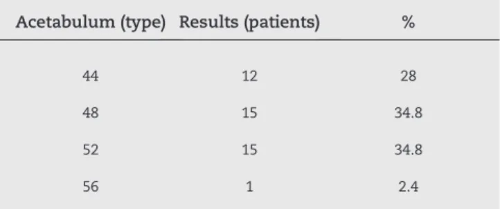
Regarding the postoperative results for the acetabular implant, number 44 was used in 12 patients, number 48 in 15 patients, number 52 in 15 patients and number 56 in one patient. Acetabular component numbers 48 and 52 were most prevalent. The percentages are presented in Table 1.
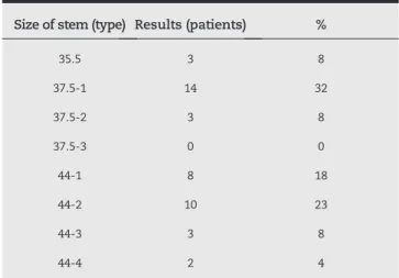
The femoral component 35.5 was used in three patients, number 37.5-1 in 14, 37.5-2 in three and 37.5-3 in none. In relation to the “offset” 44, number 44-1 was used in eight patients, 44-2 in 10, 44-3 in three and 44-4 in two. The percentages are presented in Table 2.
Regarding the plug in the femoral canal, number 10 was used in seven patients, number 12 in 13, number 14 in 14, number 16 in six, number 18 in two and number 20 in only one (Table 3).
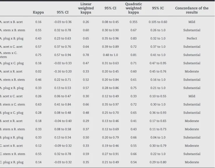
From the evaluations on the three surgeons after the postoperative results and the result from comparing the analyses between them (Table 4), using the kappa concordance test, surgeon A achieved moderate concordance in relation to the acetabular component, substantial concordance in relation to the femoral stem and substantial concordance in relation to the plug. Surgeon B achieved moderate concordance in relation to the acetabulum, moderate for the femoral stem and substantial for the plug. Surgeon C achieved moderate concordance in relation to the acetabulum, substantial for the femoral stem and moderate for the plug.
The intra-observer concordance test relating to the acetabulum showed mild concordance (Table 5). In relation to the femoral stem, the concordance was substantial. In relation to the plug, this analysis also presented substantial concordance (Table 4).
Discussion
Preoperative planning is an important step in total hip arthroplasty. Charnley11 emphasized the importance of preoperative radiographs for choosing the type of implant and appropriate size, for calculating the correct positions of the acetabular and femoral components and
Acetabulum (type) Results (patients) %
44 12 28
48 15 34.8
52 15 34.8
56 1 2.4
_ A. acet A. stem A. plug B. acet B. stem B. plug C. acet C. stem C. plug R. stem R. acet R. plug
28 44 35/5 10 44 35/5 12 44 35/5 12 35/5 44 10
29 44 35/5 10 44 35/5 12 44 35/5 12 35/5 44 10
43 48 37/5.1 10 44 37/5.1 12 48 37/5.1 12 37/5.1 44 12
33 48 35/5 12 44 35/5 12 48 35/5 12 37/5.1 48 12
11 52 37/5.2 12 48 37/5.1 12 48 37/5.1 12 37/5.1 44 12
4 52 37/5.2 12 52 44.1 12 52 44.1 12 44.1 48 10
21 52 44.3 12 52 44.3 12 52 44.3 12 44.1 48 14
12 52 44.4 12 52 44.4 12 52 44.4 12 44.2 52 12
13 48 37/5.2 14 44 37/5.1 12 44 37/5.1 12 37/5.1 44 12
41 48 37/5.1 10 52 44.1 14 48 37/5.1 12 35.5 44 12
19 48 37/5.2 12 44 37/5.1 14 48 37/5.1 12 37/5.1 44 14
40 48 37/5.1 10 44 37/5.1 12 52 37/5.1 14 37/5.1 44 12
2 48 37/5.1 10 48 37/5.2 12 48 37/5.1 14 37/5.1 44 10
6 44 44.1 10 48 37/5.1 12 48 44.1 14 44.1 48 10
35 48 37/5.1 12 44 37/5.1 12 48 37/5.1 14 44.2 48 14
36 48 37/5.1 12 44 37/5.1 12 48 37/5.1 14 44.2 48 14
26 52 44.2 12 48 44.1 12 48 37/5.1 14 44.2 28 12
34 48 37/5.1 12 48 35/5 14 48 37/5.1 14 37/5.1 44 12
9 48 37/5.2 12 48 37/5.1 14 48 37/5.1 14 37/5.1 44 10
37 52 44.1 14 44 37/5.1 14 52 37/5.1 14 44.4 52 12
20 52 37/5.2 14 52 37/5.1 14 52 37/5.2 14 37/5.1 48 14
10 48 37/5.2 14 48 37/5.2 14 48 37/5.2 14 37/5.2 44 14
25 52 44.1 14 52 44.1 14 52 44.1 14 37/5.1 48 10
24 52 44.2 14 52 44.2 14 52 44.2 14 37/5.1 52 12
30 56 44.3 12 52 44.2 12 56 44.1 16 44.1 52 14
42 52 37/5.1 14 44 37/5.1 12 56 44.1 16 44.1 48 14
15 52 44.1 14 44 37/5.1 14 48 37/5.1 16 37/5.1 52 12
3 52 37/5.2 14 52 37/5.2 14 52 37/5.1 16 44.2 48 14
39 52 44.2 14 48 37/5.1 14 52 44.1 16 44.2 48 14
22 52 44.3 14 52 44.1 14 52 44.1 16 44.3 52 16
27 56 44.4 14 52 44.2 14 52 44.1 16 44.1 52 14
8 56 44.4 16 52 44.2 14 56 44.2 16 44.3 52 14
5 56 37/5.2 14 48 37/5.1 16 52 37/5.2 16 44.2 52 16
14 52 37/5.2 14 44 37/5.1 14 52 37/5.1 18 44.1 48 14
16 52 37/5.3 18 48 37/5.2 18 52 37/5.1 18 37/5.2 50 18
31 56 44.3 16 48 37/5.2 14 56 44.2 20 44.2 52 16
7 56 44.3 16 56 44.1 14 52 44.2 20 44.2 56 14
38 56 44.3 16 52 37/5.1 16 56 44.2 20 44.1 52 16
32 56 44.3 18 44 37/5.2 16 56 44.2 20 44.2 52 16
17 52 37/5.3 18 52 37/5.2 18 44 37/5.2 20 37/5.2 18 18
18 56 44.4 18 44 37/5.2 18 48 37/5.3 20 37/5.1 52 12
1 56 44.4 18 52 44.2 20 56 44.3 20 44.4 52 16
23 56 44.3 20 52 44.1 20 52 44.1 20 44.3 52 20
A: surgeon A; B: surgeon B; C: surgeon C; R: postoperatorive results.
also for equalizing the limbs and reducing intraoperative complications.
Through preoperative planning, it is possible to reduce the duration of the operation, avoid intraoperative complications and reestablish the anatomical center of hip rotation.8
One of the important items in preoperative planning is to determine the ideal size for the prosthesis (through using templating), the correct type and position, and also the correct offset for reestablishing the hip anatomy.6,9,10,14,15
Determination of the correct offset through templating depends on correctly positioning the radiographs, through taking care to avoid rotational deviation of the pelvis and legs, lateral inclination and hip flexion, which cause errors of demarcation and could alter the results from the analysis.16 Standardized radiographs are necessary, produced with the tube centered over the pubic symphysis and with internal rotation of the legs of approximately 20 degrees. In this manner, it is possible to analyze the true “offset”, because the internal rotation provides the correction for the femoral anteversion and the appropriate size for the femoral neck.8,14
The radiographs usually present an average magnification of 20%. This percentage can be changed by increasing or decreasing the distance of the X-ray tube from the film and also the distance of the film from the patient. There is a need for standardization of radiographic techniques in order to establish the magnification of radiographs in each radiographic department, thereby avoiding errors in the templating analysis.8
Another study presented concordance between planning with preoperative templating and the postoperative result of 62% for the acetabular part and 78% for the femoral part.16 However, Carter et al.14 found that there was 95% concordance when this planning was done by an experienced surgeon.
In the present study, some intra-observer discordances occurred because of differences between the magnification of the templating and that of the radiography, such that the templating presented magnification of 20% but the mean for our study was 22.47% (15-26%). Thus, among skinny patients, this magnification could decrease to 14%, and in obese patients it could increase to 26%.15 According to Knight and Atwater,16 there is a tendency for observers to underestimate the radiographic magnification, and this is therefore one of the main factors causing errors and discordance in templating.
Thus, the radiographic magnification may change according to each individual’s physical constitution, even if the position of the radiograph and its deviations such as hip flexion have previously been corrected.14-16
The observed results from the concordance test are presented in Table 4. It can thus be noted that there was greater discordance in relation to acetabular templating, both in the inter-observer and in the intra-observer analysis. There was greater difficulty in analyzing the acetabular component than in relation to the other components of the prosthesis. It should be borne in mind that the results from the surgery were obtained all three surgeons, but none of them knew who had done the templating analysis previously.
The acetabular component presents certain particular features: it is the most difficult part of the intraoperative period; loss of control over pelvis flexion makes it difficult to do; presence of rotational deviations on the surgical table is a limiting factor in positioning errors;15 and there is no doubt that this is the part of the procedure in which each surgeon’s experience has greatest influence, with a view to maintaining the most appropriate position and also the center of hip rotation, at the level of the image of the acetabular tear. Therefore, each surgeon’s experience was a factor that produced discordance in the analysis on the acetabulum. Attention needs to be paid to the anatomical parameters in order to avoid errors relating to size and appropriate positioning.15,16
Errors in prosthesis thickness should not be taken into account as a factor for concern, given that cemented prostheses were being used. Thus, discordance in size of 2 to 3, or 3 to 4, would only lead to a small increase or decrease in the thickness of the bone cement layer, particularly in the femoral component. Diminished thickness of the cement layer, to less than 2 mm would certainly be an important factor with regard to failure and lower durability of the prosthesis.12
Size of stem (type) Results (patients) %
35.5 3 8
37.5-1 14 32
37.5-2 3 8
37.5-3 0 0
44-1 8 18
44-2 10 23
44-3 3 8
44-4 2 4
Size of plug (type) Results (patients) %
10 7 17
12 13 30
14 14 32
16 6 14
18 2 4.5
20 1 2.5
Table 2 - Size of the femoral component used in hip arthroplasty procedures.
Kappa 95% CI
Linear weighted
kappa
95% CI
Quadratic weighted
kappa
95% IC Concordance of the results
A. acet x B. acet 0.16 -0.03 to 0.36 0.26 0.08 to 0.45 0.355 0.105 to 0.60 Mild
A. stem x B. stem 0.55 0.32 to 0.78 0.60 0.30 to 0.90 0.67 0.26 to 1.0 Substantial
A. plug x B. plug 0.43 0.23 to 0.63 0.65 0.35 to 0.96 0.83 0.32 to 1.0 Perfect
A. acet x C. acet 0.57 0.37 to 0.76 0.64 0.39 to 0.89 0.72 0.37 to 1.0 Substantial A. stem x C.
stem 0.75 0.57 to 0.94 0.78 0.48 to 1.0 0.81 0.41 to 1.0 Substantial
A. plug x C. plug 0.16 -0.02 to 0.33 0.47 0.31 to 0.63 0.71 0.47 to 0.95 Substantial A. acet x R. acet 0.02 -0.16 to 0.20 0.33 0.20 to 0.45 0.60 0.45 to 0.76 Moderate A. stem x R. stem 0.46 0.22 to 0.71 0.52 0.20 to 0.84 0.61 0.16 to 1.0 Substantial A. plug x R. plug 0.33 0.13 to 0.53 0.57 0.28 to 0.86 0.75 0.21 to 1.0 Substantial
B. acet x C. acet 0.26 0.06 to 0.47 0.30 0.12 to 0.49 0.33 0.10 to 0.55 Mild
B. stem x C. stem 0.63 0.41 to 0.84 0.66 0.35 to 0.97 0.72 0.30 to 1.0 Substantial B. plug x C. plug 0.28 0.08 to 0.48 0.48 0.25 to 0.70 0.65 0.36 to 0.93 Substantial B. acet x R. acet 0.18 -0.04 to 0.40 0.29 0.13 to 0.46 0.41 0.17 to 0.65 Moderate
B. stem x R. stem 0.33 0.08 to 0.58 0.37 0.12 to 0.69 0.43 0.11 to 0.73 Moderate
B. plug x R. plug 0.33 0.13 to 0.54 0.50 0.20 to 0.79 0.66 0.04 to 1.0 Substantial C. acet x R. acet 0.12 -0.09 to 0.32 0.33 0.19 to 0.46 0.55 0.30 to 0.79 Moderate C. stem x R. stem 0.55 0.32 to 0.78 0.59 0.27 to 0.91 0.66 0.22 to 1.0 Substantial C. plug x R. plug 0.14 -0.03 to 0.32 0.35 0.21 to 0.49 0.54 0.29 to 0.80 Moderate
CI: confidence interval; A: surgeon A; B: surgeon B; C: surgeon C; R: component actually used in the surgery; acet: acetabular component. Table 4 - Comparisons between preoperative evaluations by surgeons and the results from the surgery, according to Cohen’s kappa concordance test.
Kappa 95% CI
Linear weighted
kappa
95% CI
Quadratic weighted
kappa
95% IC Concordance of the results
A. acet x B. acet 0.16 -0.03 to 0.36 0.26 0.08 to 0.45 0.355 0.105 to 0.60 Mild A. acet x C. acet 0.57 0.37 to 0.76 0.64 0.39 to 0.89 0.72 0.37 to 1.0 Substantial A. acet x R. acet 0.02 -0.16 to 0.20 0.33 0.20 to 0.45 0.60 0.45 to 0.76 Moderate
B. acet x C. acet 0.26 0.06 to 0.47 0.30 0.12 to 0.49 0.33 0.10 to 0.55 Mild
B. acet x R. acet 0.18 -0.04 to 0.40 0.29 0.13 to 0.46 0.41 0.17 to 0.65 Moderate C. acet x R. acet 0.12 -0.09 to 0.32 0.33 0.19 to 0.46 0.55 0.30 to 0.79 Moderate
In relation to the femoral component and the plug, we observed that there was greater concordance between the three surgeons, in both the inter-observer and the intra-observer analysis (Table 4). In fact, when the preoperative radiograph is in a correct position, there is no difficulty in performing the femoral templating, particularly with regard to calculating the “offset”, which in our view is an important factor for achieving greater durability of the prosthesis and for avoiding causes of joint instability.4,5,15,16
Nonetheless, these small differences in size, and not in the “offset” of the prosthesis, can and should occur in relation to the previous templating and the postoperative result, because a large number of factors, such as presence or absence of osteoporosis and young patients with thicker and harder cortical bone, influence the final result.8
One of the ways of avoiding errors of templating with conventional radiographs would be to use tomographic images. However, this would increase the exposure to radiation and the financial cost of performing examinations would be greater, thus distancing this analysis from daily practice within our setting.17-19
One good option would be to use digital templating by means of digital radiographs, which are gradually replacing conventional radiographs. In this regard, the cost of these computer graphics software programs, which is high, forms an important factor. Standardization of this type of examination is also an issue.19
It is worth highlighting that errors in image reproduction are also found in digital radiography. When the calibration of the apparatus differs greatly from the region to be analyzed, this produces a structural error in the digital correction of the magnification,18 which leads to incorrect analysis.
Bertram et al.18 presented favorable results from preoperative analysis using conventional radiographs in relation to digital radiographs, but they drew attention to the need for surgeons to bear in mind the amount of the magnification at each radiographic service.
The value of templating today in surgery done by means of computerized navigation and in digital planning is perhaps still somewhat doubtful. However, preoperative analysis using templating on conventional radiographs is a common practice in the majority of services that perform total hip arthroplasty because of the viability and practicality of this analysis and the satisfactory results.6,7,9,18
Templating should give surgeons an idea of the ideal size, position and availability of these implants in the surgical theater and an idea of the possible errors during the surgery. In other words, it should transmit security to surgeons. Small difference in implant size are irrelevant, but errors in measuring the “offset” may have greater consequences, since they may cause greater instability and wear of the prosthesis.2,4,5,8,10
Conclusions
Use of templating in preoperative planning for cemented total arthroplasty was shown to be effective. This should be particularly highlighted in relation to planning the femoral component and the plug, which presented higher concordance rates. Regarding the acetabular component, the concordance rate was lower, both in inter-observer and in intra-observer analyses.
Conflicts of interest
The authors declare that there was no conflict of interests in conducting this study.
R E F E R E N C E S
1. Harris WH, Sledge CB. Total hip and total knee replacement (1). N Engl J Med. 1990;323(11):725-31. 2. Muller ME. Lessons of 30 years of total hip arthroplasty.
Clin Orthop. 1992:274:12-21.
3. Zheng G, Marx A, Langlotz U, Widmer KH, Buttaro M, Nolte LP. A hybrid CT-free navigation system for total hip arthroplasty. Comput. Aided Surg. 2002:7(3):129-45. 4. Krushell RJ, Burke DW, HARRIS WH. Elevated-rim
acetabular components. Effect on range of motion and stability in total hip arthroplasty. J Arthroplasty. 1991;6(Suppl):S53-8.
5. Sultan PG, Tan V, Lai M, GARINO JP. Independent contribution of elevated-rim acetabular liner and femoral head size to the stability of total hip implants. J Arthroplasty. 2002;17(3):289-92.
6. Noble PC, Sugano N, Johnston JD et al. Computer
simulation: how can it help the surgeon optimize implant position? Clin Orthop Relat Res. 2003;417:242-52.
7. Vicecont M, Chiarini A, Testi D, Taddei F, Bordini B, Traina F, et al. New aspects and approaches in pre-operative planning of hip reconstruction: a computer simulation. Langenbecks Arch Surg. 2004;389(5):400-4.
8. Della Valle AG, Padgett DE, Salvati EA. Preoperative planning for primary total hip arthroplasty. J Am Acad Orthop Surg. 2005;13(7):455-62.
9. Barrack RL, Burnett SJ. Preoperative planning. 2nd ed. Lippincott (XX): Williams & Wilkins, 2006.
10. Barrack RL. Preoperative planning for revision total hip arthroplasty. Clin Orthop Relat Res. 2004;(420):32-8. 11. Charnley J. Low friction artroplasty of the hip. New York:
Springer Verlag; 1979.
12. Sandhu HS, Martim WN, Bishay M, Pozo JL Jr. Acetabular cement mantles and component positioning. Are we achieving “ideal” results? Arthroplasty. 2006;21(6):841-5. 13. Landis JR, Koch GG. The measurement of observer agreement
for categorical data. Biometrics. 1977;33(1):159-74. 14. Carter LW, Stovall DO, Young TR. Determination of
accuracy of preoperative templating of noncemented femoral prostheses. J Arthroplasty. 1995;10(4):507-13. 15. Eggli S, Pisan M, Muller ME. The value of preoperative
16. Knight JL, Atwater RD. Preoperative planning for total hip arthoplasty. Quantitating its utility and precision. J Arthroplasty. 1992;7(Suppl):403-9.
17. Sugano N, Ohzono K, Nishii T, Haraguchi K, Sakai T, Ochi T. Computed-tomography. Based computer preoperative planning for total hip arthroplasty. Comput Aided Surg. 1998;3(6):320-4.
18. The B, Diercks RL, Van Ooijen PM, Van Horn JR. Comparison of analog and digital preoperative planning in total hip and knee arthroplasties. A prospective study of 173 hips and 65 total knees. Acta Orthop. 2005;76(1):78-84.