ORIGINAL ARTICLE
Elevated plasma concentrations of S100 calcium-binding
protein B and tumor necrosis factor alpha in children with
autism spectrum disorders
Selin Aktan Guloksuz,
1Osman Abali,
2Esin Aktas Cetin,
3Sema Bilgic Gazioglu,
3Gunnur Deniz,
3Abdurrahman Yildirim,
4Ivana Kawikova,
5Sinan Guloksuz,
6,7James F. Leckman
11Child Study Center, School of Medicine, Yale University, New Haven, CT, USA.2Department of Child and Adolescent Psychiatry, Istanbul
University School of Medicine, Istanbul, Turkey.3Department of Immunology, Institute of Experimental Medicine (DETAE), Istanbul University, Istanbul, Turkey.4Department of Pediatrics, Istanbul Memorial Hospital, Istanbul, Turkey.5Department of Pediatrics, School of Medicine, Yale University, New Haven, CT, USA.6Department of Psychiatry, School of Medicine, Yale University, New Haven, CT, USA.7Department of
Psychiatry and Neuropsychology, South Limburg Mental Health Research and Teaching Network, European Graduate School of Neuroscience (EURON), School for Mental Health and Neuroscience (MHeNS), Maastricht University, Maastricht, The Netherlands.
Objective: To investigate plasma concentrations of S100B (a calcium-binding protein derived pri-marily from the glia) and inflammatory cytokines in children with autism and the relationship between S100B and cytokine concentrations.
Methods:Plasma levels of S100B, tumor necrosis factor alpha (TNF-a), interferon gamma, interleukin (IL)-1b, IL-4, IL-6, IL-10, and IL-17A were measured in 40 unmedicated children with autism and 35 normally developing healthy children. The severity of autism was assessed using the Childhood Autism Rating Scale (CARS).
Results: Concentrations of both S100B and TNF-a were higher in children with autism before and after adjusting for a priori-selected confounders (age, sex, and body mass index). S100B concen-trations were higher in children with severe autism compared to children with mild-moderate autism. However, this association remained as a trend after adjusting for confounders. S100B concentrations correlated positively with TNF-aconcentrations.
Conclusion: Our findings showing an increase in peripheral concentrations of S100B and TNF-a
provide limited support to the hypothesis about the roles of altered immune function and S100B in autism spectrum disorder (ASD). Studies of larger numbers of well-characterized individuals with ASD are needed to clarify the potential role of the immune system in the pathophysiology of this disorder.
Keywords: Autism; inflammation; glia; tumor necrosis factor; cytokine; S100B
Introduction
Autism spectrum disorder (ASD) is a neurodevelopmental disorder characterized by deficits in the development of social, communicative, and cognitive skills, accompanied by restricted, repetitive patterns of behavior or interests. The heterogeneous and complex pathoetiology of ASD remains largely unknown. Recently, the role of aberrant immune system functioning in the pathogenesis of ASD has received greater attention.1-4 Specifically, current evidence suggests that microglial activation, synaptic under-connectivity, and neuroinflammation may contri-bute to some cases of ASD.5,6
Earlier studies have documented prominent microglial activation and increased production of inflammatory cytokines and chemokines, including interferon gamma (IFN-g), interleukin (IL)-1b, IL-6, IL-12p40, tumor necrosis
factor alpha (TNF-a), and chemokine (C-C motif) ligand 2 (CCL2) in the brain tissue and cerebrospinal fluid (CSF) of some individuals with ASD.3,7 Although findings are not consistent, there is growing evidence indicating elevated plasma levels of pro-inflammatory cytokines, including TNF-a, IFN-g, IL-1b, IL-6, IL-12, IL-17A, IL-23,8-15 and decreased plasma levels of the anti-inflammatory cyto-kines IL-10 and transforming growth factor-beta (TGF-b) in children with ASD as compared to controls.1,16Although the phenotypic and neurobiological heterogeneity in ASD might be underlying factors that account for inconsistent findings, it should also be noted that important confounding factors influencing immune markers, such as medication and body mass index (BMI), have not been adequately addressed in previous research.
S100B is a calcium-binding protein. It is glial-specific and is expressed primarily by astrocytes. S100B mod-ulates the cell cycle (e.g., proliferation, differentiation, apoptosis), neurite extension, and energy metabolism in the central nervous system (CNS).17,18 Although S100B has been rigorously investigated in a number of neuro-logical and psychiatric conditions, including schizophre-nia,18mood disorders,19and Alzheimer’s disease,20only
Correspondence: Selin Aktan Guloksuz, Child Study Center, School of Medicine, Yale University, 230 South Frontage Rd., New Haven, CT, 06519, USA.
E-mail: [email protected]
Submitted Oct 21 2015, accepted Sep 18 2016, Epub Jan 12 2017.
a small number of studies have focused on ASD. Higher S100B concentrations have been reported in children with ASD compared to controls and associated with increased severity of ASD.21In addition, a recent study addressed risk genes for ASD through copy number variation anal-ysis and identified S100B as one of the genes potentially associated with ASD.22 High concentrations of S100B may be an indicator of glial cell pathology and ongoing damage. However, the scarcity of longitudinal studies prevents us from making further inferences as to whether elevated concentrations of S100B is a cause or a con-sequence of ASD.21
In this study, we aimed to determine whether there is a difference between circulating S100B and cytokines (IL-1b, IL-4, IL-6, IL-10, IL-17A, IFN-g, and TNF-a) mea-sured in peripheral plasma samples from unmedicated ASD patients compared to healthy control children (HC) after adjusting for confounding factors.
Methods
Study population
A total of 75 children were enrolled for this cross-sectional study in the summer months (July-August 2013), to reduce the probability of flu infection and seasonal allergy as possible confounders. Forty children with ASD diagnosed according to the DSM-IV criteria (mean age = 7.136
3.89 years, 10 [25%] girls) were recruited among long-term follow-up outpatients of the Autism Clinic of the Child and Adolescent Psychiatry Department at Istanbul University School of Medicine, Istanbul, Turkey. Of children with ASD, 28 (70%) were diagnosed with autistic disorder, while 12 (30%) were diagnosed with pervasive developmental disorder, not otherwise specified. Following initial screen-ing, the autism diagnosis was reconfirmed according to the DSM-IV criteria through independent clinical interviews and medical chart reviews conducted by two child psychiatrists (SAG and OA) to eliminate diagnostic reliability issues. Thirty-five typically developing HC without any Axis I/II psychiatric disorders or positive family history of ASD were recruited from the Well Child Outpatient Clinic (mean age = 6.7563.96 years, 13 [37%] girls). Detailed medical and psychiatric interviews - including developmental and family history - of HC and their parents were conducted by a child psychiatrist (SAG) and a pediatrician (AY). The study was approved by the Ethics Committee of Istanbul School of Medicine, Istanbul University, and carried out in accor-dance with the Declaration of Helsinki. All parents of the participants gave written informed consent before enroll-ment in the study. Children who were aged 12 years or older also signed the consent forms themselves.
Each participant underwent a physical examination and a medical chart review, including immunization records. Additionally, the parents of each participant were inter-viewed by medical doctors (SAG and AY) to obtain a detailed medical history. Weight and height were measured to calculate the BMI (18.1466.39 kg/m2 in ASD group; 17.0264.42 kg/m2in HC group).
All children were unmedicated (including any psycho-tropic medications) and in good health at the time of study
participation. The exclusion criteria were any known neuro-logical disorders (e.g., fragile-X syndrome, tuberous sclerosis), congenital metabolic disorders, chronic immune-related diseases (e.g., asthma, atopic dermatitis), chronic infec-tious diseases (e.g., tuberculosis), acute infecinfec-tious dis-ease within the last 4 weeks, immunization within the last 8 weeks, and use of any immune-modulating medica-tion (including non-steroidal anti-inflammatory drugs and aspirin) within the last 4 weeks.
Clinical assessments
Symptom severity in children with ASD was assessed using the Childhood Autism Rating Scale (CARS), which also aims to help screen and differentiate ASD diagnosis from other developmental disorders. CARS is rated on a four-point scale from 1 to 4 (in ½-point increments) on 15 dimensions (relationships with people, imitation, emotional response, body use, object use, adaptation to change, visual response, auditory response, near-receptor response, anxiety, verbal communication, nonverbal communication, activity level, intellectual inconsistency, and general impression), with total scores ranging from 15 to 60.23 Total scores between 30 and 36.5 indicate mild-moderate autism, whereas total scores of 37 or greater indicate severe autism. The mean CARS score of children with ASD was 39.7866.73. Of the children with ASD, 17 (42.5%) were rated as having mild-moderate and 23 (57.5%) as having severe autism.
Developmental regression - the loss of previously acquired developmental milestones such as social skills, nonverbal communication, imitation, self-care, simple pre-tend play, direct eye gaze, and orienting to name24- was assessed according to previous medical records and a clinical interview, and was found to be present in 12 (30%) children in the ASD group.
Fasting blood samples from children with ASD and HC were collected into heparin vacuum tubes (BD Bios-ciences, San Jose, CA, USA) between 8:00 a.m. and 10:00 a.m. and processed immediately after collection by centrifugation at 2,000 g for 10 min. Plasma samples were aliquoted into tubes and stored at -80o
C until analysis. The samples were not exposed to freeze-thaw cycles.
Measurement of IL-17A and S100B
this system were 2.3 pg/mL for IL-17A and 35 pg/mL for S100B.
Measurement of IL-1b, IL-4, IL-6, IL-10, IFN-g, and TNF-a
A panel for the IL-1b, IL-4, IL-6, IL-10, TNF-a, and IFN-g cytokines was run using the bead-based Milliplex MAP multiplex technology (Millipore Corporation, Billerica, MA, USA). The analytes were measured according to the manufacturer’s instructions. Antibody-labeled beads were resuspended and aliquoted into a pre-washed 96-well filter plate. Plasma samples and controls were mixed with the beads and incubated overnight at 4o
C. After overnight incubation, the plate was incubated with biotinylated detection antibodies and streptavidin-phycoerythrin solu-tion and run on the Luminex 200tinstrument (Luminex, Austin, TX, USA). Milliplex Analyst software was used for data analysis. Standard curves were generated for each analyte and the mean fluorescence intensity value was converted into concentration using a five-parameter logistic curve fit. The minimum detectable concentrations for IL1-b, IL-4, IL-6, IL-10, TNF-a, and IFN-gwere 0.6 pg/mL, 4.5 pg/mL, 0.9 pg/mL, 1.1 pg/mL, 0.7 pg/mL, and 0.8 pg/mL, respectively.
Data analysis
Data were analyzed using STATA version 12.0. Associa-tions between group (ASD vs. HC) and immune markers (IL-1b, IL-4, IL-6, IL-10, IL-17A, IFN-g, TNF-a, and S100B) were analyzed using multiple regressions with immune markers as dependent variables and a dummy variable for the groups serving as an independent variable, and the HC group serving as a reference. In children with ASD, mul-tiple regression procedures were applied to examine associations between immune markers and developmen-tal regression (no developmendevelopmen-tal regression group serving as reference) as well as autism severity (coded as mild-moderate vs. severe, with mild-mild-moderate serving as reference). As our outcome variables deviated from normality, we applied the distribution-free method of bootstrapping to each regression analysis (with n = 1,000 bootstrap resamples).25 Analyses were adjusted a priori for age, sex, and BMI. Spearman’s rank correlation was applied to determine associations between S100B and cytokine concentrations. Two-sided statistical significance was set at po0.05.
Results
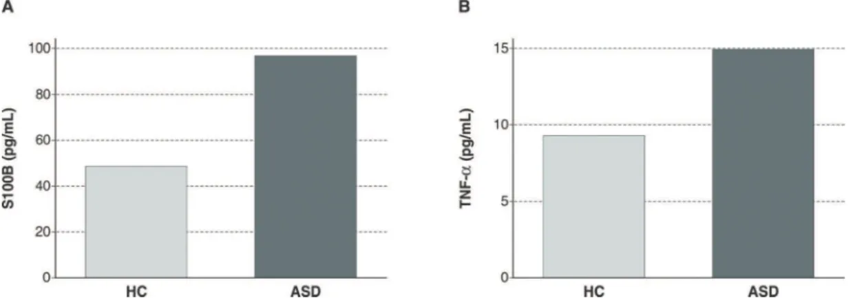
Immune marker concentrations are shown in Table 1. Even after controlling for age, sex, and BMI, there were significant elevations in plasma levels of both S100B (Figure 1A) and TNF-a(Figure 1B) in children with ASD as compared to HC (Table 2).
Plasma S100B concentrations in children with severe ASD symptoms were higher than in children with mild-moderate ASD symptoms (B = 128.26, 95%CI = 16.25-240.26, p = 0.02). However, after controlling for age, sex, and BMI, S100B concentrations were no longer signifi-cantly different between children with severe ASD and
those with less severe ASD. No other significant differen-ces were found in plasma cytokine concentrations in children with severe ASD compared to children with mild-moderate ASD (data not shown). In addition, there was no association between developmental regression and plasma immune markers (data not shown).
S100B correlated significantly with TNF-a(Spearman’s rho = 0.34, p = 0.003). There were no other correlations between S100B and cytokines.
Discussion
A role for immune involvement in ASD has long been hypothesized.4 Our findings that plasma S100B and TNF-a concentrations were higher in children with ASD confirm the findings of earlier studies and provide addi-tional limited support for this hypothesis. Studies of larger numbers of well-characterized individuals with ASD are needed to clarify the potential role of S100B and the immune system in the pathophysiology of ASD. Based on the work of Careaga et al.,26 it may be pos-sible to identify immune subphenotypes within the ASD population.4
Although S100B has predictive value in estimating neuronal damage and altered blood-brain barrier per-meability, and is easily measured in the periphery, little attention has been paid to its potential role as a peripheral marker in ASD.21As a result, our finding of elevated cir-culating levels of S100B is of particular interest. A growing body of evidence suggests the potential use of circulating S100B as a biomarker of global glial activity, which is shown to be altered in several neurological and neurop-sychiatric conditions, ranging from CNS traumatic and vascular brain injury27to psychiatric conditions, including schizophrenia, mood disorders,18and childhood trauma.28 At higher concentrations in the brain, S100B can also cause neuronal apoptosis by both microglial activation and neurotoxicity.29,30
Our findings replicated an earlier study that also reported higher circulating S100B concentrations in children with ASD compared to typically developing children, and an association between ASD severity and S100B.21 We should note that earlier studies of plasma cytokines have yielded variable results. For example, a recent meta-analysis examined 19 cytokines across 17 studies with a total sample size of 743 individuals with ASD and 592 controls. Although this meta-analysis did not evaluate S100B measurements, it is important to note that our study of Turkish children with ASD failed to confirm the presence of elevated plasma levels of IL-1b, IL-6, or IFN-g in
individuals with ASD. These discrepancies are possibly due to the existence of subsets among patients with ASD. By lumping all of the cases together in a single ASD group, these differences may constitute a major confounding factor. Combined with previous data, our findings lead us to further consider phenotypic and neurobiological hetero-geneity in ASD and the stability of immune markers over the course of the disease process. There is a significant need for longitudinal studies to help clarify the nature of the crosstalk between central neural circuits and the immune system, and the putative roles of various molecules in disease progress.
Table 1 Plasma concentrations (pg/mL) of immune markers
ASD HC
S100B 182.50 (34.61) 48.13 (13.17)
TNF-a 15.59 (7.97) 10.50 (4.77)
IFN-g 9.20 (8.07) 10.73 (14.03)
IL-1b 0.97 (0.56) 0.89 (0.85)
IL-4 19.22 (22.13) 13.86 (13.58)
IL-6 9.21 (3.93) 13.22 (14.09)
IL-10 49.87 (45.16) 71.84 (126.65)
IL-17A 2.69 (3.50) 3.26 (3.22)
Data presented as mean (standard deviation).
ASD = autism spectrum disorder; HC = healthy children; IFN-g= interferon gamma; IL = interleukin; TNF-a= tumor necrosis factor alpha.
Figure 1 A) Plasma concentrations of S100B in children with autism spectrum disorder (ASD) and typically developing healthy children (HC). B) Plasma concentrations of tumor necrosis factor alpha (TNF-a) in children with ASD and typically developing HC.Bars represent median values.
Table 2 Associations between immune markers and autism spectrum disorder
B* SE 95%CI p-value B*w SE 95%CI p-value
S100B 134.37 34.81 66.14 to 202.59 o0.001 139.99 36.99 67.47 to 212.50 o0.001
TNF-a 5.08 1.46 2.23 to 7.94 o0.001 5.81 1.41 3.06 to 8.57 o0.001
IFN-g -1.53 2.62 -6.66 to 3.59 0.56 -1.49 2.76 -6.89 to 3.92 0.59
IL-1b 0.08 0.18 -0.27 to 0.42 0.66 0.11 0.17 -0.22 to 0.44 0.52
IL-4 5.36 4.37 -3.20 to 13.91 0.22 5.78 4.40 -2.84 to 14.40 0.19
IL-6 -4.01 2.46 -8.84 to 0.82 0.10 -3.67 2.59 -8.76 to 1.41 0.16
IL-10 -21.97 22.38 -65.83 to 21.89 0.33 -20.13 24.45 -68.06 to 27.79 0.41
IL-17A -0.57 0.74 -2.03 to 0.89 0.44 -0.71 0.76 -2.21 to 0.78 0.35
95%CI = 95% confidence interval; B = regression coefficient; IFN-g= interferon gamma; IL= interleukin; SE = standard error; TNF-a= tumor necrosis factor alpha.
*Normally developing healthy children serving as the reference.
wControlled for age, sex, and body mass index.
CSF levels of S100B and other cytokines are also of potential interest. However, since astrocytes are the primary source of S100B and plasma levels of S100B have been shown to be a valid marker of CNS trauma,27 we chose to measure plasma concentrations rather than CSF levels, given the well-defined risks associated with lumbar puncture, which, in our estimation, exceeded the potential benefit. Likewise, noninvasive positron emission tomography and single-photon emission computed tomo-graphy using radioligands could be used to determine the regional specificity, if any, of CNS immune activation, including activation of microglial cells.35 Radioligands -such as [11C]PK11195 and the more specific [11C]PBR-28 - binding to 18 kDa translocator protein (TSPO), which is highly expressed by activated microglia, provide a unique opportunity to trace microglial activation in the brain.35However, just as performing lumbar puncture has associated risks, so does the use of radioactive ligands, especially in children.
Several other limitations need to be taken into account when interpreting our findings. Despite the fact that we were able to recruit children with established diagnoses of ASD who were being followed up in the specialty autism clinic of an academic medical center,36 where patients were observed by trained child psychiatrists and ulti-mately diagnosed by expert diagnosticians, collecting data by using the Autism Diagnostic Interview Revised (ADI-R) and the Autism Diagnostic Observation Schedule (ADOS) could have provided more detailed information. Unfortunately, neither instrument has been translated or adapted for use in the Turkish medical system thus far; therefore, these clinical ratings were not available for use in this study. Also, although all confirmed cases with fragile X were excluded, rigorous genetic testing was not performed, so this sample might have included children with the fragile X premutation.
In conclusion, its limitations notwithstanding, this study shed some light on the possible role of immune activation in ASD, as reflected in our findings of an increase in peri-pheral levels of S100B and TNF-a. To further elucidate the role of immune molecules in individuals with ASD, prospective longitudinal studies investigating a broad set of immune markers, both in serum and CSF, in large samples - with sufficient power to allow control for con-founders - are needed.
Acknowledgements
SG would like to acknowledge the support of the European Community’s Seventh Framework Programme under grant agreement HEALTH-F2-2009-241909 (Project EU-GEI).
Disclosure
The authors report no conflicts of interest.
References
1 Mitchell RH, Goldstein BI. Inflammation in children and adolescents with neuropsychiatric disorders: a systematic review. J Am Acad Child Adolesc Psychiatry. 2014;53:274-96.
2 Gibney SM, Drexhage HA. Evidence for a dysregulated immune system in the etiology of psychiatric disorders. J Neuroimmune Pharmacol. 2013;8:900-20.
3 Onore C, Careaga M, Ashwood P. The role of immune dysfunction in the pathophysiology of autism. Brain Behav Immun. 2012;26: 383-92.
4 McDougle CJ, Landino SM, Vahabzadeh A, O’Rourke J, Zurcher NR, Finger BC, et al. Toward an immune-mediated subtype of autism spectrum disorder. Brain Res. 2015;1617:72-92.
5 Rodriguez JI, Kern JK. Evidence of microglial activation in autism and its possible role in brain underconnectivity. Neuron Glia Biol. 2011;7:205-13.
6 Suzuki K, Sugihara G, Ouchi Y, Nakamura K, Futatsubashi M, Takebayashi K, et al. Microglial activation in young adults with autism spectrum disorder. JAMA Psychiatry. 2013;70:49-58.
7 Morgan JT, Chana G, Pardo CA, Achim C, Semendeferi K, Buckwalter J, et al. Microglial activation and increased microglial density observed in the dorsolateral prefrontal cortex in autism. Biol Psychiatry. 2010;68:368-76.
8 Ashwood P, Krakowiak P, Hertz-Picciotto I, Hansen R, Pessah IN, Van de Water J. Altered T cell responses in children with autism. Brain Behav Immun. 2011;25:840-9.
9 Ashwood P, Krakowiak P, Hertz-Picciotto I, Hansen R, Pessah I, Van de Water J. Elevated plasma cytokines in autism spectrum disorders provide evidence of immune dysfunction and are associated with impaired behavioral outcome. Brain Behav Immun. 2011;25:40-5. 10 Jyonouchi H, Sun S, Le H. Proinflammatory and regulatory cytokine
production associated with innate and adaptive immune responses in children with autism spectrum disorders and developmental regres-sion. J Neuroimmunol. 2001;120:170-9.
11 Ricci S, Businaro R, Ippoliti F, Lo Vasco VR, Massoni F, Onofri E, et al. Altered cytokine and BDNF levels in autism spectrum disorder. Neurotox Res. 2013;24:491-501.
12 Croonenberghs J, Bosmans E, Deboutte D, Kenis G, Maes M. Acti-vation of the inflammatory response system in autism. Neu-ropsychobiology. 2002;45:1-6.
13 El-Ansary A, Al-Ayadhi L. Neuroinflammation in autism spectrum disorders. J Neuroinflammation. 2012;9:265.
14 Suzuki K, Matsuzaki H, Iwata K, Kameno Y, Shimmura C, Kawai S, et al. Plasma cytokine profiles in subjects with high-functioning aut-ism spectrum disorders. PloS One. 2011;6:e20470.
15 Al-Ayadhi LY, Mostafa GA. Elevated serum levels of interleukin-17A in children with autism. J Neuroinflammation. 2012;9:158.
16 Okada K, Hashimoto K, Iwata Y, Nakamura K, Tsujii M, Tsuchiya KJ, et al. Decreased serum levels of transforming growth factor-beta1 in patients with autism. Prog Neuropsychopharmacol Biol Psychiatry. 2007;31:187-90.
17 Donato R, Sorci G, Riuzzi F, Arcuri C, Bianchi R, Brozzi F, et al. S100B’s double life: intracellular regulator and extracellular signal. Biochim Biophys Acta. 2009;1793:1008-22.
18 Rothermundt M, Ahn JN, Jorgens S. S100B in schizophrenia: an update. Gen Physiol Biophys. 2009;28 Spec No Focus: F76-81. 19 Schroeter ML, Sacher J, Steiner J, Schoenknecht P, Mueller K.
Serum S100B represents a new biomarker for mood disorders. Curr Drug Targets. 2013;14:1237-48.
20 Edwards MM, Robinson SR. TNF alpha affects the expression of GFAP and S100B: implications for Alzheimer’s disease. J Neural Transm (Vienna). 2006;113:1709-15.
21 Al-Ayadhi LY, Mostafa GA. A lack of association between elevated serum levels of S100B protein and autoimmunity in autistic children. J Neuroinflammation. 2012;9:54.
22 Egger G, Roetzer KM, Noor A, Lionel AC, Mahmood H, Schwarz-braun T, et al. Identification of risk genes for autism spectrum dis-order through copy number variation analysis in Austrian families. Neurogenetics. 2014;15:117-27.
23 Schopler E, Reichler RJ, Rochen Renner B. The Childhood Autism Rating Scale (CARS).Los Angeles: Western Psychological Services; 1988.
24 Goldberg WA, Osann K, Filipek PA, Laulhere T, Jarvis K, Modahl C, et al. Language and other regression: assessment and timing. J Autism Dev Disord. 2003;33:607-16.
26 Careaga M, Rogers S, Hansen RL, Amaral DG, Van de Water J, Ashwood P. Immune endophenotypes in children with autism spec-trum disorder. Biol Psychiatry. 2015 Sep 11. pii: S0006-3223(15) 00738-6. doi: 10.1016/j.biopsych.2015.08.036. [Epub ahead of print]. 27 Sen J, Belli A. S100B in neuropathologic states: the CRP of the
brain? J Neurosci Res. 2007;85:1373-80.
28 Falcone T, Janigro D, Lovell R, Simon B, Brown CA, Herrera M, et al. S100B blood levels and childhood trauma in adolescent inpatients. J Psychiatr Res. 2015;62:14-22.
29 Villarreal A, Aviles Reyes RX, Angelo MF, Reines AG, Ramos AJ. S100B alters neuronal survival and dendrite extension via RAGE-mediated NF-kappaB signaling. J Neurochem. 2011;117:321-32. 30 Bianchi R, Kastrisianaki E, Giambanco I, Donato R. S100B protein
stimulates microglia migration via RAGE-dependent up-regulation of chemokine expression and release. J Biol Chem. 2011;286:7214-26. 31 Adami C, Sorci G, Blasi E, Agneletti AL, Bistoni F, Donato R. S100B
expression in and effects on microglia. Glia. 2001;33:131-42.
32 Bonizzi G, Karin M. The two NF-kappaB activation pathways and their role in innate and adaptive immunity. Trends Immunol. 2004;25:280-8.
33 Young AM, Campbell EC, Lynch S, Dunn MH, Powis SJ, Suckling J. Regional susceptibility to TNF-alpha induction of murine brain inflammation via classical IKK/NF-kappaB signalling. PloS One. 2012;7:e39049.
34 O’Neill LA, Kaltschmidt C. NF-kappa B: a crucial transcription factor for glial and neuronal cell function. Trends Neurosci. 1997;20: 252-8.
35 Zurcher NR, Bhanot A, McDougle CJ, Hooker JM. A systematic review of molecular imaging (PET and SPECT) in autism spectrum disorder: current state and future research opportunities. Neurosci Biobehav Rev. 2015;52:56-73.