3 artigo 442
original article
1 – Orthopedist and Traumatologist; Specialist in Pediatric Orthopedics at Hospital Maria Amélia Lins, FHEMIG, Belo Horizonte, MG; MSc in Health Sciences (UFMG), focusing on Children’s and Adolescents’ Health.
2 – Resident Physician in Orthopedics and Traumatology, Hospital Maria Amélia Lins, FHEMIG, Belo Horizonte, MG. 3 – Resident Physician at Hospital Mater Dei, Belo Horizonte, MG.
4 – Orthopedist and Traumatologist, Hospital Maria Amélia Lins, FHEMIG, Belo Horizonte, MG.
5 – Orthopedist and Traumatologist; Specialist in Pediatric Orthopedics at Hospital Maria Amélia Lins, FHEMIG, Belo Horizonte, MG.
Work performed in the Pediatric Orthopedics and Traumatology Outpatient Clinic of Hospital Maria Amélia Lins, Belo Horizonte, MG. Correspondence: Rua Congonhas 668, Bairro Santo Antonio, 30300-100 Belo Horizonte, MG. E-mail: [email protected] Work received for publication: October 29, 2010; accepted for publication: November 4, 2011.
FLOATING ELBOW IN CHILDREN: A DESCRIPTIVE STUDY OF 31 CASES
ATTENDED IN A REFERENCE CENTER FOR PEDIATRIC TRAUMA
Dorotea Starling Malheiros1, Gustavo Henrique Silva Bárbara2, Leandro Gonçalves Mafalda2, João Lopo Madureira Júnior3, Gilberto Ferreira Braga4, Dalton Lopes Terra5
aBstract
Objective: To conduct a descriptive analysis on 31 cases of children with floating elbow who were attended at our clinic between 1994 and 2009, and to review the literature relating to this topic. Methods: Data were obtained through examining the medical records. The following variables were used: age, gender, side, mechanism, type of fracture, classification, treatment and complications. Results: Twenty-four patients (77.4%) were male and seven (22.6%) were female. The mean age was 8.5 (± 3.2) years, ranging from one to 14 years. The left side was predominantly affected (67.7%). The commonest injury mechanism was a fall from a height (74.2%). All the supracondylar fractures were Gartland type III. Distal radius fractures alone, of Salter-Harris type II, were diagnosed in 22 patients (71%). Open fractures occurred in 22 cases (71%). Closed reduction
The authors declare that there was no conflict of interest in conducting this work
this article is available online in portuguese and english at the websites: www.rbo.org.br and www.scielo.br/rbort
and application of a plaster cast for a closed fracture of the distal radius was performed in two patients (6.45%). Simultaneous conservative treatment for two fractures was not used. Sixteen supracondylar fractures (54.8%) were fixed using crossed wires, at 90° to each other, and in 14 cases (45.16%), an intramedullary wire was used together with another wire introduced through the lateral epicondyle at 45°. The following complications were observed: deformed consolidation (10%), nerve injuries (6%), compartment syndrome (3%) and pin path infection (16%). Conclusions: This is an uncommon injury that in most cases results from high-energy trauma. Surgical treatment for both fractures is recommended by most authors. Ulnar nerve injuries were correlated with the fixation method, but no neurological injuries were triggered by the initial trauma.
Keywords: Child; Elbow, Humeral Fractures; Forearm
introdUction
Fracturing of one or both bones of the forearm together with fracturing of the ipsilateral humerus represents severe injury of the upper limb in children(1). Stanitski and Micheli(2) were the first to
use the term “floating elbow” to describe associations between such injuries. They are not common, with prevalence ranging from 2 to 13%, and in most cases they are associated with high-energy trauma(2,3).
In a study on 3,472 patients attended at a pediatric traumatology reference center, Buckley et al(4) stated
that regarding the incidence of concomitant fractures, the most frequent associations were between ankle, tibial and fibular fractures (ten cases), followed by radial, ulnar and humeral fractures (nine cases).
501
association between the multiple injuries was between humeral fractures and fractures of the forearm bones (25 cases).
Exposed fractures, compartment syndrome and neurovascular abnormalities were more frequent in children with floating elbow, than in cases of supracondylar fracture of the humerus alone(6).
Although conservative treatment has been cited in the literature(7,8), most authors have considered
that percutaneous fixation using pins is funda-mental for humeral fractures, both to achieve a better functional result and to diminish the risk of neurovascular complications(2,9-11).
Templeton and Graham(12) considered that
fixa-tion for forearm fractures was advantageous, since this enabled better monitoring of the neurovascu-lar status of the affected arm and facilitated caring for skin wounds in the case of exposed fractures. The present study had the aims of conducting a descriptive analysis on 31 cases of children with floating elbow attended at our clinic between 1994 and 2007, and making a detailed review of the literature relating to this topic. This is therefore an important study, given the severity and rarity of this condition, as well as the scarcity of studies in the Brazilian literature with a similar approach.
material and methods
This was a retrospective, cross-sectional and observational study. It evaluated children who had suffered fractures in one or both forearm bones together with fracturing of the ipsilateral humerus (the injury known as floating elbow). These chil-dren were attended at the Pediatric Orthopedic and Traumatology Outpatient Clinic of Hospital Maria Amélia Lins, between June 1994 and December 2009.
The variables studied were: age, gender, side affected, trauma mechanism, type of fracture, classification, treatment administered and complications secondary to the treatment.
Data were obtained from analysis on the medical records and were transferred to the research pro-tocol. Patients with supracondylar fractures of the humerus of Gartland types I and II were excluded.
The statistical analyses were done by means of the Epi-Info 8.0 and SPSS 12.0 software. The significance level was taken to be 5%. However, because of the small sample size and the lack of a group for making comparisons among the results, no such statistical calculations could be made. Be-cause the variables were separate, analysis through comparing mean values became impossible. Thus, it was decided to undertake a descriptive study in which the profile of patients with a floating elbow could be outlined.
The present study was approved by the institu-tion’s Research Ethics Committee and was regis-tered with the Brazilian Ministry of Health, in the National Research Ethics System (SISNEP), under cover page number 221.462 and CAAE number 0076.0.287.000-08.
resUlts
Evaluations were made on 1,941 medical files of patients with fractures who were attended in our clinic between June 1994 and December 2009. Of these, 913 presented injuries in the upper limbs and 928 in the lower limbs. Thirty-two presented a floating elbow condition. One patient who pre-sented a supracondylar fracture of the humerus of Gartland type II was excluded, thus giving a study group of 31 patients.
Regarding gender, 24 patients (77.4%) were male and seven (22.6%) were female. The mean age was 8.5 years (± 3.2), with a range from one to 14 years, and 17 patients (54.83%) were between six and 10 years old.
The left side was more affected (67.7%). The commonest injury mechanism was falls from a height, in the cases of 23 patients (74.2%), mainly from fruit trees. This was followed by falls from bicycles (16.1%) and car accidents (9.7%).
All the supracondylar fractures of the humerus were classified as Gartland type III. The com-monest pattern among the forearm bone injuries consisted of fracturing of the distal radius alone (Salter-Harris type II growth plate injury), in 22 (71%) of the patients.
Exposed fractures occurred in 22 patients (71%), with the following distribution: 15 (48.4%) with exposure of the humerus and seven (22.6%) with an exposed fracture of one or both of the forearm bones. Simultaneous exposure of the humerus and forearm occurred in two patients. Regarding the classification, all of them were of Gustillo and Anderson type I (Table 1).
All the exposed fractures were treated with immediate fixation as an emergency measure, using Kirschner wires. In two patients (6.45%), it was decided to perform closed reduction followed by placement of a plaster cast extending from the axilla to the palm, to treat the closed fracture of the distal radius. Conservative treatment for both fractures was not used simultaneously for any of the patients (Table 2).
The fixation method most used for the distal radius
was two Kirschner wires (93.54%): one introduced into the radial styloid and the other dorsally through Lister’s tubercle. Regarding the supracondylar fractures of the humerus, 17 (54.8%) were fixed with crossed wires (one medially and the other laterally), while in 14 cases (45.16%), one intramedullary wire was used, followed by another that was introduced through the lateral epicondyle, crossing the first wire at an angle of 45 degrees.
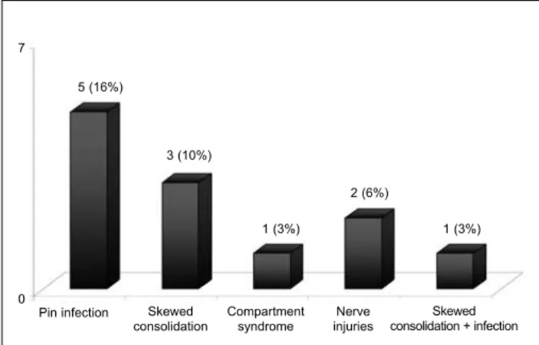
Complications occurred in 12 patients, distributed as follows: skewed consolidation (10%), nerve injury (6.0%), compartment syndrome (3.0%) and infection of the pin paths (16%) (Figure 1).
table 1 – Prevalence of the presence of exposure, in relation to
fracture location.
fracture location
total (freq%)
closed (freq%)
exposed (freq%)
Humeral fracture 31 (100%) 16 (51.6%) 15 (48.4%) Radial fracture alone 22 (71%) 19 (61.3%) 3 ( 9.7%)
Fractures in both
forearm bones 9 (29%) 5 (16.15%) 4 (12.9%) Source: AOTP, 1994/2007.
table 2 – Treatment used, according to fracture location and
presence of exposure.
type of fracture
open reduction + fixation (%)
closed reduction + fixation (%)
closed reduction + plaster (%)
total (100%)
Closed humeral
fracture 1 (6.2%) 15 (93.8%) None 16 Exposed humeral
fracture 12 (80%) 3 (20%) None 15 Closed radial
fracture alone 2 (10.5%) 15 (79.5%) 2 (10.5%) 19 Exposed radial
fracture alone 3 (100%) None None 3 Closed fractures
in both forearm bones
2 (40%) 3 (60%) None 5
Exposed fractures in both forearm
bones
4 (100%) None None 4 Source: AOTP, 1994/2007.
Open reduction of the supracondylar fracture of the humerus was performed on 13 patients (41.93%), and this gave rise to three cases of skewed consoli-dation (in varus).
Ulnar nerve injuries were diagnosed in two patients (6.5%) who had both undergone fixation with crossed wires at 90°. No neurological injuries were associated with the initial trauma.
Infection along the paths of the Kirschner wires were found in five patients (16.12%), and this problem was resolved by removing the wires.
One patient evolved with compartment syndrome, which was treated by means of forearm fasciotomy in the emergency unit.
discUssion
The large number of cases, compared with published studies so far available in the literature(1,2,4-7,12), figure 1 – Distribution of complications among patients with floating elbow.
Source: AOTP, 1994/2007
Pin infection Skewed consolidation
Compartment syndrome
Nerve injuries
Skewed consolidation + infection 5 (16%)
3 (10%)
1 (3%)
2 (6%)
1 (3%) 7
503
was due to the fact that our clinic is the biggest public reference center for pediatric traumatology in the state of Minas Gerais. Consequently, large numbers of complex injuries, like floating elbow, are referred to us from the entire state.
The gender and side predominance that we found corroborated data in the literature(2,6,12). The
predominance of the mechanism of falls from a height, especially from trees, emphasizes that associations of fractures of one or both forearm bones with supracondylar fractures of the ipsilateral humerus result from high-energy trauma in most cases, given that the force is not totally dissipated through the first fracture alone(6). Thus, the incidence of exposed
fractures that was found (71%) was much higher than among cases of single fractures. In a meta-analysis on 61 studies reporting on a total of 7,212 supracondylar fractures of the humerus, Wilkins(13) found that the
incidence of exposed fractures was just 1%.
Likewise, Harrington et al(1) affirmed that open
reduction is performed more often on patients with floating elbow. This is because the violence of the trauma involved results in fractures with large displacements, and the proximal fragment of the supracondylar fracture of humerus often becomes trapped in the anterior soft tissue, thus making closed reduction impossible. In the present study, 41.93% of the supracondylar fractures of the humerus (13 patients) required open reduction.
The forearm fracture pattern most frequently found consisted of distal radius fractures, which is in agreement with the current literature(2,6,12). Templeton
and Graham(12) affirmed that if the forearm fracture
occurs in the proximal or middle third of the forearm, the lever thus created is too small to generate the moment of force that would be needed to produce a fracture in the ipsilateral humerus.
Although conservative treatment has been cited in the literature(7,8), most authors agree that surgical
treatment is essential(2,6,12,13). Traction does not
allow anatomical reduction, and placement of a plaster cast with the arm flexed (which would be necessary to provide greater stability to supracondylar fractures) is contraindicated because of the severe edema that is present, which would increase the risk of compartment syndrome. Moreover, high incidence of cubitus varus has been associated with
conservative treatment(9). In the present study, none
of the supracondylar fractures of the humerus were treated conservatively.
Templeton and Graham(12) affirmed that they
gave priority to fixation of supracondylar fractures of the humerus, since these were associated with a greater number of complications. Also according to these authors, once such fractures have been stabilized, management of the forearm fractures becomes easier.
Two patients underwent closed reduction followed by placement of a plaster cast extending from the axilla to the palm, to treat closed fractures of the distal radius. Harrington et al(1) stated that they achieved
good results from similar treatment for four of their 12 patients with floating elbow. However, fixation for both fractures has been advocated by some authors(6,13), because in their view, this would enable
better monitoring of the neurovascular status of the affected arm. This would facilitate caring for skin wounds, in the case of exposure fractures, and would have lower incidence of loss of reduction, compared with conservative treatment.
The fixation methods used for treating the supracondylar fractures of the humerus consisted of crossed wires, either one medially and the other laterally, or one intramedullary wire with another introduced through the lateral epicondyle. There were two cases of ulnar nerve injury associated with introduction of the medial wire, and these were considered to be iatrogenic complications. Although fixation with crossed wires is more stable, there is in practice a greater risk of injuring the ulnar nerve, because the medial wire is passed through the bone blindly(11,13). In our clinic, since the year
2000, the preferred method has consisted of using an intramedullary wire in association with another lateral wire crossing the first wire at an angle of 45 degrees. This has so far produced excellent results, without any reports of neural lesions(14).
Varus consolidation of the supracondylar fracture of the humerus was found in three patients who had all initially undergone open reduction. This can be explained by the severity of the fractures, which required open reduction, along with insufficient reduction performed by the surgeon during the operation. Labelle et al(15) observed that varus
inclination of the distal fragment was the cause of the deformity in all their patients with cubitus varus following a supracondylar fracture.
Contrary to reports in the literature, no neurological lesions were triggered by the initial trauma in any of our cases. In a series of eight cases of floating elbow, Templeton and Graham(12)
reported two occurrences of lesions in the medial nerve, one in the anterior interosseous nerve and one in the ulnar nerve. According to these authors, this incidence was high because of the severity of the trauma and the resultant high-energy injury.
references
1. Harrington P, Sharif I, Fogarty EE, Dowling FE, Moore DP. Management of the floating elbow injury in children. Simultaneous ipsilateral fractures of the elbow and forearm. Arch Orthop Trauma Surg. 2000;120(3-4):205-8.
2. Stanitski CL, Micheli LJ. Simultaneous ipsilateral fractures of the arm and forearm in children. Clin Orthop Relat Res. 1980;(153):218-22.
3. Piggot J, Graham HK, McCoy GF. Supracondylar fractures of the humerus in children. Treatment by straight lateral traction. J Bone Joint Surg Br. 1986;68(4):577-83.
4. Buckley SL, Gotschall C, Robertson W Jr, Sturm P, Tosi L, Thomas M, et al. The relationships of skeletal injuries with trauma score, injury severity score, length of hospital stay, hospital charges, and mortality in children admitted to a regional pediatric trauma center. J Pediatr Orthop. 1994;14(4):449-53.
5. Malheiros DS. Estudo descritivo de lesões ortopédicas em crianças e adolescentes em centro de atendimento nível II [tese]. Belo Horizonte: Universidade Federal de Minas Gerais. Faculdade de Medicina; 2009. 6. Ring D, Waters PM, Hotchkiss RN, Kasser JR. Pediatric floating elbow. J
Pediatr Orthop. 2001;21(4):456-9.
7. Papavasiliou V, Nenopoulos S. Ipsilateral injuries of the elbow and forearm in children. J Pediatr Orthop. 1986;6(1):58-60.
8. Biyani A, Gupta SP, Sharma JC. Ipsilateral supracondylar fracture of humerus and forearm bones in children. Injury. 1989;20(4):203-7.
9. Reed FE Jr, Apple DF Jr. Ipsilateral fractures of the elbow and forearm. South Med J. 1976;69(2):149-51.
10. Williamson DM, Cole WG. Treatment of ipsilateral supracondylar and forearm fractures in children. Injury. 1992;23(3):159-61.
11. Flynn JC, Matthews JG, Benoit RL. Blind pinning of displaced supracondylar fractures of the humerus in children. Sixteen years’ experience with long-term follow-up. J Bone Joint Surg Am. 1974;56(2):263-72.
12. Templeton PA, Graham HK. The ‘floating elbow’ in children. Simultaneous supracondylar fractures of the humerus and of the forearm in the same upper limb. J Bone Joint Surg Br. 1995;77(5):791-6.
13. Wilkins KE. Fractures and dislocations of the elbow region. In: Rockwood CA, Wilkins KE, King RE, editors. Fractures in children. New York: JB Lippincott; 1991. p.509-828.
14. Terra DL, Santos MHB, Malheiros DS, Lima CLFA, Cunha FM. Nova abordagem no tratamento da fratura supracondilar do úmero instável em crianças e adolescentes. Rev Bras Ortop. 2005;40(1/2):42-51.
15. Labelle H, Bunnell WP, Duhaime M, Poitras B. Cubitus varus deformity following supracondylar fractures of the humerus in children. J Pediatr Orthop. 1982;2(5):539-46.
conclUsions
Floating elbow is an unusual injury within pediatric traumatology.
It mostly results from high-energy trauma. Surgical treatment is recommended for both fractures by most authors.
Ulnar nerve injuries in our patients were related to the fixation method, contrary to what is cited in the literature.