J of Evolution of Med and Dent Sci/ eISSN- 2278-4802, pISSN- 2278-4748/ Vol. 4/ Issue 14/ Feb 16, 2015 Page 2294
A COMPREHENSIVE STUDY ON MANAGEMENT OF SUPRACONDYLAR
FRACTURES OF THE HUMERUS IN CHILDREN
Ajith Kumar K. S1, Amardeep G2, N. J. Mani3, Muraleedharan4, Gangadharan P5, Roney Thomas6
HOW TO CITE THIS ARTICLE:
Ajith Kumar K. S, Amardeep G, N. J. Mani, Muraleedharan, Gangadharan P, Roney Thomas. A Comprehensive Study on Management of Supracondylar Fractures of the Humerus in Children. Journal of Evolution of Medical and Dental Sciences 2015; Vol. 4, Issue 14, February 16; Page: 2274-2287, DOI: 10.14260/jemds/2015/332
ABSTRACT: BACKGROUND: Supracondylar fracture of humerus is the commonest injury around elbow in children. Management of displaced Supracondylar fracture of humerus is one of the most challenging one to prevent complications. It needs accurate anatomical reduction and internal fixation. Our prospective study has evaluated that surgical fixation in type III supracondylar fractures of humerus in children (gartland classification) gives more stable fixation, better anatomical reduction with minimal complication. MATERIALS & METHODS: In our series, 28 cases of supracondylar fractures of humerus treated by various methods [including conservative & operative methods] and followed up for a mean of 6 months with radiological & clinical evaluation. RESULTS:
The results of study of 28 patients was evaluated according to Flynn’s criteria, out of which 22 patients had TYPE III fracture.18 patients (82%) with type III fractures undergone surgical fixation with k wires, 12 patients (67%) had excellent results, 5 patients (28%) good results & 1 patient (5%) had poor result. 4 patients (18%) with type III treated conservatively, 1 patient (25%) had good results, 2 patients (50%) fair & 1 patient (25%) had poor result. 4 patients had type II fracture, 3 patients (75%) treated conservatively had excellent results,1 patient (25%) treated surgically had good result. 2 patients with TYPE I fracture treated conservatively had excellent results.
CONCLUSIONS: Surgical fixation in type III supracondylar fractures of humerus in children gives more stable fixation, excellent functional and cosmetic results. The results of Conservative treatment in type III supracondylar fractures are poor. The results of Conservative treatment type I & type II supracondylar fractures are good.
KEYWORDS: Supracondylar fracture humerus, K-wire, Gartland.
INTRODUCTION: Supracondylar fracture of humerus is the commonest injury around elbow in children. It constitutes about 65.4% of all the fractures about the elbow in children. The occurrence rate increases progressively in the first five years of life to peak between 5–7 years of age.
The Supracondylar fracture of humerus demand great respect in treatment because if it is not treated properly it may give rise to serious complications such as Volkmann’s ischemic contracture, Neurovascular injury, Myositis ossificans, Stiffness of elbow and Malunion.1
The management of displaced Supracondylar fracture of the humerus is one of the most challenging one to prevent complications. It needs accurate anatomical reduction and internal fixation
There is no controversy regarding treatment of un displaced supracondylar fractures. Undisplaced supracondylar fractures require simple immobilization of the elbow in above elbow plaster of paris slab with the elbow in 90° flexion.
J of Evolution of Med and Dent Sci/ eISSN- 2278-4802, pISSN- 2278-4748/ Vol. 4/ Issue 14/ Feb 16, 2015 Page 2295 Closed reduction and pin fixation is the most common operative treatment of Supracondylar fracture humerus. An initial attempt of closed reduction is indicated for almost all displaced Supracondylar fractures that are not open.3
The treatment of TYPE II and TYPE III Supracondylar fractures of the humerus in children with closed reduction and percutaneous pinning has dramatically lowered the rate of complications from this injury. The incidence rates of malunion (cubitus varus) and compartment syndrome have both decreased.4
During early part of the century there was a reluctance to recommend open reduction of supracondylar fracture. Closed reduction with splint or cast immobilization and treatment with traction has traditionally been recommended for displaced supracondylar fractures, but difficulty in reduction, loss of reduction postoperatively or during follow up leads to malunion and elbow stiffness.5
But now a lot of changes in medical field have taken place especially in orthopedic trauma. A better understanding of bio-mechanics, quality of implants, principles of internal fixation, soft tissue care, antibiotics and asepsis have all contributed to the radical changes. Thus we have advanced from the conservative approach to open reduction and internal fixation in fractures as an acceptable mode of treatment.6
Various modalities of treatment have been proposed for the treatment of displaced supracondylar fractures of the humerus in children, such as closed reduction and plaster of paris slab application, skin traction, overhead skeletal traction, closed reduction and percutaneous pin fixation and open reduction with internal fixation.7 Hence there is a need for study to compare the outcome of various modalities of treatment.
Objectives of our study:
1) Evaluate the outcome of conservative and surgical management. 2) Compare the results of conservative & surgical management.
3) Compare the results of open & closed methods of surgical management.
4) Study the age, sex and side incidence, mode of injury and complications of supracondylar fractures of humerus in children.
MATERIAL & METHODS:In our prospective study, 28 cases of supracondylar fractures treated by various methods [including conservative & operative methods] were studied between August 2007- January 2009 at our institution and followed for an average of 6 months.
All children up to 14 years of age with supracondylar fractures of humerus closed fractures were included in the study.
All children of age more than 14 years, open fractures, patient with ipsilateral distal radius or proximal humerus fractures & poly trauma patients were excluded from the study.
Data was collected by detailed clinical history, physical examination followed by investigation.
J of Evolution of Med and Dent Sci/ eISSN- 2278-4802, pISSN- 2278-4748/ Vol. 4/ Issue 14/ Feb 16, 2015 Page 2296 X-rays were taken in two planes (anteroposterior and lateral).
All fractures were classified according to Gartland’s classification.
1. Gartland's classification8: Type I Undisplaced.
Type II Displaced (with intact posterior cortex). Type III Completely displaced (no cortical contact). a) Posteromedial.
b) Posterolateral.
All type I supracondylar fractures of humerus were treated conservatively with above elbow POP slab and majority of type II supracondylar fractures of humerus were treated conservatively with closed reduction and plaster of paris slab application.
An initial attempt of closed reduction is indicated in almost all displaced supracondylar fractures that are not open.4
Type III supracondylar fractures requiring a reduction was usually immobilized in a splint with elbow in a comfortable position of approximately 200 – 400 of flexion and avoidance of tight bandaging & splinting & the arm should then be gently elevated.
Patients were operated between 1 to 3 days of injury surgical interval and majority(86%) of the patient underwent surgery on 1st day of hospitalization. Timing of reduction of type III supracondylar humerus fractures was urgent rather than emergency unless skin tented, severe swelling, vascular compromise.
1. Conservative treatment (Closed reduction and plaster of paris slab application): Closed reduction was done under general anaesthesia after taking consent for the procedure & following strict pre –op instructions, Firstly; longitudinal traction is applied with the elbow in extension and forearm in supination. While the traction is maintained the medial or lateral displacement is corrected by applying a valgus or varus force at the distal fragment site. The posterior displacement of the distal fragment is then corrected by applying a force to its posterior aspect while the elbow is gently flexed. The adequacy of the reduction is checked under image intensification & above elbow POP slab is applied with the elbow at greater than 120° flexion.
2. Closed reduction and percutaneous `K' wire fixation:
J of Evolution of Med and Dent Sci/ eISSN- 2278-4802, pISSN- 2278-4748/ Vol. 4/ Issue 14/ Feb 16, 2015 Page 2297 migration and cut off outside the skin to allow removal in the outpatient clinic without anaesthesia. Postoperatively, the extremity is placed in a well-padded posterior splint with the elbow flexed only 90°.
3. Open reduction and internal fixation: The major indications4 for open reduction were failed closed reduction & an irreducible fracture. Operative technique: After taking consent and following strict pre-op protocol and under general anaesthesia by using standard posterior
Campbell’s approach, lateral or anteromedial approach/or anterolateral approach the fracture site was exposed. Haematoma was evacuated and saline wash was given to clearly visualize fractured site. Fracture was reduced by levering the distal end of proximal fragment posteriorly. Reduction was assessed by taking in to consideration the medial and lateral pillar anatomy. Once good reduction was confirmed, the lateral pin was placed at the center of lateral epicondyle obliquely across fracture site to engage the opposite cortex of the proximal fragment. The medial pin was placed through the apex of the medial epicondyle. The fractures were secured with 1.2 mm – 2.0mm K-wires depending upon the age of the patient at an angle of 30° in coronal plane to engage in opposite cortex on both side. Fracture stability was assessed, the elbow extended and carrying angle was measured and compared to that on the non-affected side. The pins were bent and cut off outside the skin to allow removal in the outpatient clinics without anesthesia. Wound was closed in layers. Postoperatively the extremity was placed in well-padded posterior splint with elbow flexed to 90°.
POST-OPERATIVE MANAGEMENT: Appropriate antibiotic and analgesics were used, operated limb was elevated, & Careful observations for any neurovascular deficit were observed at regular interval. Patients immediate post-operative radiographs were taken. Patients were discharged between 3 -5 days with advice to come on 10th post-operative day.
Follow up: Sutures were removed on the 10th post-operative day. [in case of open reduction & internal fixation ].Patients were called for follow up after 3 weeks the POP slab was removed.,The K. wires were removed after 4 to 6 weeks. Active range of motion exercises was encouraged. Further follow up were done at 12 weeks and 24 weeks. All patients were evaluated clinically and radiologically following treatment with conservative or surgical methods. During follow up data were analysed as per Flynn’s criteria9 for Reduction Assessment in Supracondylar fractures of humerus, parameters like carrying angle & range of motion of the elbow were recorded.
Flynn’s criteria:
RESULT RATING
COSMETIC FACTOR: CARRYING ANGLE
LOSS(degrees)
FUNCTIONAL FACTOR: MOTOIN
LOSS(degrees)
SATISFATORY
Excellent 0-5 0-5
Good 6-10 6-10
Fair 11-15 11-15
J of Evolution of Med and Dent Sci/ eISSN- 2278-4802, pISSN- 2278-4748/ Vol. 4/ Issue 14/ Feb 16, 2015 Page 2298
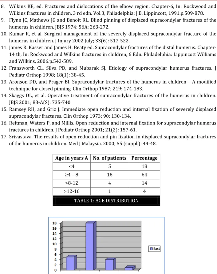
OBSERVATION & RESULTS: In our series of 28 cases of supracondylar fracture of humerus, all the fractures were closed extension type with mean age of 6 years and 18 patients (64,28%) were between 4-8 years and 15 (53.5%) were male and 13(46.5%) patients were female. 21(75%) patients sustained injury due to fall on a out stretched hand while playing , with fracture on left side in 17 (61%) patients.
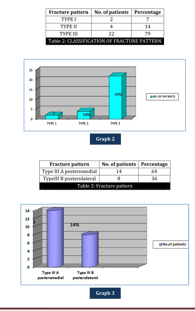
Out of 22 patients with TYPE III fracture, 14 (63.63%) patients had posteromedial displacement and 8 (36.36%) patients had posterolateral displacement. Majority of patients i.e. 85% were treated on 1st day of hospitalization and discharged on 3rd post-operative day.
The final results of present study of 28 patients, 22(79%) patients had TYPE III fracture, 4(14%) had TYPE II fracture & 2(7%) had TYPE I fracture.
Out of 22 patients with TYPE III fracture, 18 (82%) undergone surgical fixation with k wires with 12 patients treated with percutaneous k wire fixation & 6 patients undergone open reduction & internal fixation with k wires.
12(67%) patients had excellent results, 5(28%) patients had good results & 1(5%) patient had poor results.
Out of 12 patients treated with percutaneous k wire fixation, 8(67%) patients had excellent results, 4(33%) patients had good results.
Out of 6 patients treated with open reduction & k wire fixation, 4(67%) patients had excellent results, 1(17%) patient had good results & 1(17%) patients had poor results.
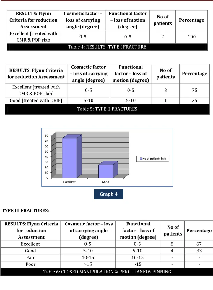
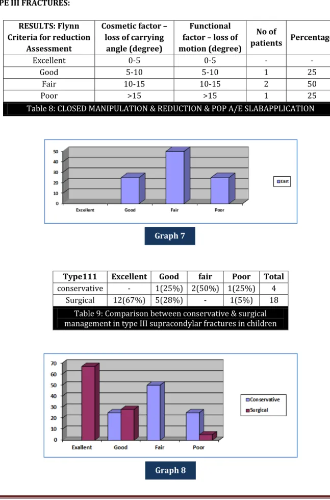
The remaining 4 (18%) patients with type III fracture was treated conservatively with closed manipulation & reduction above elbow pop slab application in flexion , out of which 1(25%)patient good results,2(50%) patients had fair results & 1(25%) patient had poor results. Out of 4 patients with type II fracture, 3(75%) patients were treated conservatively in a above elbow pop slab & all had excellent results, 1(25%) patient was treated with open reduction & internal fixation with k wires had good result. Out of 2 patients with TYPE I fracture all were treated conservatively in a above elbow pop slab & all had excellent results according to Flynn’s criteria.
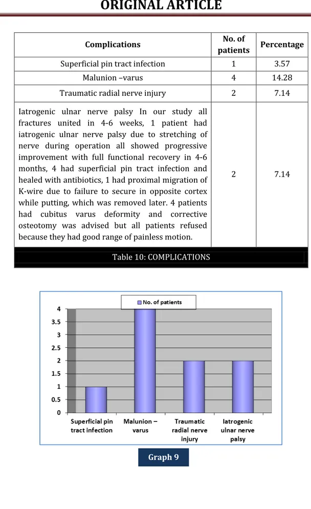
In our study all fractures united in 4-6 weeks, 2(7.14%) patients had traumatic radial nerve palsy(pre-operatively), 2(7.14%)patients had iatrogenic ulnar nerve palsy & both showed progressive improvement with full functional recovery in 4-6 months follow up, 1(3.5%) patients had superficial pin tract infection and it healed with curettage & antibiotics, 4 (14.28%) patients had cubitus varus deformity.
DISCUSSION: Supracondylar fracture of humerus is the commonest injury around elbow in children. It demand great respect in treatment because if not treated properly may give rise to neurovascular compromise, difficulty in obtaining or maintaining reduction and poor late results because of stiffness of elbow or malunion.
In our series majority of 18 (64.28%) of patients were found to be between age group of 4-8years. These numbers where comparable with studies done by Kumar R et al10& Wilkin KE et al.11
J of Evolution of Med and Dent Sci/ eISSN- 2278-4802, pISSN- 2278-4748/ Vol. 4/ Issue 14/ Feb 16, 2015 Page 2299 In the present study of 28 patients, majority of our patients 21 (75%) sustained fractures due to fall while playing. Fransworth CL et al,12 in her series 70% of cases sustained fracture due to fall while playing.
The present study of 28 patients, 17 (61%) patients sustained fracture on left side and 11patients (39%) on right side comparable with study by Wilkins KE,et al8 in which 60.8% patients sustained fracture on left side and 39.2% on right side.
In our study of 28 patients, 22 patients sustained type III fracture out of which 14(64%) had posteromedial displacement & 8(36%) poster lateral displacement in comparison with study by Aronson et al13 in which 75% had posteromedial displacement & 25% poster lateral displacement.
In our study of 28 patients, 24 patients (85.71 %) operated on 1st day of hospitalization comparable with study by David L Skaggs14 et al, of 204 patient’s average interval of time of injury and operation was 1.4 days.
In our series of 28 patients, about 20 patients (71.42 %) discharged on 3rd post-operative day comparable with study by Ramsay et al15 average duration of hospital stay in was 3.66 days
In our study of 28 patients out of which 22 patients with TYPE III fracture, 18 (82%) undergone surgical fixation with k wires among them 12 patients were treated with Closed reduction & percutaneous k wire fixation out of which 8(67%) patients had excellent results, 4 (33%) patients had good results in comparison with study by Flynn et al9 of 52 cases treated with Closed reduction & percutaneous k-wire fixation 42 (80%) patients had excellent, 7(14%) patients had good results, 2(4%) patient had fair result & 1(2%) patient had poor result.
In our study out of 22 patients with TYPE III fracture 6 patients with open reduction & internal fixation with k wires,. 4(67%) patients had excellent results, 1 (17%) patient had good results &1 (17%) patient had poor results in comparison with study by Reitman et al16 85 cases treated with open reduction & internal fixation with k wires 18 (55%) patients had excellent, 8(24%) patients had good results, 3(9%) patient had fair result & 4(12%) patient had poor result.
In our study the remaining 4 (18%) patients with type III fracture was treated conservatively with Closed reduction & cast application 1(25%)patient had good results, 2(50%) patients had fair results & 1(25%) patient had poor results in comparison with study by of Pirone et al2 100 cases treated conservatively with Closed reduction & cast application 50 (50%) patients had excellent, 27(27%) patients had good results, 3(3%) patient had fair result & 20(20%) patient had poor result.
In our study of 28 patients, 17patients (61%) had loss of range of motion of 0-5°, 7 patients (25%) had 6-10°, 2 patients (7%)had 11-15° and 2 patients(7%) had >15° of range of motion in comparison with Andrew et al1 series of 52 patients, five patients suffered a moderate loss in range of motion. 1 patient had extension loss of <10°, and 3 had flexion loss of less than 10° and last patient had flexion and extension loss of >10°.
In our study of 28 patients at the final follow up, 17patients (61%) had loss of carrying angle of 0-5°, 7 patients (25%)hadloss of carrying angle of 6-10°, 2 patients (7%)had loss of carrying angle of 11-15° and 2 patients(7%) had >15° of loss of carrying angle In our study , 4 cases (14.28%) had cubitus varus deformity in comparison with Andrew1 et al, study of 52 patients, five patients had varus angulation of <10°, 6 had 10-20° and two had varus deformity of >20°.
J of Evolution of Med and Dent Sci/ eISSN- 2278-4802, pISSN- 2278-4748/ Vol. 4/ Issue 14/ Feb 16, 2015 Page 2300 and median nerve and one each of radial, ulnar and median. All patients recovered in 2 weeks postoperatively
In our series, 1 case (3.57%) had superficial pin tract infection. after removal of the K-wire in comparison with Ramsey RH et al15 study of 15 cases, one patient had pin tract infection. In Srivastava17 study group of 42 patients about 14% had superficial pin tract infection.
CONCLUSION: Surgical fixation with either closed reduction & percutaneous pinning or open reduction and internal fixation with k wires in type III supracondylar fractures of humerus in children gives more stable fixation, better anatomical reduction with minimal complication. So it is safe and effective method of fixation. It gives excellent functional and cosmetic results when done at appropriate time for displaced supracondylar fracture of humerus in children.
In our study the results of surgical fixation with either closed reduction & percutaneous pinning or open reduction and internal fixation with k wires were comparable with one another as there was around 66% excellent results (according to Flynn criteria) in both techniques with excellent functional and cosmetic results.
Cubitus varus deformity is less, compared to other methods due to better anatomical reduction and stable fixation.
Elbow stiffness is less compared to other modalities of treatment due to early mobilization of elbow.
The results of Conservative treatment with closed manipulation & reduction with above elbow pop slab application in type III fracture supracondylar fractures of humerus in children are poor & the rate of complications like loss of reduction, malunion & restriction of movement is high with conservative treatment.
The results of Conservative treatment type I & type II supracondylar fractures of humerus in children are good & complications rate are low.
REFERENCES:
1. Andrew J. Weiland, et al. Surgical treatment of displaced supracondylar fractures of the humerus in children. JBJS 1978; 60A: 657-661.
2. Andrew J. Weiland, Stephen Meyer, Leistal, John Hopkins Hospital, Baltimore: journal of bone & joint surgery 1978: 6: 39-43.
3. Reza Omid, Paul D. Choi and David L. N. Skaggs. Supracondylar fracture of the humerus in children. J B J S Am 2008; 90: 1121-1132.
4. Norman Y Otsuka & James R Krasser: Supracondylar fracture of the humerus in children. J Am Acad Orthop Surg 1997; 5: 19-26.
5. Yusof, et al. Displaced supracondylar fracture of humerus in children – comparative study of the results of closed and open reduction. Med J Malasia 1998; 53 (Supp. A): 52-58.
6. Fleuriau-Chateau, McIntyre, Letts. To review with irreducible supracondylar fractures requiring open reduction in children and to propose guidelines for an open approach to supracondylar fractures. Can J Surg 1998 April; 41(2): 112-118.
J of Evolution of Med and Dent Sci/ eISSN- 2278-4802, pISSN- 2278-4748/ Vol. 4/ Issue 14/ Feb 16, 2015 Page 2301 8. Wilkins KE, ed. Fractures and dislocations of the elbow region. Chapter-6, In: Rockwood and
Wilkins fractures in children, 3 rd edn. Vol.3, Philadelphia: J.B. Lippincott, 1991.p.509-878. 9. Flynn JC, Mathews JG and Benoit RL. Blind pinning of displaced supracondylar fractures of the
humerus in children. JBJS 1974; 56A: 263-272.
10.Kumar R, et al. Surgical management of the severely displaced supracondylar fracture of the humerus in children. J Injury 2002 July; 33(6): 517-522.
11.James R. Kasser and James H. Beaty ed. Supracondylar fractures of the distal humerus. Chapter-14 th, In: Rockwood and Wilkins fractures in children, 6 Edn. Philadelphia: Lippincott Williams and Wilkins, 2006.p.543-589.
12.Fransworth CL. Silva PD, and Mubarak SJ. Etiology of supracondylar humerus fractures. J Pediatr Orthop 1998; 18(1): 38-45.
13.Aronson DD, and Prager BI. Supracondylar fractures of the humerus in children – A modified technique for closed pinning. Clin Orthop 1987; 219: 174-183.
14.Skaggs DL, et al. Operative treatment of supracondylar fractures of the humerus in children. JBJS 2001; 83-A(S): 735-740
15.Ramsey RH, and Griz J. Immediate open reduction and internal fixation of severely displaced supracondylar fractures. Clin Orthop 1973; 90: 130-134.
16.Reitman, Waters P, and Millis. Open reduction and internal fixation for supracondylar humerus fractures in children. J Pediatr Orthop 2001; 21(2): 157-61.
17.Srivastava. The results of open reduction and pin fixation in displaced supracondylar fractures of the humerus in children. Med J Malaysia. 2000; 55 (suppl.): 44-48.
Age in years A No. of patients Percentage
<4 5 18
≥4 – 8 18 64
>8-12 4 14
>12-16 1 4
TABLE 1: AGE DISTRIBUTION
J of Evolution of Med and Dent Sci/ eISSN- 2278-4802, pISSN- 2278-4748/ Vol. 4/ Issue 14/ Feb 16, 2015 Page 2302
Fracture pattern No. of patients Percentage
TYPE I 2 7
TYPE II 4 14
TYPE III 22 79
Table 2: CLASSIFICATION OF FRACTURE PATTERN
Fracture pattern No. of patients Percentage
Type III A posteromedial 14 64
TypeIII B posterolateral 8 36
Table 3: Fracture pattern
7
%
14%
Graph 2
J of Evolution of Med and Dent Sci/ eISSN- 2278-4802, pISSN- 2278-4748/ Vol. 4/ Issue 14/ Feb 16, 2015 Page 2303
RESULTS: Flynn Criteria for reduction
Assessment
Cosmetic factor – loss of carrying
angle (degree)
Functional factor
– loss of motion (degree)
No of
patients Percentage
Excellent [treated with
CMR & POP slab 0-5 0-5 2 100
Table 4: RESULTS -TYPE I FRACTURE
RESULTS: Flynn Criteria for reduction Assessment
Cosmetic factor
– loss of carrying angle (degree)
Functional factor – loss of motion (degree)
No of
patients Percentage
Excellent [treated with
CMR & POP slab] 0-5 0-5 3 75
Good [treated with ORIF] 5-10 5-10 1 25
Table 5: TYPE II FRACTURES
TYPE III FRACTURES:
RESULTS: Flynn Criteria for reduction
Assessment
Cosmetic factor – loss of carrying angle
(degree)
Functional factor – loss of motion (degree)
No of
patients Percentage
Excellent 0-5 0-5 8 67
Good 5-10 5-10 4 33
Fair 10-15 10-15 - -
Poor >15 >15 - -
Table 6: CLOSED MANIPULATION & PERCUTANEOS PINNING
J of Evolution of Med and Dent Sci/ eISSN- 2278-4802, pISSN- 2278-4748/ Vol. 4/ Issue 14/ Feb 16, 2015 Page 2304
TYPE III FRACTURES:
RESULTS: Flynn Criteria for reduction
Assessment
Cosmetic factor – loss of carrying
angle (degree)
Functional factor – loss of motion
(degree)
No: of
patients Percentage
Excellent 0-5 0-5 4 66
Good 5-10 5-10 1 7
Fair 10-15 10-15 - -
Poor >15 >15 1 17
Table 7: Open reduction & k wire fixation
Graph 5
J of Evolution of Med and Dent Sci/ eISSN- 2278-4802, pISSN- 2278-4748/ Vol. 4/ Issue 14/ Feb 16, 2015 Page 2305
TYPE III FRACTURES:
RESULTS: Flynn Criteria for reduction
Assessment
Cosmetic factor – loss of carrying angle (degree)
Functional factor – loss of motion (degree)
No of
patients Percentage
Excellent 0-5 0-5 - -
Good 5-10 5-10 1 25
Fair 10-15 10-15 2 50
Poor >15 >15 1 25
Table 8: CLOSED MANIPULATION & REDUCTION & POP A/E SLABAPPLICATION
Type111 Excellent Good fair Poor Total
conservative - 1(25%) 2(50%) 1(25%) 4
Surgical 12(67%) 5(28%) - 1(5%) 18
Table 9: Comparison between conservative & surgical management in type III supracondylar fractures in children
Graph 7
J of Evolution of Med and Dent Sci/ eISSN- 2278-4802, pISSN- 2278-4748/ Vol. 4/ Issue 14/ Feb 16, 2015 Page 2306
Complications No. of
patients Percentage
Superficial pin tract infection 1 3.57
Malunion –varus 4 14.28
Traumatic radial nerve injury 2 7.14
Iatrogenic ulnar nerve palsy In our study all fractures united in 4-6 weeks, 1 patient had iatrogenic ulnar nerve palsy due to stretching of nerve during operation all showed progressive improvement with full functional recovery in 4-6 months, 4 had superficial pin tract infection and healed with antibiotics, 1 had proximal migration of K-wire due to failure to secure in opposite cortex while putting, which was removed later. 4 patients had cubitus varus deformity and corrective osteotomy was advised but all patients refused because they had good range of painless motion.
2 7.14
Table 10: COMPLICATIONS
J of Evolution of Med and Dent Sci/ eISSN- 2278-4802, pISSN- 2278-4748/ Vol. 4/ Issue 14/ Feb 16, 2015 Page 2307
AUTHORS:
1. Ajith Kumar K. S. 2. Amardeep G. 3. N. J. Mani 4. Muraleedharan 5. Gangadharan P. 6. Roney Thomas
PARTICULARS OF CONTRIBUTORS:
1. Senior Resident, Department of Orthopaedics, Baby Memorial Hospital, Calicut, Kerala.
2. Senior Resident, Department of Orthopaedics, Baby Memorial Hospital, Calicut, Kerala.
3. Senior Consultant & HOD, Department of Orthopaedics, Baby Memorial Hospital, Calicut, Kerala.
FINANCIAL OR OTHER
COMPETING INTERESTS: None
4. Consultant, Department of Orthopaedics, Baby Memorial Hospital, Calicut, Kerala. 5. Consultant, Department of Orthopaedics,
Baby Memorial Hospital, Calicut, Kerala. 6. Consultant, Department of Orthopaedics,
Baby Memorial Hospital, Calicut, Kerala.
NAME ADDRESS EMAIL ID OF THE CORRESPONDING AUTHOR:
Dr. Ajith Kumar K. S, # HF-49, Hutha Colony, Bhadravathi, Shimoga-577301, Karnataka, India.
E-mail: [email protected] [email protected]