A method for histopathological study of
the multifocal nature of spinal cord lesions
in murine experimental autoimmune
encephalomyelitis
Katherine N. Gibson-Corley, Alexander W. Boyden,
Mariah R. Leidinger, Allyn M. Lambertz, Georgina Ofori-Amanfo, Paul W. Naumann, J. Adam Goeken and Nitin J. Karandikar
Department of Pathology, University of Iowa, Iowa City, Iowa, United States
ABSTRACT
Experimental autoimmune encephalomyelitis (EAE) is a well-established mouse model for multiple sclerosis and is characterized by infiltration of mononuclear cells and demyelination within the central nervous system along with the clinical symptoms of paralysis. EAE is a multifocal and random disease, which sometimes makes histopathologic analysis of lesions difficult as it may not be possible to predict where lesions will occur, especially when evaluating cross sections of spinal cord. Consequently, lesions may be easily missed due to limited sampling in traditional approaches. To evaluate the entire length of the spinal cord while maintaining anatomic integrity, we have developed a method to section the cord within the decalcified spinal column, which allows for the study of the multifocal nature of this disease and also minimizes handling artifact. HE and Luxol fast blue staining of these spinal cord sections revealed a paucity of lesions in some areas, while others showed marked inflammation and demyelination. The percentage of spinal cord affected by EAE was evaluated at four separate areas of longitudinally sectioned cord and it varied greatly within each animal. Immunohistochemical staining of in situ spinal cords which had undergone decalcification was successful for key immuno-markers used in EAE research including CD3 for T cells, B220 for B cells and F4/80 for murine macrophages. This method will allow investigators to look at the entire spinal cord on a single slide and evaluate the spinal cord with and without classic EAE lesions.
Subjects Neurology, Pathology, Histology
Keywords Mice, Histopathology, Spinal cord, EAE
INTRODUCTION
Multiple sclerosis (MS) is a debilitating autoimmune disease characterized by cellular inflammation into–and the progressive demyelination of–the central nervous system (CNS), subsequently leading to a multitude of clinical symptoms (Sospedra & Martin, 2005;Steinbach & Merkler, 2014;Trapp & Nave, 2008). Experimental autoimmune encephalomyelitis (EAE) is a well-studied mouse model of MS-like disease due not only to its convenience for immune system manipulation, but importantly because it
Submitted25 November 2015
Accepted23 December 2015
Published26 January 2016
Corresponding author
Katherine N. Gibson-Corley, [email protected]
Academic editor
Marı´a A´ngeles Esteban
Additional Information and Declarations can be found on page 8
DOI10.7717/peerj.1600
Copyright
2016 Gibson-Corley et al.
Distributed under
recapitulates various hallmarks of human disease such as CNS inflammation and paralysis (Constantinescu et al., 2011;Duffy, Lees & Moalem-Taylor, 2014;McCarthy, Richards & Miller, 2012).
EAE can be induced by an assortment of immunizations, but most often includes the subcutaneous injection of peptides derived from neuroantigens (such as myelin basic protein, myelin proteolipid protein, or myelin oligodendrocyte glycoprotein (MOG)) as part of an emulsion with Complete Freund’s Adjuvant (CFA). In this study, we
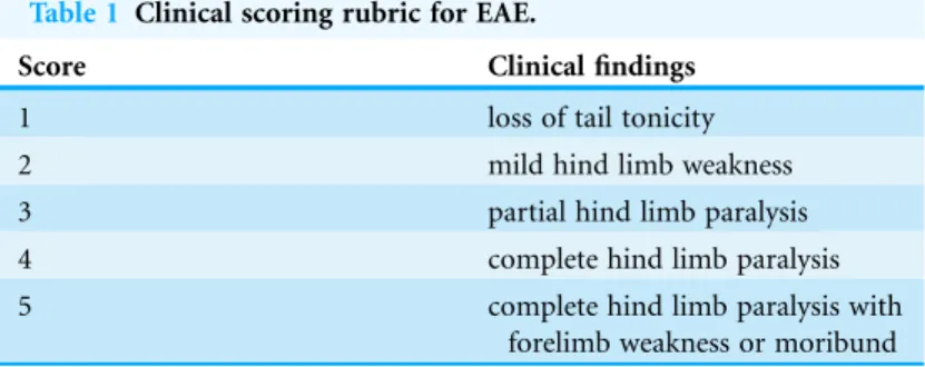
induced EAE disease in C57BL/6 mice with the commonly utilized MOG35–55peptide. Upon disease onset of this classic model, the manifestation of clinical symptoms ensues, which is characterized by a grading scale of ascending paralysis (Ortega et al., 2013; York et al., 2010) (Table 1). Inflammatory cell infiltration and demyelination of the CNS in MS/EAE drive clinical disease symptoms and therefore histological assessment of the spinal cord can be crucial in evaluating EAE disease, as well as any past and future therapeutics in this model. Indeed, researchers’ ability to efficiently and accurately evaluate MS-like disease within the EAE model has been and will continue to be productive in pushing the MS and CNS inflammatory disease fields forward.
Key histopathologic findings associated with EAE include inflammatory cell infiltration and axonal loss (Steinbach & Merkler, 2014) which can be identified using routine techniques (Klopfleisch, 2013). These include hematoxylin and eosin (HE) and Luxol fast blue (LFB) stains as well as immunohistochemical staining for important inflammatory cell markers such as T cells, B cells and macrophages (Steinbach & Merkler, 2014). This type of histopathologic analysis is commonly performed on EAE-diseased murine spinal cord, which is classically sectioned in a cross-wise (coronal) fashion. While this allows researchers to visualize the entire cord including both white and gray matter, it is at a single level and only approximately 5mm thick. This type of sectioning can be and often is
enlightening, however the vast number of spinal cord cross sections makes examination cumbersome. Furthermore, EAE is a multifocal and random disease (Day, 2005) and thus it is impossible to predict where lesions will occur throughout the spinal cord, even within areas of expected lesion localization. To histologically evaluate the entire murine spinal cord, we have developed a method to section the cord longitudinally within the decalcified spinal column allowing us to identify and study the multifocal nature of EAE disease.
Table 1 Clinical scoring rubric for EAE.
Score Clinical findings
1 loss of tail tonicity
2 mild hind limb weakness
3 partial hind limb paralysis
4 complete hind limb paralysis
5 complete hind limb paralysis with
MATERIALS AND METHODS
MiceC57BL/6 mice (females, 6–8 weeks old) were purchased from The Jackson Laboratory (Bar Harbor, ME, USA). Mice were allowed chow and water ad libitum, maintained on a 12-hour light/dark cycle, and housed in specific pathogen-free barrier facilities at the University of Iowa. All animal and tissue work was approved by the University of Iowa Institutional Animal Use and Care Committee.
Immunizations and EAE evaluation
On day 0, mice were immunized subcutaneously with 50mg of a myelin oligodendrocyte
glycoprotein peptide (MOG35–55) emulsified 1:1 volume in CFA supplemented with 4 mg/mL Mycobacterium tuberculosis(H37Ra; Difco Laboratories, Detroit, MI, USA). Mice were additionally injected intraperitoneally on days 0 and 2 with 250 ng of pertussis toxin (List Biological Laboratories, Campbell, CA, USA). Clinical EAE disease scores were monitored using the grading scale as follows: 1) loss of tail tonicity; 2) mild hind limb weakness; 3) partial hind limb paralysis; 4) complete hind limb paralysis; 5) complete hind limb paralysis with forelimb weakness or moribund/death (Table 1). Specific mice were chosen at various scores for histological evaluation.
Tissue preparation and histology
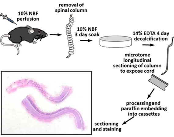
Mice were humanely euthanized by carbon dioxide asphyxiation in accordance with NIH & ACURF guidelines. Rapid fixation was achieved by whole body perfusion with the use of a simple gravity flow device utilizing 10% neutral buffered formalin (NBF) (Leica Biosystems, Wetzlar, Germany). A 60 cc syringe barrel with attached stopcock & 3 mm diameter tubing was mounted on a ring stand with the syringe 80 cm above the working surface. The syringe and tubing were flushed with 37C PBS prior to and after each use.
Following euthanasia, the thoracic cavity was immediately opened exposing the heart, the right atrium severed, and a 22 g needle attached to the gravity flow tubing inserted into the left ventricle. Flow was initiated and the animal perfused with an initial 5 ml of PBS at 37C to clear the vasculature of blood followed by immediate perfusion with 40 ml of
10% NBF at 37C.
Once perfused, the entire spinal column, including the vertebrae and enclosed spinal cord, were removed, epaxial muscles dissected off, and placed in 200 ml of 10% NBF for 3 days at room temperature on an orbital shaker set at 100 RPM for immersion fixation. After 3 days in NBF, the spines were briefly washed with tap water and placed in 200 ml of 14% EDTA (Sigma ED-EDTA, pH 7.3) for decalcification with continuous shaking. The spines were in 14% ED-EDTA for 4 days before removal, washed thoroughly with tap water for 3 hours, and sections grossed into cassettes with the use of a microtome blade. The entire spinal column was sectioned in half into longitudinal sections thus exposing the centrally located spinal cord and marking dye was used on the samples to maintain appropriate orientation. Tissues were placed back into 10% NBF, routinely processed, embedded in paraffin, and consecutive sections at 5mm thickness were cut for subsequent
Routine HE and Luxol fast blue (LFB) staining was performed on all sections. Digital images were collected with a DP73 camera and CellSens software (Olympus, Tokyo, Japan). HE-stained, longitudinally sectioned spinal cord sections were evaluated for lesions of EAE, including demyelination and inflammatory cell infiltration. Four separate areas along each spinal cord were identified and in each area the percentage of spinal cord with lesions was estimated visually (using a scale of 0, 10, 20: : :.100% affected) at 20 magnification by a board-certified veterinary pathologist.
Immunohistochemistry
To validate that immunohistochemistry would be successful using this method of tissue preparation, staining was performed for key inflammatory cells common in EAE (CD3 for T cells, B220 for B cells and F4/80 for macrophages) (Table 2). All immunohistochemical staining was performed manually using peroxidase methods and Dako Envision systems (Glostrup, Denmark).
RESULTS
HE and LFB staining
HE and LFB staining of longitudinally sectioned spinal cord sections (Fig. 2) revealed the multifocal to coalescing nature of the lesions associated with EAE. In some areas, the spinal cord can appear relatively normal while in other areas there is significant pathology
(Figs. 2Aand2D). When a section of spinal cord from a similarly affected mouse is cut cross-wise, it can be taken from an area with a paucity of lesions (Figs. 2Band2E), this would lend an investigator to think the cord was relatively unaffected. In contrast, when a cross section is taken within an area of pathology, which includes infiltration of
inflammatory cells and demyelination (Figs. 2Cand2F), the severity of disease could be overestimated.
Prior to euthanasia, each animal was given a clinical score based on their symptoms of EAE (Table 1). The clinical scores ranged from 2 (mild hind limb weakness) to 4 (complete hind limb paralysis) (Fig. 3A). Following necropsy, the percentage of spinal cord affected by EAE lesions was identified at four separate and equidistant areas of the longitudinally sectioned, HE-stained spinal cord sections to determine the variation in lesion severity. Interestingly, the percentage of spinal cord affected by EAE lesions varied greatly—even within a single sample and that, as expected, the most severe lesions
Table 2 Primary antibodies and their commercially available sources, catalog numbers, dilutions and specific antigen retrieval conditions utilized in the study.
Marker Antibody Dilution Source Conditions
CD3 Cat# RM-9107-5 1:200 Neomarkers HIER, citrate buffer (pH 6.0) B220 Cat# MCA1258G 1:6000 Serotec HIER, citrate buffer (pH 6.0) F4/80 Cat# MCAP497 1:6400 Serotec HIER, citrate buffer (pH 6.0)
appear to be in more caudal spinal cord (Fig. 3B) (Nathoo, Yong & Dunn, 2014). This illustrates the utility of longitudinally sectioning EAE spinal cords to increase the odds of identifying lesions.
Immunohistochemical staining
Immunohistochemical staining was performed on these longitudinally sectioned, decalcified specimens (Fig. 4). Specific immunomarkers (Table 2) commonly used
Figure 3 Clinical EAE score and quantification of the percentage of spinal cord affected by EAE using the method of longitudinal sectioning. (A) Clinical EAE score (Table 1) of 12 different mice with EAE ranging in age from 7–11 weeks of age. (B) Four separate areas (divided equally cranial to caudal) of longitudinally sectioned spinal cord from the same 12 mice, which were evaluated to determine what percentage of the cord at that area was affected by EAE lesions.
in EAE research were selected, including CD3 for T cells (Fig. 4B), F4/80 for macrophages (Fig. 4C) and B220 for B cells (Fig. 4D). All successfully stained the decalcified tissues and were able to identify these key immune cells within the EAE spinal cord lesions.
DISCUSSION
Due to the multifocal nature of EAE within the spinal cord, we developed a novel way of looking at the entire length of the spinal cord in situ, within the vertebrae, allowing us to visualize the multifocal nature of lesions (Fig. 1). Using both HE and LFB staining, we show that in some areas of the spinal cord there can be significant pathology but in other areas the cord can appear relatively normal (Fig. 2). The histopathologic score of the spinal cords somewhat followed the clinical score of these mice antemortem (Fig. 3). Interestingly, some animals (mouse 1 and 11) had markedly variable histopathology but had fairly high clinical scores, indicating that clinically there was significant damage to the cord that might be difficult to identify histologically if one did not section the area(s) of cord affected. It is also important to note that this technique, which uses a decalcification step, also allows for routine immunohistochemical staining for key inflammatory cell infiltrates. Decalcification can affect some immunohistochemical assays but in this case we were able to assess for the key immune cells in EAE; T cells, B cells and macrophages (Fig. 4) (Bussolati & Leonardo, 2008).
This technique will allow EAE researchers to quickly assess the spinal cord using routine histopathology and without having to make numerous slides of spinal cord cross sections. It is also important to note that the spinal cord itself does not have to be flushed from the spinal column, which not only saves time, but also preserves the integrity of the cord and minimizes handling artifacts within the tissue. Of course, there are limitations. With longitudinal sectioning, one can only visualize a single plane of the cord unlike cross sectioning where all of the white and gray matter can be assessed. If the histotechnologist does not cut deep enough into the tissue, only the white matter will be visible as the deeper gray matter can be missed. It is also difficult to visualize the central canal using longitudinal sectioning. Another challenge is working with the decalcified vertebrae surrounding the softer spinal cord. The spinal column isn’t completely flat so the undulating nature of the vertebrae can pose challenges to the histotechnologist during cutting to make sure the spinal cord isn’t folded or shredded.
CONCLUSIONS
In summary, longitudinal sectioning of the decalcified spinal column in mice is a useful technique to study the multifocal and oftentimes random nature of spinal cord lesions encountered in EAE. This technique will be useful to the researcher who would like to assess the entire length of the spinal cord without having to remove the
spinal cord from the vertebrae and without having to make numerous spinal cord cross sections.
ADDITIONAL INFORMATION AND DECLARATIONS
Funding
This work was supported, in part, by grant awards to NJK from the NIH and National MS Society. The funders had no role in study design, data collection and analysis, decision to publish, or preparation of the manuscript.
Competing Interests
The authors declare that they have no competing interests.
Author Contributions
Katherine N. Gibson-Corley conceived and designed the experiments, performed the experiments, analyzed the data, contributed reagents/materials/analysis tools, wrote the paper, prepared figures and/or tables, reviewed drafts of the paper.
Alexander W. Boyden performed the experiments, analyzed the data, contributed reagents/materials/analysis tools, wrote the paper, prepared figures and/or tables, reviewed drafts of the paper.
Mariah R. Leidinger performed the experiments, contributed reagents/materials/ analysis tools, reviewed drafts of the paper.
Allyn M. Lambertz performed the experiments, contributed reagents/materials/analysis tools, reviewed drafts of the paper.
Georgina Ofori-Amanfo performed the experiments, contributed reagents/materials/ analysis tools, reviewed drafts of the paper.
Paul W. Naumann performed the experiments, contributed reagents/materials/analysis tools, reviewed drafts of the paper.
J. Adam Goeken performed the experiments, contributed reagents/materials/analysis tools, reviewed drafts of the paper.
Nitin J. Karandikar conceived and designed the experiments, analyzed the data, contributed reagents/materials/analysis tools, wrote the paper, prepared figures and/or tables, reviewed drafts of the paper.
Animal Ethics
The following information was supplied relating to ethical approvals (i.e., approving body and any reference numbers):
Data Deposition
The following information was supplied regarding data availability: The research in this article did not generate any raw data.
Supplemental Information
Supplemental information for this article can be found online athttp://dx.doi.org/ 10.7717/peerj.1600#supplemental-information.
REFERENCES
Bussolati G, Leonardo E. 2008.Technical pitfalls potentially affecting diagnoses in immunohistochemistry.Journal of Clinical Pathology61(11):1184–1192
DOI 10.1136/jcp.2007.047720.
Constantinescu CS, Farooqi N, O’Brien K, Gran B. 2011.Experimental autoimmune
encephalomyelitis (EAE) as a model for multiple sclerosis (MS).British Journal of Pharmacology
164(4):1079–1106DOI 10.1111/j.1476-5381.2011.01302.x.
Day MJ. 2005.Histopathology of EAE. In: Ehud Lavi CSC, ed.Experimental Models of Multiple Sclerosis. New York: Springer, pp. 25–43.
Duffy SS, Lees JG, Moalem-Taylor G. 2014.The contribution of immune and glial cell types in experimental autoimmune encephalomyelitis and multiple sclerosis.Multiple Sclerosis International2014(2014):285245DOI 10.1155/2014/285245.
Klopfleisch R. 2013.Multiparametric and semiquantitative scoring systems for the evaluation of mouse model histopathology–a systematic review.BMC Veterinary Research9:123
DOI 10.1186/1746-6148-9-123.
McCarthy DP, Richards MH, Miller SD. 2012.Mouse models of multiple sclerosis: experimental autoimmune encephalomyelitis and theiler’s virus-induced demyelinating disease.Methods in Molecular Biology900:381–401DOI 10.1007/978-1-60761-720-4_19.
Nathoo N, Yong VW, Dunn JF. 2014.Understanding disease processes in multiple sclerosis through magnetic resonance imaging studies in animal models.Neuroimage Clinical4:743–756 DOI 10.1016/j.nicl.2014.04.011.
Ortega SB, Kashi VP, Tyler AF, Cunnusamy K, Mendoza JP, Karandikar NJ. 2013.The disease-ameliorating function of autoregulatory CD8 T cells is mediated by targeting of
encephalitogenic CD4 T cells in experimental autoimmune encephalomyelitis.Journal of Immunology191(1):117–126DOI 10.4049/jimmunol.1300452.
Sospedra M, Martin R. 2005.Immunology of multiple sclerosis.Annual Review of Immunology
23(1):683–747DOI 10.1146/annurev.immunol.23.021704.115707.
Steinbach K, Merkler D. 2014.Neuropathological techniques to investigate CNS pathology in Experimental Autoimmune Encephalomyelitis (EAE).Methods in Molecular Biology
1304:189–209DOI 10.1007/7651_2014_110.
Trapp BD, Nave KA. 2008.Multiple sclerosis: an immune or neurodegenerative disorder?Annual Review of Neuroscience31:247–269DOI 10.1146/annurev.neuro.30.051606.094313.