Lipo so me s: fro m bio physics to
the de sign o f pe ptide vaccine s
Departamento de Fisiologia e Biofísica, Instituto de Ciências Biológicas, Universidade Federal de Minas Gerais, Belo Horizonte, MG, Brasil F. Frézard
Abstract
Liposomes (lipid-based vesicles) have been widely studied as drug delivery systems due to their relative safety, their structural versatility concerning size, composition and bilayer fluidity, and their ability to incorporate almost any molecule regardless of its structure. Lipo-somes are successful in inducing potent in vivo immunity to incorpo-rated antigens and are now being employed in numerous immuniza-tion procedures. This is a brief overview of the structural, biophysical and pharmacological properties of liposomes and of the current strat-egies in the design of liposomes as vaccine delivery systems.
Co rre spo nde nce F. Frézard
Departamento de Fisiologia e Biofísica, ICB, UFMG Av. Antônio Carlos, 6627 30161-901 Belo Horizonte, MG Brasil
Fax: + 55-31-499-2924 E-mail: frezard@ mono.icb.ufmg.br
Presented at the International Symposium “The Third Revolution on Vaccines: DNA Vaccines”, Belo Horizonte, MG, Brasil, November 3-7, 1997.
Research supported by CNPq (No. 521010/97), FAPEMIG (No. CBS1721/95) and PRO NEX (No. 3075).
Received November 6, 1998 Accepted November 27, 1998
Ke y words
·Liposome ·Immunoadjuvant ·Immunostimulant ·Vaccine
·Encapsulation
Intro ductio n
Almost 75 years ago, Paul Ehrlich estab-lished the concept of the magic bullet envisioning a drug delivery mechanism that would target drugs directly to diseased cells. It was not long after liposomes were first constructed by A.D. Bangham in the early 1960s, that it was demonstrated that a wide variety of molecules could be encapsulated within the aqueous spaces of liposomes or inserted into their membranes. It was gener-ally assumed at that time that since lipo-somes were primarily made of natural phos-phatidyl choline, liposome-encapsulated an-tigen would avoid the mononuclear phago-cyte system (MPS) clearance and would not be recognized as a particulate antigen. How-ever, it was rapidly realized that eventually liposomes would be deposited at high con-centrations in the MPS organs, particularly in fixed macrophages (1,2), regardless of the composition or size of the vesicles. Later, the capture of liposomes by macrophages was recognized as the main mechanism by which liposomes potentiate immune
re-sponses to entrapped antigens. The pioneer work of Allison and Gregoriadis (3), that demonstrated the immunoadjuvant proper-ties of liposomes, was followed by a multi-tude of related animal immunization studies (4-7). Finally, liposomes as adjuvants have come of age, with the first liposome-based vaccine (against hepatitis A) having been licensed for use in humans (8).
Structure and biophysical prope rtie s of liposome s
hydrocar-bon chains) and with water (hydrophilic in-teractions, hydrophobic effect) lead to spon-taneous formation of closed bilayers.
Liposomes can differ in size: they can range from the smallest vesicle obtainable on theoretical grounds (diameter ~20 nm) to liposomes which are visible under the light microscope, with a diameter of 1 µm or greater, equal to the dimensions of living cells. They can also differ in terms of lipid composition and structural organization, cor-responding to uni-, oligo- or multi-lamellar vesicles. Liposomes are built in such a way that the solute can be encapsulated in the aqueous compartment (polar solutes) or em-bedded in the lipid bilayers (lipophilic or amphiphilic solutes).
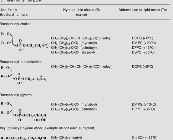
The properties of liposomes and their subsequent applicability depend on the physi-cal and physico-chemiphysi-cal characteristics of the liposomal membrane. Usually, a zwitter-ionic or non-zwitter-ionic lipid is used as the basic lipid for the preparation of liposomes (Table 1). The net surface charge of liposomes can be modified by the incorporation of posi-tively charged lipids, such as stearylamine, or negatively charged lipids, such as dicetyl-phosphate, phosphatidyl glycerol (Table 1) or phosphatidyl serine. Bilayer elasticity is related to elastic properties such as tensile strength, compressibility and bending. This property has been used to understand the response of bilayers to mechanical stress and to manufacture liposomes. The fluidity of the liposomal bilayer, when it is made from a single lipid, depends on the lipid phase transition temperature (Tc) and its relative position compared to ambient tem-perature. When ambient temperature is in-creased and reaches Tc, the membrane passes from a solid gel phase, where the lipid hydrocarbon chains are in an ordered state, to a fluid liquid-crystal phase, a disor-dered state, where molecules have more free-dom of movement (2). Hence, depending on lipid Tc (Table 1), different membranes com-posed of distinct lipids can exhibit different
fluidity levels at the same temperature. The bilayer permeability is a measure of the flux or rate at which a solute works its way from an aqueous compartment, through a bilayer, and out into the aqueous compartment on the other side. It depends on the membrane flu-idity and on the nature of the solute. Mem-brane permeability is highest at the phase transition temperature, and is lower in the gel phase than in the fluid phase. A general sequence of hydrophilic solute permeability is: water > small non-electrolytes > anions > cations @ large non-electrolytes > large poly-electrolytes (2).
The fusion properties of liposomal mem-branes have recently gained special atten-tion with the discovery of lipid composiatten-tions that allow for the delivery of macromol-ecules into the cytoplasm of cells (9,10). Membrane fusion is promoted by bilayer dehydration (11), by acidification when the membrane contains pH-sensitive lipids, such as DOPE (12), and by the presence of cat-ionic lipids or of ancat-ionic lipids with divalent cations.
Me thods for liposome pre paration and solute e ncapsulation
aqueous phase of vesicles, adsorption onto their surface, or partition into the bilayers (2). Among available methods for peptide or protein encapsulation, three are outstanding for higher encapsulating efficiency. The re-verse-phase evaporation technique, the first to use water-in-oil emulsions (15), encap-sulates up to 50% of solute. Preparation of reverse-phase evaporation vesicles (REV) consists of a rapid injection of aqueous solu-tion into an organic solvent which contains the lipids dissolved. Thus, following the for-mation of water droplets (water-in-oil emul-sion) by bath sonication of the two-phase mixture, the emulsion is dried down to a semi-solid gel in a rotary evaporator. The next step is to subject the gel to vigorous mechanical shaking to induce a phase change from a water-in-oil emulsion to a vesicle suspension. In these circumstances, some
water droplets collapse, and these droplets attach to adjacent, intact vesicles to form the outer leaflet of the bilayer of a large unila-mellar liposome (diameter in the range of 0.1 to 1 µm).
Another method (16) that produces de-hydration-rehydration vesicles (DRV) is both simple and easy to scale up, and usually gives high yields of solute entrapment (up to 80%) (16-20). Another advantage of the DRV method compared to the REV method is that it does not expose the solute to potentially denaturating organic solvents and/or to soni-cation. Preparation of DRV consists of mix-ing an aqueous solution of the solute with a suspension of empty (water-containing) li-posomes and freeze-drying the resulting mix-ture. The intimate contact of flattened lipo-somal membrane structures and solute mol-ecules in a dry environment and the fusion of
Table 1 - Examples of bilayer-forming lipids used to prepare liposomal vaccines.
Tc, Transition temperature.
Lipid family Hydrophobic chains (R) Abbreviation of lipid name (Tc)
Structural formula (name)
Phosphatidyl choline
CH3-(CH2)7-CH=CH-(CH2)7-C(O)- (oleyl) DOPC (<0oC)
CH3-(CH2)12-C(O)- (myristoyl) DM PC (+23oC)
CH3-(CH2)14-C(O)- (palmitoyl) DPPC (+42oC)
CH3-(CH2)16-C(O)- (stearoyl) DSPC (+55oC)
Phosphatidyl ethanolamine
CH3-(CH2)7-CH=CH-(CH2)7-C(O)- (oleyl) DOPE (<0oC)
Phosphatidyl glycerol
CH3-(CH2)12-C(O)- (myristoyl) DM PG (+13oC)
CH3-(CH2)14-C(O)- (palmitoyl) DPPG (+35oC)
Alkyl polyoxyethylene ether (example of non-ionic surfactant)
membranes caused by dehydration facili-tates the incorporation of solute during the controlled rehydration steps. Separation of solute-containing DRV from unentrapped solute can be carried out easily if needed (by centrifugation, for example). Vesicles formed by the dehydration-rehydration technique are multilamellar with heterogeneous sizes (di-ameters varying from 0.1 to 2.0 µm).
The third method requires the use of deter-gent (21). Lipids are first solubilized with an aqueous solution of the detergent that also contains the protein(s) to be encapsulated. The detergent should have a high critical mi-celle concentration (CMC) so that it is easily removed, for instance by dialysis. During de-tergent removal, relatively small liposomes (mean diameter in the range of 0.08 to 0.2 µm) with a narrow size distribution will be pro-duced. This latter method was found to be particularly suitable for the reconstitution of membrane proteins in liposomes.
Stability o f lipo so me pre paratio ns
The most important use of liposomes results from their ability to retain solutes for long periods of time. Other important as-pects of the stability of liposomes refer to the maintenance of their size distribution and to the chemical stability of their con-tent. Liposome stability has to be consid-ered under storage and physiological condi-tions. Under physiological conditions, sol-ute leakage depends on membrane perme-ability and also on the interaction with com-ponents of biological fluids. In serum, the lipid molecule can be transferred from the liposomal membrane to plasma high density lipoprotein. This is particularly true in the case of fluid liposomes, such as those made from dioleyl-phosphatidylcholine (DOPC), which disintegrate and release their contents within few minutes after their in-travenous administration. Following the sub-stitution of DOPC by high phase transition
temperature lipids such as distearoyl-phos-phatidylcholine (DSPC), the bilayer becomes rigid at 37oC, and consequently resistant
to lipoprotein attack. Membrane fluidity can also be controlled quite accurately by supple-menting the lipid bilayer with cholesterol, a mechanism that results in enhanced mem-brane stability (1,2), by mixing two or more lipids, or by manipulating the hydrophobic/ lipophobic character of the bilayers, for ex-ample with the use of fluorinated lipids (22-24). The rate of solute leakage also depends on the lamellarity of liposomes, multilamel-lar vesicles being more stable than unila-mellar ones (16). When given by the oral route, liposomes have to survive the deter-gent effect of bile salts and phospholipase activity. Only the most rigid liposomes were found to resist these extreme condi-tions (25).
In vivo fate of liposome s
Most of our knowledge concerning the behavior of liposomes in vivo has been
ob-tained using intravenous injections. Lipo-somes given intravenously normally interact with at least two distinct groups of plasma proteins (28): i) the plasma high density lipoproteins and ii) the so-called opsonins which, by adsorbing onto the surface of vesicles, mediate their endocytosis by MPS macrophages. The rate of liposome clear-ance from the blood circulation will there-fore depend on the ability of opsonins to bind to the liposome surface and can be manipulated through the appropriate selec-tion of liposome characteristics (1,4). For instance, fluid vesicles are removed more rapidly from blood circulation than rigid ones. It was suggested that opsonins did not adsorb as avidly on vesicles with rigid bilay-ers. Clearance from the blood stream is also influenced by surface charge (29) and by vesicle size. The longest half-life is obtained when liposomes are relatively small (diam-eter <0.05 µm) and carry no net surface charge. However, regardless of the time of liposome persistence within the vascular sys-tem, much of an injected dose is taken up by MPS macrophages, ending up in the lysoso-mal apparatus. There, liposomes are dis-rupted within the lysosomes, the solute is released locally and, depending on its na-ture, can either remain in the lysosome, be transferred into the cytoplasm, or diffuse out from cells.
The behavior of preparations given by alternative parenteral routes, such as the in-traperitoneal, subcutaneous or intramuscu-lar route, is influenced by the distribution of liposome size and their lipid composition and will also depend on the route of injection (30). A proportion of liposomes enter the lymphatic system and, eventually, the blood circulation where they behave as if given intravenously. However, whereas liver,
spleen and bone marrow take up nearly all liposomes given by the intravenous route, they will account for a much smaller propor-tion if the subcutaneous or intramuscular route is used. The remainder (up to 80% of liposomes) is retained at the site of injection and attacked by infiltrating macrophages or intercepted in the local lymph nodes.
Prolonged survival of liposomes in the circulation is required when these are de-signed to act on non-MPS tissues, within the vascular system, extravascularly through leaky capillaries or as circulating slow drug release systems. Recent works have shown that coating the liposome surface with ethyleneglycol and other hydrophilic poly-mers (31), or chemically modifying the hy-drophobic part of phospholipids (23,32), substantially prolongs the half-life of somes in the blood. Furthermore, these lipo-somes have a propensity to accumulate in implanted tumors at levels compared to other non-MPS tissues.
Liposome s as immunological adjuvants
liposome-based vaccine (against hepatitis A) being licensed for use in humans (8). Vaccines based on novasomes (8,13,33) (nonphos-pholipid liposomes formed from single-chain amphiphiles, with or without other lipids) have been licensed for the immunization of fowl against Newcastle disease virus and avian rheovirus. Other liposomal or nova-some-based vaccines against bacterial and viral infections are under development. Li-posomes offer a number of advantages as carriers of vaccines (4) in that they are bio-degradable and nontoxic, can elicit both HI and CMI (7), and can be prepared entirely synthetically. Furthermore, they are highly versatile in their structural characteristics, which allow for the precise manipulation of their immunoadjuvant properties. A number of structural variables of liposomes can in-fluence adjuvanticity (4,6): the lipid to anti-gen mass ratio, bilayer fluidity, vesicle size, surface charge (34), and the mode of antigen association with the vesicles. Manipulation of these variables usually induces variation in the immune response level of a maximal
factor of three (7). To boost the response further, different immunostimulants have been tested. Among them are avridine, mu-ramyl-dipeptide (MDP) and MDP-lipid con-jugates, nonionic block polymer surfactants, aluminum salts, IL-2, IL-6 and lipid A. It is noteworthy that the impact of the incorpora-tion of these immunostimulants into lipo-somes was not only an increase of their immunological action, but also a reduction of their toxic side effects (5). Because of the complexity of the interrelationship between the above-mentioned parameters, it is diffi-cult, at the present time, to establish general rules to maximize the immunogenicity of liposomal vaccines or to direct immune re-sponses. However, the insights into the ma-nipulation of immune responses by the choice of liposome characteristics are growing.
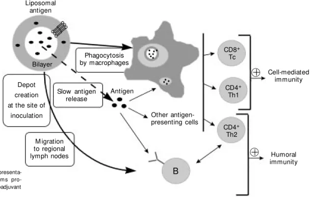
A lot of work has been done to study the interaction of liposomes with cells of the immune systems and to elucidate the mech-anism of induction of immune reactions (4). It is generally accepted that a physical asso-ciation between liposomes and antigen (as
Liposomal antigen
Bilayer
Depot creation at the site of
inoculation
Phagocytosis by macrophages
Slow antigen release
Antigen
Other antigen-presenting cells
M igration to regional lymph nodes
B
CD4+
Th2 CD4+
Th1 CD8+
Tc
Humoral immunity Cell-mediated
immunity
opposed to their simple mixing) is a prereq-uisite for adjuvanticity to occur. The current presumed mode of action of liposomes is illustrated in Figure 1. The enhancement of HI to antigens by means of their incorpora-tion into liposomes can be attributed to the generation of a depot at the site of injection which prolongs the release and interaction of free or liposome-associated antigens with antigen-presenting cells (APC). Among APC, macrophages were shown to play a major role, because of their unique ability to phag-ocytize liposomes. These cells are also ex-pected to invade the depot area in response to local inflammation. Finally, a fraction of liposomal antigen will migrate to areas in the regional lymph nodes containing T cells. This fate of liposomes, in addition to their ability to efficiently present antigens in a hydrophobic environment, may be respon-sible for the stimulation of CMI (6). It is noteworthy that liposomes, in addition to promoting immunity to antigens injected through a variety of parenteral routes, also increase IgA immunity to antigens given orally, probably because of vesicle interac-tion with the gut lymphoid tissue (25). Re-cently, more detailed information has be-come available on the fate of liposomes after uptake by cells and the subsequent induction of cytotoxic T lymphocyte responses. Lipo-somes made from dioleyl-phosphatidyletha-nolamine (DOPE) were found to be pH sen-sitive (9,12). Upon protonation of DOPE at a pH below 6.5, DOPE-containing bilayers undergo a transition from the bilayer phase to the hexagonal phase. Under these condi-tions, liposomes are destabilized and be-come fusogenic. Using sensitive and pH-insensitive (classical) liposomes, it has been possible to selectively trigger class I- or class II-restricted immune responses, at least
in vitro (35). Liposomes were taken up by
endocytosis. Acid-sensitive vesicles were de-stabilized in the acidic endosome, fused with the endosomal membrane and released lipo-somal antigen into the cytoplasm. They were
then transported to the endoplasmic reticu-lum where they combined with class I mol-ecules. On the other hand, acid-resistant vesicles were delivered to the lysosome, where they were degraded and transported to the late endosome. There, they combined with class II molecules. In vivo, no
differ-ence in immune response between the two types of liposomes could be discerned. Both pH-sensitive and -insensitive liposomes were able to induce class I-restricted immune re-sponses in vivo. Therefore, under the chosen
conditions, pH sensitivity for liposomes does not seem to be absolutely necessary.
Concluding re marks
Liposomal adjuvanticity appears to de-pend on several of the structural characteris-tics of the system which are known to deter-mine its fate in vivo and, thus, the mode of interaction with APC. Adjuvanticity is fur-ther improved by the presence of ofur-ther adju-vants. To date, the story of liposomes as vaccine-delivery systems appears to be a success. It has come about as a result of the accumulated knowledge of their interaction with in vivo systems, which has permitted
incorpora-tion of antigen-carrying liposomes into mi-crospheres, that permits single dose immuni-zation with built-in booster effects, is ex-pected to increase the immunogenicity of the preparation (36). Finally, an exciting new field in vaccinology is the use of nucleic acids as (pre-)antigens (37).
Ackno wle dgm e nts
I thank Dr. Thais V. de Freitas for her help and suggestions about the preparation of the manuscript.
Re fe re nce s
1. Ostro M J (1987). Introduction. In: Ostro M J (Editor), Liposomes: from Biophysics to Therapeutics. M arcel Dekker, Inc., New York and Basel.
2. New RRC (1990). Introduction. In: New RRC (Editor), Liposomes - a Practical Ap-proach. IRL Press, Oxford, New York, To-kyo.
3. Allison AC & Gregoriadis G (1974). Lipo-somes as immunological adjuvants. Na-ture, 252: 252.
4. Gregoriadis G (1990). Immunological ad-juvants: a role for liposomes. Immunol-ogy Today, 11: 89-97.
5. Alving CR (1991). Liposomes as carriers of antigens and adjuvants. Journal of Im-munological M ethods, 140: 1-13. 6. Gregoriadis G (1993). Liposome as
immu-nological adjuvants for peptide and pro-tein antigens. In: Gregoriadis G, Florence AT & Patel AM (Editors), Liposomes in Drug Delivery. Harw ood Academic Pub-lishers, Sw itzerland.
7. Kersten GFA & Crommelin DJA (1995). Liposomes and ISCOM S as vaccine for-mulations. Biochimica et Biophysica Acta, 1241: 117-138.
8. Gregoriadis G (1995). Engineering lipo-somes for drug delivery: progress and problems. Trends in Biotechnology, 13: 527-537.
9. Connor J, Yatvin M & Huang L (1984). PH-sensitive liposomes: acid induced fusion. Proceedings of the National Academy of Sciences, USA, 81: 1715-1720.
10. Legendre J-Y & Szoka Jr FC (1995). Lipo-somes for gene therapy. In: Puisieux F, Couvreur P, Delattre J & Devissaguet JP (Editors), Liposomes, New Systems and New Trends in Their Applications. Editions de Santé, France.
11. Crow e JH, Hoekstra FA & Crow e LM (1992). Anhydrobiosis. Annual Review of Physiology, 54: 579-599.
12. Allen TM , Hong K & Papahadjopoulos D (1990). M embrane contact fusion and
hexagonal (HII) transitions in phosphatidyl ethanolamine liposomes. Biochemistry, 29: 2976-2985.
13. Gregoriadis G (1993). Liposome Technol-ogy. 2nd edn. CRC Press Inc., Boca Raton, FL.
14. Friede M , van Regenm ort el M HV & Schuber F (1993). Lyophilized liposomes and shelf items for the preparation of im-munogenic liposome-peptide conjugates. Analytical Biochemistry, 211: 117-122. 15. Szoka F & Papahadjopoulos D (1978).
Pro-cedure for preparation of liposomes w ith large internal aqueous space and high cap-ture by reverse-phase evaporation. Pro-ceedings of the National Academy of Sci-ences, USA, 75: 4194-4198.
16. Kirby C & Gregoriadis G (1984). Dehydra-tion-rehydration vesicles: a simple method for high yield drug entrapment in lipo-somes. Biotechnology, 2: 979-984. 17. Shew RL & Deamer DW (1985). A novel
method for encapsulation of macromol-ecules in liposomes. Biochimica et Bio-physica Acta, 816: 1-8.
18. Tan L, Loyter A & Gregoriadis G (1989). Incorporation of reconstituted influenza virus envelopes into liposomes: studies of the immune response in mice. Bio-chemical Society Transactions, 17: 129-130.
19. Freitas TV & Frézard F (1997). Encapsula-tion of native crotoxin in liposomes: a safe approach for the production of antivenom and vaccination against Crotalus durissus terrificus venom. Toxicon, 35: 91-100. 20. Frézard F, Toledo VPCP, Elias RM ,
Tavares CAP, Da Costa CA, Genaro O & M ayrink W (1995). Vaccination of C57BL/ 10 mice against cutaneous leishmaniasis by liposomencapsulated antigens. M e-mórias do Instituto Osw aldo Cruz, 90 (Suppl I): 194 (Abstract IM -091). 21. Brunner J, Skrabal P & Hauser H (1976).
Single bilayer vesicles prepared w ithout sonication. Physico-chemical properties.
Biochimica et Biophysica Acta, 455: 322-331.
22. Frézard F, Santaella C, M ontisci M J, Vierling P & Riess JG (1994). Fluorinated phospholipid-based liposomes: H+/Na+
permeability, active doxorubicin encapsu-lation and stability in human serum. Bio-chimica et Biophysica Acta, 1194: 61-68. 23. Riess JG, Frézard F, Greiner J, Kraft M P,
Santaella C, Vierling P & Zarif L (1995). M embranes, vesicles and other supramo-lecular systems made from fluorinated amphiphiles. In: Barenholz Y & Lasic D (Editors), Handbook of Non-M edical Ap-plications of Liposomes. CRC Press Inc., Boca Raton, FL.
24. Frézard F, Santaella C, Vierling P & Riess JG (1994). Permeability and stability in buffer and in human serum of fluorinated phospholipid-based liposomes. Biochimi-ca et BiophysiBiochimi-ca Acta, 1192: 61-70. 25. Fattal E, Ramaldes GA & Ollivon M (1995).
Oral delivery of vaccines by means of lipo-som es. In: Puisieux F, Couvreur P, Delattre J & Devissaguet JP (Editors), Li-posomes, New Systems and New Trends in Their Applications. Editions de Santé, France.
26. Crow e LM , Crow e JH, Rodolph A, Womersley C & Appel L (1985). Preserva-tion of freeze-dried liposomes by treha-lose. Archives of Biochemistry and Bio-physics, 242: 240-247.
27. Crow e LM & Crow e JH (1995). Freeze-dried liposomes. In: Puisieux F, Couvreur P, Delattre J & Devissaguet JP (Editors), Liposom es, New Syst em s and New Trends in Their Applications. Editions de Santé, France.
28. Devine DV & M arjan JM J (1997). The role of immuno-proteins in the survival of lipo-somes in the circulation. Critical Review s in Therapeutic Drug Carrier Systems, 14: 105-131.
Biochimi-ca et BiophysiBiochimi-ca Acta, 1278: 5-11. 30. Oussoren C, Zuidema J, Crommelin DJ &
Storm G (1997). Lymphatic uptake and biodistribution of liposomes after subcu-taneous injection. II. Influence of liposo-mal size, lipid composition and lipid dose. Biochimica et Biophysica Acta, 1328: 261-272.
31. Lasic DD & Papahadjopoulos D (1995). Liposomes revisited. Science, 267: 1275-1276.
32. Santaella C, Frézard F, Vierling P & Riess JG (1993). Extended in vivo blood circula-tion time of fluorinated liposomes. Fed-eration of European Biochemical
Socie-ties Letters, 336: 481-484.
33. Gupta RK, Varanelli CL, Griffin P, Wallach DF & Sibber GR (1997). Adjuvant proper-t ies of non-phospholipid liposom es (Novasomes) in experimental animals for human vaccine antigens. Vaccine, 14: 219-225.
34. Nakanishi T, Kunisaw a J, Hayashi A, Tsut sum i Y, Kubo K, Nakagaw a S, Fujiw ara H, Hamaoka T & M ayumi T (1997). Positively charged liposomes func-tion as an efficient immunoadjuvant in in-ducing immune responses to soluble pro-teins. Biochemical and Biophysical Re-search Communications, 240: 793-797.
35. Reddy R, Zhou F, Nair S, Huang L & Rouse BT (1992). In vivo cytotoxic T lympho-cytes induction w ith soluble proteins ad-ministered in liposomes. Journal of Im-munology, 148: 1585-1589.
36. Cohen S & Langer R (1995). Long-term protein delivery from microencapsulated liposom e syst em s. In: Puisieux F, Couvreur P, Delattre J & Devissaguet JP (Editors), Liposomes, New Systems and New Trends in Their Applications. Editions de Santé, France.