Analysis of trophic responses in lesioned
brain: focus on basic fibroblast growth
factor mechanisms
1Laboratório de Fatores Neurotróficos e Plasticidade Neuronal,
Departamento de Anatomia, Instituto de Ciências Biomédicas, Universidade de São Paulo, São Paulo, SP, Brasil
2Department of Neuroscience, Division of Cellular and Molecular
Neurochemistry, Karolinska Institute, Stockholm, Sweden G. Chadi1
and K. Fuxe2
Abstract
The actions of fibroblast growth factors (FGFs), particularly the basic
form (bFGF), have been described in a large number of cells and
include mitogenicity, angiogenicity and wound repair. The present
review discusses the presence of the bFGF protein and messenger
RNA as well as the presence of the FGF receptor messenger RNA in
the rodent brain by means of semiquantitative radioactive
in situ
hybridization in combination with immunohistochemistry. Chemical
and mechanical injuries to the brain trigger a reduction in
neurotrans-mitter synthesis and neuronal death which are accompanied by astroglial
reaction. The altered synthesis of bFGF following brain lesions or
stimulation was analyzed. Lesions of the central nervous system
trigger bFGF gene expression by neurons and/or activated astrocytes,
depending on the type of lesion and time post-manipulation. The
changes in bFGF messenger RNA are frequently accompanied by a
subsequent increase of bFGF immunoreactivity in astrocytes in the
lesioned pathway. The reactive astrocytes and injured neurons
synthe-size increased amount of bFGF, which may act as a paracrine/autocrine
factor, protecting neurons from death and also stimulating neuronal
plasticity and tissue repair.
Correspondence
G. Chadi
Departamento de Anatomia ICB, USP
Av. Prof. Lineu Prestes, 2415 05508-900 São Paulo, SP Brasil
Fax: 55 (011) 813-0845 E-mail: gerchadi@usp.br Presented at the 5th International Symposium on Radioautography, São Paulo, SP, Brasil, August 24-26, 1997. Research supported by FAPESP (No. 94/3858-3), CNPq (No. 523005/94-6) and Sandoz Foundation for Gerontological Research (LA-94-2-08).
Received December 29, 1997 Accepted January 5, 1998
Key words •Radioautography
•
In situ
hybridization •Basic fibroblast growthfactor
•FGF receptor •Astrocyte •Image analysis
•Central nervous system •Brain lesion
Intracellular and intercellular signals
rep-resented by protein growth factors are
re-sponsible for important cellular functions
like mitosis, growth, chemotaxis and
mainte-nance of cells in vitro and in vivo (1).
Neu-rotrophic factors are substances with
mito-genic activity on neurons and glial cells (2).
Neurotrophic factors may maintain survival
of neurons
in vitro and protect embryonic
and mature neurons from neurotoxicity
in
vitro and in vivo (3).
of development or maturity of the nervous
tissue as well as to the stage of neuronal
activation or lesion (6). Division,
differen-tiation and maturation of neurons and glial
cells are important actions of neurotrophic
factors (7-9) during embryonic life.
Further-more, neurotrophic proteins expressed by
the developmental central nervous system
induce neuronal sprouting and outgrowth of
fibers (10). Neurotrophic factors play a role
also during adult life by inducing
morpho-logical and chemical changes in mature
neu-rons (11-13).
Growth factors are grouped into families
such as neurotrophins (10,14-17), epidermal
growth factor family (18,19) and fibroblast
growth factors (FGFs) (20). The protein and
messenger RNA of neurotrophic molecules
have been described in most areas of the
central nervous system (21) and many of
them show trophic properties towards
neu-rons (22). Some features concerning the role
of neurotrophic molecules are their actions
on specific neuronal groups (12,23) or
neu-rons from different regions of the central
nervous system (24). They can also
modu-late different intracellular messenger
sys-tems (25,26) and the expression of
immedi-ate or secondary response genes (27,28).
Furthermore, neurotrophic factors promote
single or multifunctional actions on neurons
and participate in intercellular
communica-tion events, which may lead to a modulacommunica-tion
of their final effects in a neuronal cell
popu-lation (26,29).
Nerve growth factor was the first
de-scribed and to date is the best understood
member of the neurotrophins. Nerve growth
factor and the other members of its family,
i.e., brain-derived neurotrophic factor and
neurotrophin-3, have been found in the
cen-tral nervous system (3,30-32). Insulin-like
growth factor, transforming growth factor,
epidermal growth factor and glial-derived
neurotrophic factor are also examples of
neurotrophic factors present in the central
nervous system which show neurotrophic
activity towards neurons (1,12,18,21,22,25,
30,33,34).
Trowell and collaborators (35) in the
1930’s described the presence of high
mito-genic activity of pituitary and brain extracts
on fibroblasts. Purification procedures soon
revealed the presence of two compounds, an
acid one and a basic one, sharing 60%
ho-mology (36-38) which were called acidic
(aFGF) and basic (bFGF) fibroblast growth
factors, respectively. These molecules exert
their effects on a large spectrum of cells,
comprising fibroblasts and endothelial cells
(39) and on nerve tissue including neurons
and glial cells (40).
Immunohistochemistry and
in situ
hy-bridization demonstrated the presence of
aFGF mainly located in specific nerve cell
groups of the central nervous system, like
peripheral somatic motor neurons (41). bFGF
has also been described in many brain
re-gions (42-45).
The spectrum of biological actions of
bFGF includes mitogenicity and cell
prolif-eration, differentiation, maintenance,
migra-tion and repair (46). In view of the
wide-spread distribution and multifunctional
ac-tions of bFGF, many reports regarding the
role of this neurotrophic molecule in the
central nervous system have been published
in the last eight years.
It is known that bFGF is able to stimulate
the proliferation of neuronal and glial
pro-genitors isolated from adult rat brain (47) as
well as to induce survival and differentiation
of transplanted adult neuronal progenitor
cells (48). bFGF has been shown to enhance
neurite outgrowth in cultured rat spinal cord
and hippocampal neurons (49,50), which
can be blocked by heparitinase or tyrosine
inhibitors (49). Furthermore, bFGF can
in-hibit apoptotic cell death of cultured cortical
neurons from embryonic rats induced by
Ca
2+ionophores (51).
trophic agent for embryonic, neonate and
adult dopamine cells
in vitro and in vivo
(19,52,53), while aFGF requires heparin for
its effects on fetal midbrain neurons (19).
Cells genetically modified to produce bFGF
have potent growth-promoting effects and
function on fetal dopamine neurons
im-planted into rats with experimental
parkin-sonism (54). Although bFGF
immunoreac-tivity is not modified in dopaminergic
neu-rons of the substantia nigra during normal
aging in humans, it is reduced in this cell
group of patients with Parkinson’s disease
(55), which suggests its role in
neurodegen-erative disorders.
Mappings of the bFGF protein and
mes-senger RNA and its receptor mesmes-senger RNA
as well as its altered synthesis following
experimental brain lesions have been
ana-lyzed by immunohistochemistry and in situ
hybridization radioautography in order to
evaluate the role of bFGF in the adult brain.
Immunohistochemistry and
in situ
hybrid-ization are frequently employed in these
analyses since they can be easily measured
and permit evaluation of the responses at the
cell level.
bFGF immunohistochemistry employing
a bFGF polyclonal antibody shows bFGF
immunoreactivity in the cytoplasm of
neu-rons and in the nuclei of glial cells
(42-44,56-59) widely distributed in the
develop-ing and mature rodent central nervous
sys-tem (60). However, the use of monoclonal
bFGF antiserum has revealed the protein in
the nuclei of glial cells throughout the
fore-brain and the midfore-brain as well as in the
nuclei of some neuronal cells like those of
layer II of retrosplenial granular cortex, the
CA2 region of the hippocampus, the
indu-sium griseum and the fasciola cinerea (45,61).
bFGF messenger RNA has been mapped
in the embryonic and adult brain (58,61-63).
In the normal adult rat, hybridization of
35S-labelled bFGF cRNA densely S-labelled
neu-rons in few discrete areas, including the
tenia tecta, indusium griseum, and
hippo-campal stratum pyramidale of regions CA2,
as illustrated in Figure 1A,B. Diffuse
distri-bution of radioautographic labelling
through-out the forebrain is suggestive of
localiza-tion in glial cells (63).
The FGFs function through high affinity
plasma membrane receptors with tyrosine
kinase activity (49,64,65). Different subtypes
of FGF receptors have been described in the
central nervous system (66,67), explaining
the multifunctional action of bFGF (67).
A large number of nerve cell populations
in the adult rat brain were found to express
FGF receptor messenger RNA using a rat
FGF receptor cRNA probe which showed
moderate labelling in the dopamine cell
bod-ies of the substantia nigra and ventral
teg-mental area and other aminergic nerve cell
groups in the brainstem (43,68). Strong
la-belling was detected over pyramidal cells in
all subregions of the cornus Amon,
particu-larly in the CA2 subregion, as well as in the
dentate gyrus (43,68). Figure 1F shows high
expression of the FGF receptor signal in the
pyramidal cell layer of the hippocampal
for-mation and in the dentate gyrus in a coronal
section of the adult mouse brain by means of
in situ hybridization radioautography using
a riboprobe for the full length (flg) of the
FGF receptor. Subtypes of the FGF receptor
have just started to be mapped and FGF
receptor type 1 messenger RNA was
de-scribed in the embryonic and adult rat
cen-tral nervous system (62).
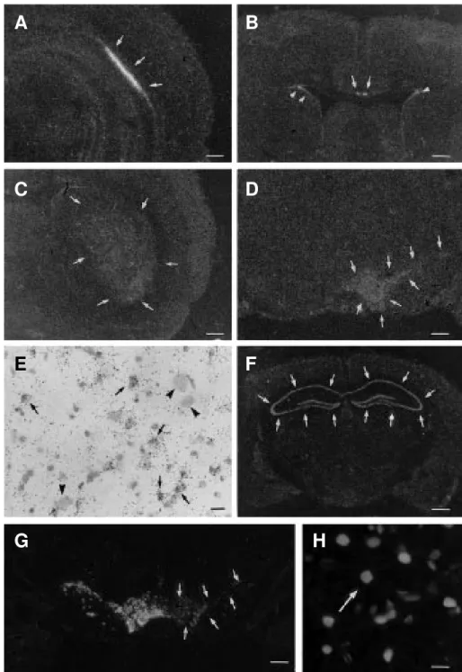
Figure 1 - Photomicrographs of film autoradiograms (A-D,G) and bright-field microscopy (E) of coronal sections of rat brain showing in situ hybridization of bFGF mRNA (A-D) and tyrosine hydroxylase mRNA (G) after unilateral nigral injection of saline (A-B) or 6-hydroxydopamine (6-OHDA) (C,D,G). The bFGF mRNA signal is seen in the CA2 region of the hippocampal formation (arrows in A) as well as in the indusium griseum (arrows) and subventricular regions (arrowheads) (B). The ipsilateral caudate putamen nucleus (C) and the ipsilateral pars compacta of the substantia nigra and ventral tegmental area (D) show a strong increase in bFGF mRNA 24 h and 7 days after the neurotoxin injection, respectively. After film emulsion dipping of the hybridized section, bright-field microscopy of the bFGF mRNA signal shows silver grains over cresyl violet-counterstained small putative glial nuclei (arrows) in the sections from the pars compacta (E) of a 6-hydroxydopamine (7 days)-injected rat. Clusters of silver grains are not found over putative neuronal profiles (arrowheads) in the above cited regions (E). Disappearance of tyrosine hydroxylase mRNA is observed in the region of dopaminergic lesions (arrows in G). Double immunofluorescence shows bFGF immunoreactivity in the nuclei of glial fibrillary acidic protein-immunoreactive astrocytes (H). Figure F illustrates the presence of the FGF receptor (flg) mRNA signal in the film autoradiograms localized in the hippocampal formation of mouse brain (arrows). Scale bars = 1000 µm (A-D,F,G), 50 µm (E), 25 µm (H).
A
B
C
D
E
F
related to different functional properties of
the molecule (69). Nuclear bFGF has been
considered to be involved in the regulation
of nuclear events such as gene expression
and transcription (65,71). Furthermore, the
synthesis of bFGF proteins could play a role
in cell function by modifying the
intracellu-lar signaling pathway, by modulating both
adenylate cyclase activity and the regulatory
G-proteins and also by interfering with cAMP
to regulate cell proliferation (72). The
differ-ent molecular weight forms of bFGF also
show functional differences on FGF
recep-tor function. It was reported that the
22.5-kDa isoform of the bFGF acts by increasing
FGF receptor type 1 messenger RNA
stabil-ity, thus enhancing cell responses to
exog-enous bFGF (73).
It has been postulated that intracellular
bFGF is released by alternative mechanisms
(18). The extracellular matrix represents a
natural reservoir for various
heparin-bind-ing growth factors (66). bFGF interacts with
heparin-like polysaccharides and the
hepa-ran sulfate proteoglycans of the extracellular
matrix may also be involved in the
presenta-tion of bFGF to its cell surface high affinity
receptor located in the neuroglia and
neu-rons, even though this is not essential for
high affinity binding to occur (74,75). This
interaction may facilitate the signal
trans-duction pathway of the molecule (76), thus
providing a regulatory mechanism for its
paracrine biological activity (77,78). After
binding to heparan sulfate proteoglycans or
to FGF tyrosine kinase receptors, bFGF is
internalized by different pathways (79).
Heparin-binding growth factors, i.e.
bFGF, are thought to play a role in the
pro-cesses of tissue homeostasis, regeneration or
repair following injury. These factors are
active upon release from neighboring
in-flammatory or lesioned cells, as well as upon
release from heparan sulfate associated with
the extracellular matrix (80). It is known that
the cells of a mechanically injured tissue
release biologically active substances and
that the conditioned medium from injured
cultured cells is highly mitogenic probably
due to the presence of growth factors (18).
Since bFGF has been reported to induce its
own gene expression (71), once released it
might be responsible for the increase in bFGF
messenger RNA in the neighborhood of the
lesioned cell. Sublethal injury triggers the
increases in the transcriptional activation of
the bFGF gene which may be related to the
recovery from cell injury (81).
Regulation of the bFGF protein and
mes-senger RNA at the cellular level following
experimental lesions applied to the rat brain
has been analyzed in order to evaluate the
trophic responses of the adult central
ner-vous system. 6-Hydroxydopamine is known
to induce lesion in the ascending dopamine
pathway and to promote experimental
par-kinsonism in rats (61). Figure 1G shows a
radioautogram of in situ hybridization of the
tyrosine hydroxylase messenger RNA to
dem-onstrate the disappearance of the signal in
the pars compacta of the substantia nigra and
ventral tegmental area following injury to
dopamine cells of a rat that received a
stereo-taxic nigral injection of 6-hydroxydopamine
and was sacrificed 7 days later.
6-hydroxydopamine. The degree of the changes
in the bFGF messenger RNA signal in the
hybridized sections was quantitated by means
of image analysis and the results obtained
for the pars compacta of the substantia nigra/
ventral tegmental area and for the
neostria-tum are shown in Figure 2. Bright-field
mi-croscopy demonstrated an increased number
of putative glial cells expressing the bFGF
messenger RNA signal (Figure 1E). bFGF
immunohistochemistry revealed that as early
as 2 h after drug injection, the density of glial
bFGF immunoreactive profiles was increased
in the pars compacta of the substantia nigra
and the ventral tegmental area. bFGF
immu-noreactivity was increased in the nuclei of
astrocytes only within the ipsilateral
sub-stantia nigra and ventral tegmental area at 72
h, and 1 week after the injection of
6-hy-droxydopamine (61). Figure 1H shows a
double immunofluorescence for the
simul-taneous detection of the bFGF and glial
fibril-lary acidic protein (a marker for astrocytes)
immunoreactivity in the pars compacta of
the substantia nigra of a
7-day-6-hydroxydo-pamine-lesioned rat. Reactive astrocytes
show hypertrophied cytoplasm and processes
and increased bFGF immunoreactivity.
Mechanical lesions of the central
ner-vous system also trigger increases of bFGF
and its messenger RNA in reactive
astro-cytes mostly localized close to the wound.
The upregulated synthesis of the astroglial
bFGF, as seen by the increase in bFGF
mes-senger RNA and protein after mechanical
lesion, was described following
implanta-tion of a microdialysis probe into the rat
hippocampus (59).
The trophic responses of bFGF have been
also evaluated in the forebrain of the rat
submitted to exotoxicity (58,82).
Radioac-tive in situ hybridization revealed a fast (6
h), strong (300-400% of control) and
wide-spread increase of bFGF messenger RNA
after kainate-induced epileptic seizures (58).
Transcript analysis at the cellular level by
nonradioactive
in situ hybridization revealed
a marked increase of messenger RNA in the
nuclei of astrocytes in several forebrain
re-gions. These changes were accompanied by
an enhancement of bFGF immunoreactivity
in the nuclei of reactive astrocytes (58).
The expression of bFGF messenger RNA
is not only triggered by strong lesions like
the one induced by 6-hydroxydopamine on
dopamine cells (61) but is highly enhanced
following the acute stage of focally evoked
convulsive seizures (83) and cerebral
contu-sion (84). Brief exposure to hypoxia also
induces increases in bFGF messenger RNA
and protein which are considered to protect
rat cortical neurons from prolonged hypoxic
stress (85). It is interesting that perinatal
asphyxia during birth triggers increases in
bFGF gene expression that are accompanied
by an increased number of dopamine nerve
cell bodies in the mesencephalon of the rat
(86). Hypoxia of the mature nervous tissue
also increases bFGF messenger RNA (87).
Bilateral recurrent seizure activity
in-duced by a focal stainless-steel wire in the
hippocampal hilus increases bFGF
messen-ger RNA in astrocytes throughout the
fore-brain and also in neurons of the dentate
gyrus and olfactory cortex (63).
Further-more, it was also shown that limbic motor
seizures produced by kainate injection
trig-ger a fast elevation of bFGF gene expression
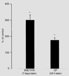
Figure 2 - Effects of a nigral 6-hydroxydopamine (6-OHDA) in-jection on the basic fibroblast growth factor (bFGF) mRNA lev-els in the ipsilateral pars com-pacta of the substantia nigra (SNC), ventral tegmental area (VTA) and caudate putamen (CP) nucleus 7 days and 24 h after the injection, respectively, using in situ hybridization procedures combined with image analysis. The numbers represent the per-cent values of the specific opti-cal density values obtained in the respective regions of control sa-line-injected animals (N = 3-6). *P<0.05 compared to respective control (Mann-Whitney U-test).
% of control
400
300
200
100
0
SNC/VTA (7 days later)
CP (24 h later)
*
in rat hippocampus and striatum. In the
hip-pocampus, a fast increase of bFGF
messen-ger RNA was localized in the neurons of the
dentate gyrus and the CA1 layer, whereas
the messenger RNA levels remained
un-changed in astrocytes. It is also possible that
the neuronal activity may regulate the bFGF
gene expression in an autocrine and intracrine
fashion at an early time, whereas delayed
expression is related to paracrine trophic
actions taking place in the activated
astro-cytes following neuronal injury (82). It is
possible that the expression of bFGF
mes-senger RNA in neurons is related to the
degree of neuronal activation since enhanced
noradrenergic tone evoked by the lipophilic
beta-adrenergic receptor agonist clenbuterol
elicited only an increase in the levels of glial
bFGF messenger RNA as seen in the
cere-bral cortex and hippocampus (32).
The increased synthesis of bFGF
follow-ing lesions of the central nervous system is
accompanied by an increased gene
expres-sion of the FGF receptor as seen by in situ
hybridization technique (88).
Expression of c-fos messenger RNA
af-ter cortical ablation in the rat brain is
modu-lated by bFGF and the NMDA receptor is
also involved in the expression of this
imme-diate early gene (89). Furthermore, bFGF
may maintain the homeostasis of the
intra-cellular Ca
2+concentration and thus protect
against glutamate-induced neurotoxicity (64).
It is well known that the interactions
between glial cells and neurons are
impor-tant for neuronal trophism in the central
nervous system (8,34,52,90); however, the
neurotrophic factor-mediated paracrine
trophic actions of glial cells on neurons are
only now beginning to be investigated.
Astroglial cells express neurotrophic and/
or neurite growth-promoting factors that are
regulated during development and/or after
central nervous system lesions and exert
pro-tective or regenerative effects on neurons
following lesions (8,91).
The glia-promoted neuronal trophism via
trophic factors has been described in several
neurotransmitter systems. It is known that
bFGF reduces the number of dopamine cells
undergoing apoptotic cell death and also
alters motor function and improves the
sur-vival of fetal ventral mesencephalic grafts,
actions that depend on the proliferation of
glial cells since they were blocked by
cy-tosine arabinoside (34,92). Furthermore,
bFGF can influence the survival of
cholin-ergic neurons by stimulating and increasing
glia, which may produce factor(s) that are
necessary for cholinergic neuron survival
(93). It was speculated that trophic responses
to multiple growth factors may compensate
for the endogenous deficiency in glial
fac-tors and/or the presence of glial inhibitory
factors in the central nervous system (33).
The interactions between glial cells may
modulate the paracrine trophic actions of
these cells to neurons. Interleukins released
by reactive microglia following brain injury
are capable of stimulating the synthesis of
neurotrophic factors by astrocytes (90). In
fact, it was demonstrated that interleukin-1
beta increases bFGF messenger RNA
ex-pression in adult rat brain and organotypic
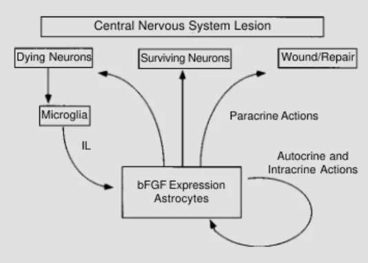
hippocampal cultures (94). Figure 3
illus-trates some possible autocrine, intracrine and
paracrine mechanisms of the upregulated
glial bFGF following a central nervous
sys-tem lesion.
Although regeneration of injured
neu-rons does not occur after trauma in the
cen-tral nervous system, functional recovery is
commonly observed. The increased trophic
Central Nervous System Lesion
Surviving Neurons Wound/Repair Dying Neurons
Microglia Paracrine Actions
Autocrine and Intracrine Actions bFGF Expression
Astrocytes IL
activity in the injured central nervous system
and the properties of neurotrophic factors in
neuronal growth and maintenance suggest
that these polypeptides are probably involved
in recovery of function by stimulating
neu-rite outgrowth in the surviving neurons or in
the related/neighbor pathway, needed for
reorganization of the injured nerve tissue.
Neurotrophic factors also stimulate
neu-rotransmitter synthesis probably to
compen-sate for the reduced neuronal actions, to
sustain survival of injured neurons and to
stimulate revascularization and glial
re-sponses to injury (31).
Acknowledgment
We thank Ms. Maria S.P. Abel for
techni-cal assistance and Mr. Andrés E.P. Ruiz for
the photography work.
References
1. Loughlin SE & Fallon JH (1993). Neu-rotrophic Factors. Academic Press, Inc., San Diego.
2. Hefti F, Denton TL, Knusel B & Lapchak PA (1993). Neurotrophic factors: what are they and what are they doing? In: Loughlin SE & Fallon JH (Editors), Neurotrophic Factors. Ac-ademic Press, Inc., San Diego, 25-49. 3. Thoenen H & Barde YA (1980).
Physiolo-gy of nerve growth factor. Physiological Reviews, 60: 1284-1335.
4. Levi-Montalcini R & Hamburger V (1953). A diffusible agent of mouse sarcoma, pro-ducing hyperplasia of sympathetic ganglia hyperneurotization of viscera in the chick embryo. Journal of Experimental Zoology, 123: 233-287.
5. Cohen S, Levi-Montalcini R & Hamburger V (1954). A nerve growth-stimulating fac-tor isolated from snake venom. Proceed-ings of the National Academy of Sciences, USA, 40: 1014-1018.
6. Hefti F, Hartikka J & Knusel B (1989). Function of neurotrophic factors in the adult and aging brain and their possible use in the treatment of neurodegenera-tive diseases. Neurobiology of Aging, 10: 515-533.
7. Barotte C, Eclancher F, Ebel A, Labourdette G, Sensenbrenner M & Will B (1989). Effects of basic fibroblast growth factor (bFGF) on choline acetyl-transferase activity and astroglial reaction in adult rats after partial fimbria transec-tion. Neuroscience Letters, 101: 197-202. 8. Baumann N, Baron-Van Evercooren A, Jacque C & Zalc B (1993). Glial biology and disorders. Current Opinion in Neurol-ogy and Neurosurgery, 6: 27-33. 9. Noble M & Wolswijk G (1992).
Develop-ment and regeneration in the O-2A lin-eage. Journal of Neuroimmunology, 40: 287-294.
10. Thoenen H, Bandtlow C & Heumann R (1987). The physiological function of nerve growth factor in the central nervous sys-tem: Comparison with the periphery. Re-views of Physiology, Biochemistry and Pharmacology, 109: 145-178.
11. Kamegai M, Niijima K, Kunishita T, Nishizawa M, Ogawa M, Araki M, Ueki A, Konishi Y & Tabira T (1990). Interleukin-3 as a trophic factor for central cholinergic neurons in vitro and in vivo. Neuron, 2: 429-436.
12. Knusel B, Michel PP, Schwaber JS & Hefti F (1990). Selective and nonselective stim-ulation of central cholinergic and dopami-nergic development in vitro by nerve growth factor, basic fibroblast growth fac-tor, epidermal growth facfac-tor, insulin and the insulin-like growth factors I and II. Journal of Neuroscience, 10: 558-570. 13. Korr H, Siewert E, Bertram C, Labourdette
G & Sensenbrenner M (1992). Autoradio-graphic studies of rat astroglial cell prolif-eration in vitro with and without treat-ment with basic fibroblast growth factor. Cell Proliferation, 25: 605-622.
14. Barde YA, Edgar D & Thoenen H (1982). Purification of a new neurotrophic factor from mammalian brain. EMBO Journal, 1: 549-553.
15. Ernfors P, Lonnerberg P, Ayer LC & Persson H (1990). Developmental and re-gional expression of basic fibroblast growth factor mRNA in the rat central nervous system. Journal of Neuroscience Research, 27: 10-15.
16. Hallbook F, Ibanez CF & Persson H (1991). Evolutionary study of nerve growth factor family reveals a new member abundantly expressed in Xenopus ovary. Neuron, 6: 845-858.
17. Berkemeier LR, Winslow JW, Kaplan DR, Nikolics K, Goeddel DV & Rosenthal A (1991). Neurotrophin-5: A novel neu-rotrophic factor that activates trk and trkB. Neuron, 7: 857-866.
18. Crowley ST, Ray CJ, Nawaz D, Majack RA & Horwitz LD (1995). Multiple growth fac-tors are released from mechanically in-jured vascular smooth muscle cells. American Journal of Physiology, 269: 1641-1647.
19. Mena MA, Casarejos MJ, Gimenez GG & Garcia DYJ (1995). Fibroblast growth fac-tors: structure-activity on dopamine neu-rons in vitro. Journal of Neural Transmis-sion, Parkinsons Disease Dementia Sec-tion, 9: 1-14.
20. Baird A & Klagsbrun M (1991). The fibro-blast growth factor family: An overview. In: Baird A & Klagsbrun M (Editors), The Fibroblast Growth Factor Family. Annals of the New York Academy of Sciences, New York, xi.
21. Fallon JH, Annis CM, Gentry LE, Twarzik DR & Loughlin SE (1990). Localization of cells containing transforming growth fac-tor alfa precursor immunoreactivity in the basal ganglia of the adult rat brain. Growth Factors, 2: 241-250.
22. Nakao N, Odin P, Lindvall O & Brundin P (1996). Differential trophic effects of ba-sic fibroblast growth factor, insulin-like growth factor-1, and neurotrophin-3 on striatal neurons in culture. Experimental Neurology, 138: 144-157.
23. Roher H (1990). The role of growth fac-tors in the control of neurogenesis. Euro-pean Journal of Neuroscience, 2: 1005-1015.
25. Krupinski J, Rajaram R, Lakonishok M, Benovic JL & Cerione RA (1988). Insulin-dependent phosphorylation of GTP-bind-ing proteins in phospholipid vesicles. Jour-nal of Biological Chemistry, 263: 12333-12341.
26. Hendry IA & Crouch MF (1993). Synergy, retrograde transport and cell death. In: Loughlin SE & Fallon JH (Editors), Neu-rotrophic Factors. Academic Press, Inc., San Diego, 51-87.
27. Sharp FR, Gonzalez MF, Hizanaga K, Mobley WC & Sagar SM (1989). Induction of the c-fos gene product in the rat fore-brain following cortical lesions and NGF injections. Neuroscience Letters, 100: 117-122.
28. Gizang-Ginsberg E & Ziff EB (1990). Nerve growth factor regulates tyrosine hydroxy-lase gene transcription through a nucle-oprotein complex that contain c-fos. Genes and Development, 4: 477-491. 29. Sporn MB & Roberts AB (1988). Peptide
growth factors are multifunctional. Na-ture, 332: 217-219.
30. Fallon JH & Loughlin SE (1993). Func-tional implications of the anatomical local-ization of neurotrophic factors. In: Loughlin SE & Fallon JH (Editors), Neu-rotrophic Factors. Academic Press, Inc., San Diego, 1-24.
31. Mocchetti I & Wrathall JR (1995). Neu-rotrophic factors in central nervous sys-tem trauma. Journal of Neurotrauma, 12: 853-870.
32. Hayes VY, Isackson PJ, Fabrazzo M, Follesa P & Mocchetti I (1995). Induction of nerve growth factor and basic fibro-blast growth factor mRNA following clenbuterol: contrasting anatomical and cellular localization. Experimental Neurol-ogy, 132: 33-41.
33. Faber EA, Solomon A, Abraham JA, Marikovsky M & Schwartz M (1996). In-volvement of wound-associated factors in rat brain astrocyte migratory response to axonal injury: in vitro simulation. Journal of Clinical Investigation, 97: 162-171. 34. Zawada WM, Kirschman DL, Cohen JJ,
Heidenreich KA & Freed CR (1996). Growth factors rescue embryonic dopa-mine neurons from programmed cell death. Experimental Neurology, 140: 60-67.
35. Trowell OA, Chir B & Willmer EN (1939). Growth of tissues in vitro. IV. The effects of some tissue extracts on the growth of periosteal fibroblasts. Journal of Experi-mental Biology, 16: 60-70.
36. Gospodarowicz D (1987). Isolation and characterization of acidic and basic fibro-blast growth factor. Methods in Enzymol-ogy, 147: 106-119.
37. Gospodarowicz D, Cheng J, Lui G-M, Baird A & Böhlen P (1984). Isolation of brain fibroblast growth factor by heparin-Sepharose affinity chromatography: iden-tity with pituitary fibroblast growth factor. Proceedings of the National Academy of Sciences, USA, 81: 6963-6967.
38. Lobb RR, Harper JW & Fett JW (1986). Purification of heparin-binding growth fac-tors. Analytical Biochemistry, 154: 1-14. 39. Gospodarowicz D, Neufeld G &
Schweigerer L (1987). Fibroblast growth factor: structural and biological properties. Journal of Cellular Physiology, 5 (Suppl): 15-26.
40. Lamb TM & Harland RM (1995). Fibro-blast growth factor is a direct neural in-ducer, which combined with noggin gen-erates anterior-posterior neural pattern. Development, 121: 3627-3636. 41. Kuzis K, Reed S, Cherry NJ, Woodward
WR & Eckenstein FP (1995). Develop-mental time course of acidic and basic fibroblast growth factors’ expression in distinct cellular populations of the rat cen-tral nervous system. Journal of Compara-tive Neurology, 358: 142-153.
42. Chadi G, Rosen L, Cintra A, Tinner B, Zoli M, Pettersson RF & Fuxe K (1993). Corti-costerone increases FGF-2 (bFGF) immu-noreactivity in the substantia nigra of the rat. Neuroreport, 4: 783-786.
43. Fuxe K, Chadi G, Agnati LF, Tinner B, Rosén L, Janson AM, Møller A, Cintra A, Cao Y, Goldstein M, Lindahl U, David G, Ögren S, Toffano G, Baird A & Pettersson RF (1993). Fibroblast growth factor-2, gan-glioside GM1 and the trophic regulation of the basal ganglia. Focus on the nigro-striatal dopamine neurons. In: Fuxe K, Agnati LF, Bjelke B & Ottoson D (Editors), Trophic Regulation of the Basal Ganglia. Focus on Dopamine Neurons. Pergamon Press, Oxford, Stockholm, Sweden, 1-41. 44. Matsuda S, Okumura N, Yoshimura H, Koyama Y & Sakanaka M (1992). Basic fibroblast growth factor-like immunoreac-tivity in Purkinje cells of the rat cerebel-lum. Neuroscience, 50: 99-106. 45. Woodward WR, Nishi R, Meshul CK,
Wil-liams TE, Coulombe M & Eckenstein FP (1992). Nuclear and cytoplasmic localiza-tion of basic fibroblast growth factor in astrocytes and CA2 hippocampal neurons. Journal of Neuroscience, 12: 142-152.
46. Kilpatrick TJ & Bartlett PF (1995). Cloned multipotential precursors from the mouse cerebrum require FGF-2, whereas glial re-stricted precursors are stimulated with ei-ther FGF-2 or EGF. Journal of Neurosci-ence, 15: 120-127.
47. Temple S & Qian X (1995). bFGF, neurotrophins, and the control or cortical neurogenesis. Neuron, 15: 249-252. 48. Gage FH, Coates PW, Palmer TD, Kuhn
HG, Fisher LJ, Suhonen JO, Peterson DA, Suhr ST & Ray J (1995). Survival and dif-ferentiation of adult neuronal progenitor cells transplanted to the adult brain. Pro-ceedings of the National Academy of Sci-ences, USA, 92: 11879-11883.
49. Aoyagi A, Nishikawa K, Saito H & Abe K (1994). Characterization of basic fibroblast growth factor-mediated acceleration of ax-onal branching in cultured rat hippocampal neurons. Brain Research, 661: 117-126. 50. Iwasaki Y, Shiojima T, Ikeda K, Tagaya N,
Kobayashi T & Kinoshita M (1995). Acidic and basic fibroblast growth factors en-hance neurite outgrowth in cultured rat spinal cord neurons. Neurological Re-search, 17: 70-72.
51. Takei N, Ogaki H & Endo Y (1995). Basic fibroblast growth factor inhibited Ca2+
ionophore-induced apoptotic cell death of cultured cortical neurons from embryonic rats. Neuroscience Letters, 192: 124-126. 52. Chadi G, Moller A, Rosen L, Janson AM, Agnati LA, Goldstein M, Ogren SO, Pettersson RF & Fuxe K (1993). Protec-tive actions of human recombinant basic fibroblast growth factor on MPTP-lesioned nigrostriatal dopamine neurons after intraventricular infusion. Experimen-tal Brain Research, 97: 145-158. 53. Kirschner PB, Henshaw R, Weise J,
Trubetskoy V, Finklestein S, Schulz JB & Beal MF (1995). Basic fibroblast growth factor protects against excitotoxicity and chemical hypoxia in both neonatal and adult rats. Journal of Cerebral Blood Flow and Metabolism, 15: 619-623.
54. Takayama H, Ray J, Raymon HK, Baird A, Hogg J, Fisher LJ & Gage FH (1995). Ba-sic fibroblast growth factor increases do-paminergic graft survival and function in a rat model of Parkinson’s disease. Nature Medicine, 1: 53-58.
56. Matsuyama A, Iwata H, Okumura N, Yoshida S, Imaizumi K, Lee Y, Shiraishi S & Shiosaka S (1992). Localization of basic fibroblast growth factor-like immunoreac-tivity in the rat brain. Brain Research, 587: 49-65.
57. Chadi G, Tinner B, Agnati LF & Fuxe K (1993). Basic fibroblast growth factor (bFGF, FGF-2) immunoreactivity exists in the noradrenaline, adrenaline and 5-HT nerve cells of the rat brain. Neuroscience Letters, 160: 171-176.
58. Humpel C, Lippoldt A, Chadi G, Ganten D, Olson L & Fuxe K (1993). Fast and wide-spread increase of basic fibroblast growth factor messenger RNA and protein in the forebrain after kainate-induced seizures. Neuroscience, 57: 913-922.
59. Humpel C, Chadi G, Lippoldt A, Ganten D, Fuxe K & Olson L (1994). Increase of ba-sic fibroblast growth factor (bFGF, FGF-2) messenger RNA and protein following implantation of a microdialysis probe into rat hippocampus. Experimental Brain Re-search, 98: 229-237.
60. Eckenstein FP, Kuzis K, Nishi R, Wood-ward WR, Meshul C, Sherman L & Ciment G (1994). Cellular distribution, subcellular localization and possible functions of ba-sic and acidic fibroblast growth factors. Biochemical Pharmacology, 47: 103-110. 61. Chadi G, Cao Y, Pettersson RF & Fuxe K
(1994). Temporal and spatial increase of astroglial basic fibroblast growth factor synthesis after 6-hydroxydopamine-in-duced degeneration of the nigrostriatal dopamine neurons. Neuroscience, 61: 891-910.
62. Grothe C & Meisinger C (1995). Fibroblast growth factor (FGF)-2 sense and anti-sense mRNA and FGF receptor type 1 mRNA are present in the embryonic and adult rat nervous system: specific detec-tion by nuclease protecdetec-tion assay. Neuro-science Letters, 197: 175-178.
63. Gall CM, Berschauer R & Isackson PJ (1994). Seizures increase basic fibroblast growth factor mRNA in adult rat forebrain neurons and glia. Brain Research, Molec-ular Brain Research, 21: 190-205. 64. Mattson MP, Lovell MA, Furukawa K &
Markesbery WR (1995). Neurotrophic fac-tors attenuate glutamate-induced accu-mulation of peroxides, elevation of intra-cellular Ca2+ concentration, and
neurotox-icity and increase antioxidant enzyme ac-tivities in hippocampal neurons. Journal of Neurochemistry, 65: 1740-1751.
65. Hawker JJ & Granger HJ (1994). Tyrosine kinase inhibitors impair fibroblast growth factor signaling in coronary endothelial cells. American Journal of Physiology, 266: 107-120.
66. Soulet L, Chevet E, Lemaitre G, Blanquaert F, Meddahi A & Barritault D (1994). FGFs and their receptors, in vitro and in vivo studies: new FGF receptor in the brain, FGF-1 in muscle, and the use of functional analogues of low-affinity hepa-rin-binding growth factor receptors in tis-sue repair. Molecular Reproduction and Development, 39: 49-54.
67. Baird A (1994). Fibroblast growth factors: activities and significance of non-neu-rotrophic growth factors. Current Opinion in Neurobiology, 4: 78-86.
68. Wanaka A, Johnson EM & Milbrandt J (1990). Localization of FGF receptor mRNA in the adult rat central nervous system by in situ hybridization. Neuron, 5: 267-281.
69. Rifkin DB, Moscatelli D, Roghani M, Nagano Y, Quarto N, Klein S & Bikfalvi A (1994). Studies on FGF-2: nuclear localiza-tion and funclocaliza-tion of high molecular weight forms and receptor binding in the absence of heparin. Molecular Reproduction and Development, 39: 102-104.
70. Florkiewicz RZ, Baird A & Gonzalez AM (1991). Multiple forms of bFGF: differen-tial nuclear and cell surface localization. Growth Factors, 4: 265-275.
71. Flott-Rahmel B, Gerdes W, Pechan PA, Brysch W, Schlingensiepen K-H & Seifert W (1992). bFGF induces its own gene expression in astrocytic and hippocampal cell cultures. Neuroreport, 3: 1077-1080. 72. Estival A, Robberecht P, Fanjul M, Rouot
B, Hollande E, Vaysse N & Clemente F (1996). Cells retrovirally transfected with fibroblast growth factor-2 isoforms exhibit altered adenylate cyclase activity and G-protein functionality. Biochemical Journal, 315: 619-624.
73. Estival A, Monzat V, Miquel K, Gaubert F, Hollande E, Korc M, Vaysse N & Clemente F (1996). Differential regulation of fibro-blast growth factor (FGF) receptor-1 mRNA and protein by two molecular forms of basic FGF. Modulation of FGFR-1 mRNA stability. Journal of Biological Chemistry, 271: 5663-5670.
74. Brickman YG, Ford MD, Small DH, Bartlett PF & Nurcombe V (1995). Heparan sul-fates mediate the binding of basic fibro-blast growth factor to a specific receptor on neural precursor cells. Journal of Bio-logical Chemistry, 270: 24941-24948.
75. Murphy PR & Knee RS (1995). Basic fibro-blast growth factor binding and process-ing by human glioma cells. Molecular and Cellular Endocrinology, 114: 193-203. 76. Faham S, Hileman RE, Fromm JR,
Linhardt RJ & Rees DC (1996). Heparin structure and interactions with basic fi-broblast growth factor. Science, 271: 1116-1120.
77. Laslett AL, McFarlane JR, Hearn MT & Risbridger GP (1995). Requirement for heparan sulphate proteoglycans to medi-ate basic fibroblast growth factor (FGF-2)-induced stimulation of Leydig cell ste-roidogenesis. Journal of Steroid Biochem-istry and Molecular Biology, 54: 245-250. 78. Venkataraman G, Sasisekharan V, Herr
AB, Ornitz DM, Waksman G, Cooney CL, Langer R & Sasisekharan R (1996). Prefer-ential self-association of basic fibroblast growth factor is stabilized by heparin dur-ing receptor dimerization and activation. Proceedings of the National Academy of Sciences, USA, 93: 845-850.
79. Gleizes PE, Noaillac DJ, Amalric F & Gas N (1995). Basic fibroblast growth factor (FGF-2) internalization through the hepa-ran sulfate proteoglycans-mediated path-way: an ultrastructural approach. Euro-pean Journal of Cell Biology, 66: 47-59. 80. Muthukrishnan L, Warder E & McNeil PL
(1991). Basic fibroblast growth factor is efficiently released from a cytosolic stor-age site through plasma membrane dis-ruptions of endothelial cells. Journal of Cellular Physiology, 148: 1-16.
81. Ku PT & D’Amore PA (1995). Regulation of basic fibroblast growth factor (bFGF) gene and protein expression following its release from sublethally injured endothe-lial cells. Journal of Cellular Biochemistry, 58: 328-343.
82. Riva MA, Donati E, Tascedda F, Zolli M & Racagni G (1994). Short- and long-term induction of basic fibroblast growth factor gene expression in rat central nervous system following kainate injection. Neu-roscience, 59: 55-65.
83. Riva MA, Gale K & Mocchetti I (1992). Basic fibroblast growth factor mRNA in-creases in specific brain regions following convulsive seizures. Brain Research, Mo-lecular Brain Research, 15: 311-318. 84. Iwamoto Y, Yamaki T, Murakami N,
85. Sakaki T, Yamada K, Otsuki H, Yuguchi T, Kohmura E & Hayakawa T (1995). Brief exposure to hypoxia induces bFGF mRNA and protein and protects rat cortical neu-rons from prolonged hypoxic stress. Neu-roscience Research, 23: 289-296. 86. Andersson K, Blum M, Chen Y, Eneroth
P, Gross J, Herrera MM, Bjelke B, Bolme P, Diaz R, Jamison L & Fuxe K (1995). Perinatal asphyxia increases bFGF mRNA levels and DA cell body number in the mesencephalon of rats. Neuroreport, 6: 375-378.
87. LaManna JC, Boehm KD, Mironov V, Hudetz AG, Hritz MA, Yun JK & Harik SI (1994). Increased basic fibroblastic growth factor mRNA in the brains of rats exposed to hypobaric hypoxia. Advances in Experi-mental Medicine and Biology, 361: 497-502.
88. Yuguchi T, Kohmura E, Yamada K, Wanaka A, Otsuki H, Sakaguchi T, Yamashita T, Tohyama M & Hayakawa T (1994). Mes-senger RNA and protein expression of basic fibroblast growth factor receptor af-ter cortical ablation. Brain Research, Mo-lecular Brain Research, 25: 50-56. 89. Kohmura E, Yuguchi T, Yamada K,
Sakaguchi T, Wanaka A & Hayakawa T (1995). Expression of c-fos mRNA after cortical ablation in rat brain is modulated by basic fibroblast growth factor (bFGF) and the NMDA receptor is involved in c-fos expression. Brain Research, Molecu-lar Brain Research, 28: 117-121. 90. Giulian D, Vaca K & Corpuz M (1993).
Brain glia release factors with opposing actions upon neuronal survival. Journal of Neuroscience, 13: 29-37.
91. Muller HW, Junghans U & Kappler J (1995). Astroglial neurotrophic and neu-rite-promoting factors. Pharmacology and Therapeutics, 65: 1-18.
92. Zeng BY, Jenner P & Marsden CD (1996). Altered motor function and graft survival produced by basic fibroblast growth fac-tor in rats with 6-OHDA lesions and fetal ventral mesencephalic grafts are associ-ated with glial proliferation. Experimental Neurology, 139: 214-226.
93. Perkins LA & Cain LD (1995). Basic fibro-blast growth factor (bFGF) increases the survival of embryonic and postnatal basal forebrain cholinergic neurons in primary culture. International Journal of Develop-mental Neuroscience, 13: 51-61. 94. Rivera S, Gold SJ & Gall CM (1994).