In vitro
antim icro bial activity o f a
ne w se rie s o f 1,4-naphtho quino ne s
1Departamento de Ciências dos Alimentos, Instituto de Ciências e Tecnologia de Alimentos, and 2Departamento de Q uímica O rgânica, Instituto de Q uímica, Universidade Federal do Rio
Grande do Sul, Porto Alegre, RS, Brasil A. Riffel1, L.F. Medina1,
V. Stefani2, R.C. Santos2,
D. Bizani1 and A. Brandelli1
Abstract
The antibacterial activity of a series of 1,4-naphthoquinones was demonstrated. Disk diffusion tests were carried out against several Gram-positive and Gram-negative bacteria. The compound 5-amino-8-hydroxy-1,4-naphthoquinone was the most effective, presenting inhibition zones measuring 20 mm against staphylococci, streptococci and bacilli at 50 µg/ml. Methicillin-resistant Staphylococcus aureus
and several clinical isolates of this bacterium were also inhibited. Naphthazarin, 5-acetamido-8-hydroxy-1,4-naphthoquinone, and 2,3-diamino-1,4-naphthoquinone were the next most active compounds. The minimal inhibitory concentration of the active compounds was determined against S. aureus, ranging from 30 to 125 µg/ml. All compounds presented a minimal bactericidal concentration higher than 500 µg/ml, indicating that their effect was bacteriostatic. The EC50, defined as the drug concentration that produces 50% of maximal effect, was 8 µg/ml for 5-amino-8-hydroxy-1,4-naphthoquinone against
S. aureus, S. intermedius, and S. epidermidis. These results indicate an effective in vitro activity of 5-amino-8-hydroxy-1,4-naphthoquinone and encourage further studies for its application in antibiotic therapy.
Co rre spo nde nce
A. Brandelli ICTA, UFRGS
Av. Bento Gonçalves, 9500
91501-970 Porto Alegre, RS Brasil
Fax: + 55-51-316-7048 E-mail: abrand@ vortex.ufrgs.br
Received April 11, 2001 Accepted April 17, 2002
Ke y words
• 5-Amino-8-hydroxy-1,4-naphthoquinone
•Antimicrobial agent
•Staphylococcus aureus
•Q uinone
•Naphthazarin
• 5-Acetamido-8-hydroxy-1,4-naphthoquinone
• 2,3-Diamino-1,4-naphtho-quinone
Intro ductio n
Naphthoquinones are widely distributed in plants, fungi, and some animals. Their biological activities have been studied in-cluding their effects on prokaryotic and eu-karyotic cells (1,2).
Plumbagone, juglone and lawsone are naturally occurring naphthoquinones of plant origin that have antibacterial effects on sev-eral species of both aerobic and anaerobic organisms (3), and toxins derived from naphthazarin (5,8-dihydroxy-1,4-naphtho-quinone) are produced by Fusarium solani
and attack plants, other fungi and bacteria (4). The antimicrobial activity of the natural naphthoquinone products alkannin and shi-konin and their derivatives has been investi-gated. In general, they are active against Gram-positive bacteria such as Staphylococ-cus aureus, Enterococcus faecium, and Ba-cillus subtilis, but they are inactive against Gram-negative bacteria (5).
(2-[trans-4-(4-chloro- phenyl)-cyclohexyl]-3-hydroxy-1,4-naphtho-quinone) is approved as the drug of choice by the FDA (9). Antibacterial activity has been described for isoxazolylnaphthoqui-nones (10), and some hydroxyquiisoxazolylnaphthoqui-nones and their metal complexes (11,12). The fungi-toxicity of naphthoquinones (8), particularly against several species of Candida (13-15), and the inhibitory activity of some naphtho-quinones on HIV-1 protease have also been described (16,17).
The mechanism of action of naphtho-quinones has not been completely elucidated. Atovaquone, an analog of ubiquinone, acts by interfering with the electron transport chain of mitochondria at site bc1 (complex III) in Plasmodium species. This conse-quently inhibits nucleic acid and ATP syn-thesis (9,18). Indeed, ß-lapachone (3,4-dihydro-2,2-dimethyl-2H-naphtho[1,2] pyran-5,6-dione) is an antimicrobial naph-thoquinone that caused increased generation of superoxide anion and hydrogen peroxide in Trypanosoma cruzi (19). Recent studies indicate that both antibacterial isoxazolyl naphthoquinones and naphthazarin toxins uncouple the electron transport chain (20,21). The development of new antimicrobial agents is a research area of the utmost impor-tance. Antimicrobial resistance among key microbial pathogens continues to grow at an alarming rate worldwide. Resistance among strains of S. aureus, Pseudomonas spp, Strep-tococcus spp and Enterobacteriaceae has been described (22-24). The increased preva-lence of antibiotic-resistant bacteria emerg-ing from the extensive use of antibiotics may render the current antimicrobial agents in-sufficient to control at least some bacterial infections.
The challenge of synthesizing derivatives of natural antimicrobial naphthoquinones to improve their pharmaceutical properties has been accepted by several laboratories. In-deed, the synthesis and evaluation of antimi-crobial activity of bioactive analogs of kigeli-none (25), alkannin (5), and lapachol (26)
has been reported. In the present study, we describe the antibacterial activity of a new series of 1,4-naphthoquinones.
Mate rial and Me thods
Bacte ria
The strains screened for antibacterial ac-tivity were clinical isolates obtained at the Faculdade de Veterinária, UFRGS. Strains of Listeria spp were food isolates. Strains of methicillin-resistant S. aureus were isolated at Hospital de Pronto Socorro de Porto Ale-gre, RS. S. aureus ATCC 25923, S. epider-midis ATCC 14990, S. saprophyticus ATCC 15305, and E. coli ATCC 25922 were from our collection. Stock cultures of the bacterial strains were maintained on nutrient agar plates (Difco Laboratories, Detroit, MI, USA) at 4ºC. Every two months, the bacteria were grown twice overnight in fresh nutrient broth (Oxoid, Basingstoke, UK), then streaked on fresh nutrient agar plates, again grown over-night, checked for purity, and then stored at 4ºC. Strains of Streptococcus were submit-ted to susceptibility tests after isolation on 5% sheep blood agar.
Naphtho quino ne s
The twelve naphthoquinones studied here were synthesized as described elsewhere (27,28), and their structures are shown in Figure 1. The degree of purity was deter-mined by standard procedures (thin layer chromatography, melting point, elemental analysis). The structures were confirmed by proton nuclear magnetic resonance (1H-NMR), 13C-NMR, infrared spectroscopy, UV-visible
spectroscopy, and mass spectrometry.
Growth de te rmination
di-luted at 10-1 to 10-8 in phosphate-buffered
saline, samples were homogenized, and then loaded in triplicate amounts of 20 µl onto nutrient agar plates. Plates were incubated for 24 h at 37ºC, and counting was done on plates containing 30 to 100 colonies.
Susce ptibility te sts
The susceptibility tests were performed according to the NCCLS recommendations (30,31). Screening tests regarding the inhibi-tion zone were carried out by the filter paper disk method (30,32). The inoculum suspen-sion was prepared from colonies grown over-night on an agar plate, and inoculated into Mueller-Hinton broth (Merck, Darmstadt, Germany) to yield a 0.5 McFarland turbidity standard solution. A sterile swab was im-mersed in the bacterial suspension and used to inoculate Mueller-Hinton agar plates. The disks were applied to the surface of inocu-lated plates using a sterile forceps.
The compounds were dissolved in di-methylsulfoxide (DMSO) or dimethylform-amide (DMF), and then applied to the disks. The inhibition zone was measured around each disk after 24 h at 37ºC. Strains of Streptococcus pyogenes and Enterococcus faecalis were tested on Mueller-Hinton agar plates supplemented with 5% sheep blood (32). Controls using DMSO and DMF were adequately done. To assess the minimal in-hibitory concentration (MIC) defined as the drug concentration at which no growth was visible, 96-well sterile microplates (Corning Inc., New York, NY, USA) were filled with 0.1 ml of serial two-fold dilutions (500 to 0.12 µg/ml) of the different naphthoquinones. The compounds were diluted in Mueller-Hinton broth from a 10 mg/ml stock solution in DMSO. A standardized number of bacte-ria (0.1 ml of a 106 CFU/ml suspension in
Mueller-Hinton broth) were inoculated into each well. A growth well (broth plus inocu-lum) and a sterility control well (broth only) were included in each panel. Microplates
were incubated at 37ºC for 24 h, and then MIC was determined as the last dilution at which no increase in visual turbidity was observed (31,32). MIC values were deter-mined in the absence or presence of 1% bovine serum albumin. The minimal bacteri-cidal concentration (MBC), defined as the concentration producing a ≥99.9% reduc-tion in CFU number in the initial inoculum, was determined by subculture on nutrient agar as previously described (31,32).
Compound R1 R2 R3 R4 R5 R6
I H H OH H H OH
II H H NH2 H H OH
IIa H H NHCOCH3 H H OH
IIb OH H NH2 H H OH
IIc H H NH2 Br H OH
IId H H NHCOC9H19 H H OH
IIe H H NHCOC13H27 H H OH
IIf Br Br NH2 Br Br OH
IIIa Cl Cl H H H H
IIIb NH2 NH2 H H H H
IIIc N3 N3 H H H H
R6 R5
R1
R2 R4
R3 O
O
OH
OH O NH
OH
A
B
De te rmination of EC50
EC50 is the drug concentration that
pro-duces 50% of maximal effect (33). This procedure was carried out for the compound 5-amino-8-hydroxy-1,4-naphthoquinone. The microorganisms were grown in Mueller-Hinton broth to yield a suspension contain-ing 106 CFU/ml, and then added to sterile
tubes containing the naphthoquinone to ob-tain final concentrations of 1, 10, 30, 100, and 1000 µg/ml. Tubes were incubated at 37ºC for 24 h, and CFU were then counted for each tube.
Re sults
The antibacterial effect observed for this family of naphthoquinones against S. aureus is presented in Table 1. Compound II (5-amino-8-hydroxy-1,4-naphthoquinone) pre-sented the widest inhibition zones. The ac-tivity of related compounds was in the fol-lowing order: II>I, IIa>IIIb>IIb, IIIa, IIIc>IId>IIc, IIe. Inhibitory activity was not observed for compounds IIf, which contains four bulky bromo atoms, and IV, which has an amino group replacing the keto group at position 1. The ATCC strain presented ex-pected inhibition zones against antimicro-bial agents that are used therapeutically (32). The effect of these compounds on S. aureus strains of clinical origin or of collection ori-gin (ATCC) was similar (Table 1). Methicil-lin-resistant Staphylococcus aureus (MRSA) strains were also inhibited by some naphtho-quinones tested, whereas they were resistant to all the comercially available antimicrobial agents tested, except vancomycin.
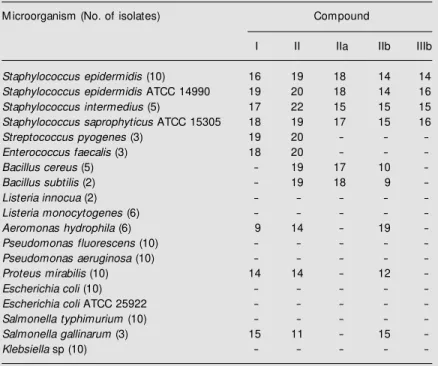
The most effective naphthoquinones were tested against other microorganisms listed in Table 2. Compound II inhibited the growth of several species of staphylococci, bacilli, streptococci, and some Gram-negative bac-teria such as Proteus mirabilis, Salmonella gallinarum, and Aeromonas hydrophila. Compound IIb inhibited the growth of
staph-Table 1. Susceptibility of Staphylococcus aureus to naphthoquinones. Compound Concentration Inhibition zone (mm)
(µg/disk)
ATCC Clinical origin M RSA
I 50 19 15 15
II 50 21 17 15
IIa 50 19 16 11
IIb 50 14 15 10
IIc 50 9 9 9
IId 50 10 10
-IIe 50 9 9
-IIf 50 - -
-IIIa 50 15 14 12
IIIb 50 15 16 17
IIIc 50 14 14
-IV 50 - -
-Ampicillin 10 32 20
-Streptomycin 10 18 22
-Novobiocin 5 22 24 11
Vancomycin 30 18 18 12
Gentamicin 10 24 26
-Tetracycline 10 30 28 11 Results are reported for S. aureus ATCC 25923, 12 clinical isolates, and 5 methicillin-resistant S. aureus (M RSA) isolates. Data are the mean of five determinations for each isolate. M RSA isolates w ere tested by previously described methods (30,32).
Table 2. Antimicrobial susceptibility testing of a family of naphthoquinones. M icroorganism (No. of isolates) Compound
I II IIa IIb IIIb
Staphylococcus epidermidis (10) 16 19 18 14 14
Staphylococcus epidermidis ATCC 14990 19 20 18 14 16
Staphylococcus intermedius (5) 17 22 15 15 15
Staphylococcus saprophyticus ATCC 15305 18 19 17 15 16
Streptococcus pyogenes (3) 19 20 - -
-Enterococcus faecalis (3) 18 20 - -
-Bacillus cereus (5) - 19 17 10
-Bacillus subtilis (2) - 19 18 9
-Listeria innocua (2) - - - -
-Listeria monocytogenes (6) - - - -
-Aeromonas hydrophila (6) 9 14 - 19
-Pseudomonas fluorescens (10) - - - -
-Pseudomonas aeruginosa (10) - - - -
-Proteus mirabilis (10) 14 14 - 12
-Escherichia coli (10) - - - -
-Escherichia coli ATCC 25922 - - - -
-Salmonellatyphimurium (10) - - - -
-Salmonella gallinarum (3) 15 11 - 15
ylococci and bacilli. P. mirabilis, A. hydro-phila and S. gallinarum were also inhibited by compounds I and IIb.
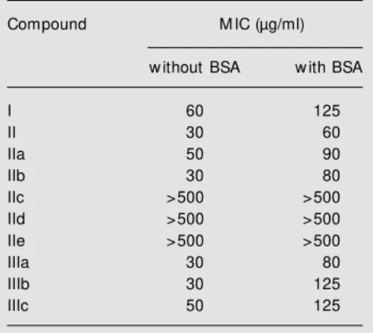
The MIC of naphthoquinones was deter-mined against S. aureus (Table 3). The com-pounds presented MIC values in the 30-125 µg/ml range, except for compounds IIc, IId, and IIe, which showed MIC values higher than 500 µg/ml. These compounds also pre-sented lower inhibition zones in disk diffu-sion tests (Table 1). Lower MIC values were observed for molecules bearing most hydro-philic groups. The presence of 1% (w/v) serum albumin in culture media often caused increased MIC values (Table 3). Similar re-sults were obtained against ATCC and MRSA strains.
In tests for determination of MBC, bacte-rial growth was higher than 0.1% the CFU in the initial inoculate. This resulted in MBC values higher than 500 µg/ml for all com-pounds tested. The MBC values obtained for the naphthoquinones tested indicate their bacteriostatic activity.
The dose-response effect of compound II was tested against different species of Staph-ylococcus. Similar dose-response curves were observed for S. aureus, S. epidermidis, and S. intermedius, presenting an EC50 of
ap-proximately 8 µg/ml (Figure 2).
D iscussio n
The search for new antimicrobial agents is an important line of research because of the resistance acquired by several patho-genic microorganisms. The prevalence of strains of S. aureus resistant to conventional antibiotics has increased to high levels in some hospitals (34,35). This bacterium is one of the most prevalent microorganisms in nosocomial infections worldwide and me-thicillin-resistant strains represent 15-45% of all S. aureus isolates (36). MRSA isolates carry the mec gene, a 2130-bp DNA frag-ment of non-staphylococcal origin encoding a low antibiotic-affinity penicillin-binding
protein (37). Since methicillin resistance was first identified in a clinical isolate of S. au-reus, its incidence has increased significant-ly, MRSA has spread globalsignificant-ly, and the mec gene has also been detected in several other staphylococcal species (37,38). Specifical-ly, S. aureus growth, including MRSA, was effectively inhibited by compound II and other derivatives, suggesting the applicabil-ity of these naphthoquinones against staphy-lococci.
The antimicrobial effect of some naph-thoquinones has been reported, including
Table 3. M inimal inhibitory concentration (M IC) of naphthoquinones against Staphylococcus aureus. Compound M IC (µg/ml)
w ithout BSA w ith BSA
I 60 125
II 30 60
IIa 50 90
IIb 30 80
IIc >500 >500 IId >500 >500 IIe >500 >500
IIIa 30 80
IIIb 30 125
IIIc 50 125
Data are representative of five independent deter-minations. BSA, 1% (w /v) bovine serum albumin in medium.
L
o
g
C
F
U
/m
l
10
8
6
4
2
0
0 1 10 100 1000 Naphthoquinone (µg/ml)
Figure 2. Dose-response effect of 5-amino-8-hydroxy-1,4-naphtho-quinone (compound II) against
Staphylococcus aureus (circles),
S. epidermidis (squares), and S. intermedius (triangles). The mi-croorganism suspension con-taining 106 CFU/ml w as added
their structure-activity relationship. Some studies indicate that the antibacterial activity of a family of isoxazolylnaphthoquinones requires a free keto group at position 1, and the substituent at position 2 must be a hy-droxyl group (10). The naphthoquinones tested in the present study have a free keto group, but the most effective ones do not have a hydroxyl group at position 2. Com-pound IIb, which has a hydroxyl group at this position, showed lower activity than com-pound II. Indeed, comcom-pound IV did not cause inhibition, suggesting that the free keto group at position 1 is required for activity.
Another study of the antimicrobial activ-ity of 1,4-naphthoquinones indicated that active compounds must possess at least a substitution at position 2 or 3, which is either an releasing or weaker electron-withdrawing group (14). Consequently, the hydrogen bonding capacity is enhanced and allows the compound to bind more strongly to its site of action. Moreover, excess hydro-philicity or lipohydro-philicity causes a loss of activity (14) because the oil/water partition coefficient must be adequate for the activity of the compound. These effects were ob-served in the compounds tested in the pres-ent study. A substitution at position 2 or 3 was not always present in these compounds, as exemplified by compounds I and II, which were the most active. When substitutions are present at these positions, they must be elec-tron-releasing, as in IIIb, for increased activ-ity. If the substitution is electron-withdraw-ing as in IIc, IIIa, and IIIc, there is a decrease in activity. The addition of a hydrophilic group, hydroxyl at position 2 (IIb), caused a decrease in activity. The excess of lipophi-licity, as observed in compounds with
n-alkyl substituents with ten or more carbons (IId and IIe), resulted in loss of activity.
Chemically related compounds were also effective against staphylococci, presenting similar MIC values (3,10,39). Particularly, isoxazolylnaphthoquinones show MIC rang-ing from 16 to 64 µg/ml against different clinical isolates of S. aureus, suggesting their potential application as antimicrobial agents (10). These compounds also protected mice infected with S. aureus, inhibiting septice-mia in vivo (40). Among the naphthoqui-nones tested in the present study, compound II presented the highest inhibition zone and a MIC of 30-60 µg/ml. In addition, it was active against other Gram-positive and some Gram-negative bacteria, suggesting effec-tive antibacterial activity.
Juglones are naturally occurring naph-thoquinones of plant origin that present anti-microbial activity (12), showing a ten-fold increase in MIC values in the presence of serum albumin (3). The antimicrobial activ-ity of the compounds tested appears to be minimally altered by albumin, indicating a more effective activity compared to juglones. Plant extracts containing these quinones of plant origin are used for various diseases in India (12), suggesting the absence of adverse physiological effects. Preliminary toxicity and protection studies indicate that com-pound II has no adverse effects on rats (data not shown).
Re fe re nce s
1. Perry NB, Blunt JW & M unro M HG (1991). A cytotoxic and antifungal 1,4-naphtho-quinone and related compounds from a New Zealand brow n alga, Landsburgia quercifolia. Journal of Natural Products, 54: 978-985.
2. Thomson RH (1971). Naturally Occurring Quinones. Academic Press, London, Eng-land.
3. Didry N, Pinkas M & Dubreil L (1986). Activité antibacterienne de naphthoqui-nones d’origine végetale. Annales Phar-maceutiques Françaises, 44: 73-78. 4. Phelps DC, Nemec S, Baker R & M ansel
R (1990). Immunoassay for naphthazarin phytotoxin produced by Fusarium solani.
Phytopathology, 80: 298-302.
5. Papageorgiou VP, Assim opoulou AN, Couladouros EA, Hepw orth D & Nicolaou KC (1999). The chemistry and biology of alkannin, shikonin, and related naphtha-zarin nat ural product s. Angew andt e Chemie International, 38: 270-300. 6. Touraire C, Caupole R, Payard M ,
Commenges G, Bessieres M H, Bories C, Loiseal PM & Gayral P (1996). Synthesis and protozoocidal activities of quinones.
European Journal of M edicinal Chemis-try, 31: 507-511.
7. Bullock FJ, Tw eedie JF, M cRitchie DD & Tucker M A (1970). Antiprotozoal qui-nones: 2-am ino-1,4-napht hoquinone-imines potential antimalarials. European Journal of M edicinal Chemistry, 13: 550-552.
8. Hoover RE & Day AR (1954). Preparation of some imidazole derivatives of 1,4-naph-thoquinone. Journal of the Am erican Chemical Society, 76: 4148-4152. 9. United States Pharmacopeia (1997). Drug
Information for the Health Care Profes-sional. Vol. 1. 17th edn. United States Pharmacopeial Convention, Rockville, VA, USA.
10. Bogdanov PM , Albesa I, Sperandeo NR & De Bertorello M M (1993). Actividad anti-bacteriana in vitro de isoxazolilnafto-quinonas. Revista Argentina de M icrobio-logia, 25: 119-128.
11. Bakola-Christianopoulou M N, Ecaterinia-dou LB & Sarris KJ (1986). Evaluation of the antimicrobial activity of a new series of hydroxy-quinone chelates of some tran-sition metals. European Journal of M e-dicinal Chemistry, 21: 385-390.
12. Joshi CR (1986). M etal chelates of ju-glones and their antimicrobial activity. In-dian Journal of Pharmaceutical Sciences,
48: 101-104.
13. Gaf ner S, W olf ender JL, Nianga M , St oceckli-Evans H & Host et t m ann K (1996). Antifungal and antibacterial naph-t hoquinones f rom New bouldia leavis
roots. Phytochemistry, 42: 1315-1320. 14. Gershon H & Shanks L (1975).
Fungitoxic-ity of 1,4-naphthoquinones to Candida al-bicans and Trichophyt on m ent hagro-phytes. Canadian Journal of M icrobiology, 21: 1317-1320.
15. Gupta HP, Singh R, Srivastava OP, Khana JM , M athur IS & Gupta SK (1981). Anti-fungal property of some substituted naph-thoquinones, anthraquinone and imidazol-quinones. Indian Journal of M icrobiology, 21: 57-59.
16. Brinw orth RI & Fairlie DP (1995). Hydroxy-quinones are competitive non-peptide in-hibitors of HIV-1 proteinase. Biochimica et Biophysica Acta, 1253: 5-8.
17. M azumder A, Shaomeng W, Neamati N, Nicklaus M , Sunder S, Chen J, M ilne G, Rice W & Pommier Y (1996). Antiretroviral agents as inhibitors of both human immu-nodeficiency virus type 1 integrase and protease. Journal of M edicinal Chemistry, 39: 2472-2481.
18. Fry M & Pudney M (1992). Site of action of the antimalarial hydroxynaphthoqui-none, 2-[trans-4-(4' -chlorophenyl) cyclo-hexyl]-3-hydroxy-1,4-napht hoquinone (566C80). Biochemical Pharmacology,43: 1545-1553.
19. Docampo R, Cruz FS, Boveris A, M uniz RPA & Esquivel DM S (1978). Lipid peroxi-dation and the generation of free radicals, superoxide anion, and hydrogen peroxide in ß-lapachone-treated Trypanosoma cru-zi epimastigotes. Archives of Biochemis-try and Biophysics, 186: 292-297. 20. Bogdanov PM , Albesa I, Sperandeo NR,
Luna C & De Bertorello M M (1996). Anti-bacterial effect of 2-hydroxy-N-(3,4-di- methyl-5-isoxazolyl)-1,4-naphthoquinone-4-imine on Staphylococcus aureus. Expe-rientia, 52: 600-604.
21. Rohnert U, Heiser I, Nemec S, Baker R, Ossw ald W & Elstner EF (1998). Diapho-rase-mediated oxygen activation and un-coupling of mitochondrial electron trans-port by naphthazarin toxins produced by
Fusarium solani. Journal of Plant Physiol-ogy, 153: 684-692.
22. Chopra I, Haw key PM & Hinton M (1992). Tetracyclines, molecular and clinical as-pects. Journal of Antimicrobial Chemo-therapy, 29: 245-277.
23. Kresken M & Wiedemann B (1988). De-velopment of resistance to nalidixic acid and the fluoroquinolones after introduc-tion of norfloxacin and ofloxacin. Antimi-crobial Agents and Chemotherapy, 32: 1285-1288.
24. Bhavnani SM & Ballow CH (2000). New agents for Gram-positive bacteria. Current Opinion in M icrobiology, 3: 528-534. 25. Nagata K, Hirai KI, Koyama J, Wada Y &
Tamura T (1998). Antimicrobial activity of novel furanonaphthoquinone analogs. An-timicrobial Agents and Chemotherapy, 42: 700-702.
26. Oliveira CGT, M iranda FF, Ferreira VF, Freitas CC, Rabello RF, Carballido JM & Correa LCD (2001). Synthesis and antimi-crobial evaluation of 3-hydrazino-naphtho-quinones as analogs of lapachol. Journal of the Brazilian Chemical Society, 12: 339-345.
27. Fariña F, M artinez-Utrilla M , Paredes M & Stefani V (1985). Synthesis of 5-amino-8-hydroxy-1,4-naphthoquinone and deriva-tives. Synthesis, 8: 781-784.
28. Franceschini FC, Alonso FS & Stefani V (1995). Reaction of 5-amino-8-hydroxy-1,4-naphthoquinone w ith aliphatic and aromatic amines. Theoretical study of its reactivity and tautom eric equilibrium .
Annali di Chimica, 91: 453-460.
29. M illes AAL & M isra SS (1938). The esti-mation of the bacterial prow ler of blood.
Journal of Hygiene, 38: 732-749. 30. National Committee for Clinical
Labora-tory Standards (1990). Performance Stan-dards for Antimicrobial Disk Susceptibility Test s. 4t h edn. Approved St andards NCCLS Document M 2-A4, NCCLS, Villa-nova, PA, USA.
31. National Committee for Clinical Labora-tory Standards (1993). M ethods for Dilu-tion Antimicrobial Susceptibility Tests for Bacteria that Grow Aerobically. 3rd edn. Approved Standards NCCLS Document M 7-AE, NCCLS, Villanova, PA, USA. 32. Hindler JA, How ard BJ & Keiser JF (1994).
Antimicrobial agents and susceptibility testing. In: How ard BJ (Editor), Clinical and Pathogenic M icrobiology. M osby-Year Book Inc., St. Louis, M O, USA. 33. Katzung BG (1995). Basic and Clinical
Pharm acology. 6t h edn. Applet on & Lange, Norw alk, CT, USA.
35. Shalit I, Berger SA, Gorea A & Frimerman H (1989). Widespread quinolone resis-tance among methicillin-resistant Staphy-lococcus aureus isolates in a general hos-pital. Antimicrobial Agents and Chemo-therapy, 33: 181-184.
36. Emori TG & Gaynes RP (1993). An over-view of nosocomial infections including the role of the microbiology laboratory.
Clinical M icrobiology Review s, 6: 428-442.
37. M atsuhashi M , Song M D, Ishino F, Wachi M , Doi M , Inoue M , Ubukata K, Yamashita
N & Konno M (1986). M olecular cloning of the gene of a penicillin binding protein supposed to cause high resistance to beta-lactam antibiotics in Staphylococcus aureus. Journal of Bacteriology, 167: 975-980.
38. Ubukat a K, Nonoguchi R, Song M D, M atsuhashi M & Konno M (1990). Homol-ogy of mecA gene in methicillin-resistant
Staphylococcus haemolyticus and Staphy-lococcus simulans to that of Staphylococ-cus aureus. Antimicrobial Agents and Chemotherapy, 34: 170-172.
39. Guiraud P, Steiman R, Campos-Takaki GM , Seigle-M urandi F & Buochberg M S (1994). Comparison of antibacterial and antifungal activity of lapachol and ß-lapachone. Planta M edica, 60: 373-374. 40. Albesa I, Bogdanov PM , Eraso A,