A brain microdialysis study on 5-HT
release in freely moving rat lines
selectively bred for differential 5-HT
1A
receptor function
1Departamento de Fisiologia, Universidad de Los Andes, Mérida, Venezuela 2Bowles Center for Alcohol Studies, School of Medicine,
University of North Carolina at Chapel Hill, Chapel Hill, NC, USA L.E. Gonzalez1,
M.A. Parada1, S. Tucci1, L. Teneud1, D.H. Overstreet2 and L. Hernandez1
Abstract
Breeding for high and low hypothermic responses to systemic admin-istration of a serotonin1A (5-HT1A) receptor agonist (8-hydroxy-2-(di-n-propylamino)tetralin, 8-OH-DPAT) has resulted in high DPAT-sensitive (HDS) and low DPAT-DPAT-sensitive (LDS) lines of rats, respec-tively. These lines also differ in several behavioral measures associ-ated with stress. In the present microdialysis study we observed that basal 5-HT concentrations in the prefrontal cortex and dorsal hippo-campus did not differ significantly between HDS and LDS rats. Thus, behavioral differences between the HDS and LDS lines might not be attributed to differences in basal 5-HT release. However, both lines had lower basal levels of 5-HT release than their randomly bred control group (random DPAT-sensitive, RDS) in the prefrontal cortex (mean ± SEM, pg/20 µl, was 3.0 ± 0.4 for LDS, 3.8 ± 0.3 for HDS and 6.4 ± 0.6 for RDS; F(2,59) = 5.8, P<0.005). The administration of (±)-fenfluramine (10 mg/kg) induced a greater increase in hippocampal 5-HT levels in HDS rats (500%) as compared with LDS (248%) or RDS (243%) rats (P<0.0001). There were no significant differences in the prefrontal cortex among lines, with a fenfluramine-induced 5-HT increase of about 900% in the three groups. This differential response to fenfluramine may be due to functional alterations of hippocampal 5-HT reuptake sites in the HDS line.
Correspondence
L.E. Gonzalez Apartado, 109 Merida 5101-A Venezuela
Fax: +58-274-263-8304 E-mail: [email protected]
Research supported by CONICIT (No. G-97000820) and CDCHT-ULA (No. M653-9903A).
Received August 20, 2002 Accepted November 18, 2002
Key words
·Brain microdialysis ·Serotonin ·5-HT1A rat lines ·Fenfluramine ·8-OH-DPAT
Introduction
The prototypical serotonin1A (5-HT1A) re-ceptor agonist 8-hydroxy-2-(di-n-propyl-amino)tetralin (8-OH-DPAT) induces hypo-thermic responses in the rat. Selective breed-ing for high and low 8-OH-DPAT sensitivity to this hypothermic response has led to the establishment of the high DPAT-sensitive (HDS) and low DPAT-sensitive (LDS) lines
than the LDS line but, again, there was no difference in locomotor activity (4). Overall, these studies suggest that these lines respond differently to stress. It is remarkable that the HDS line is more stressed or anxious and also exhibits greater susceptibility to behav-ioral despair compared to the LDS line (1-4). Autoradiographic studies of the medial pre-frontal and cingulated cortices have revealed that HDS rats had more 5-HT1A binding sites than LDS rats (1). Administration of 8-OH-DPAT into the dorsal hippocampus decreased social interaction in the LDS line but not the HDS line, suggesting functional differences in the dorsal hippocampal 5-HT system. However, intrahippocampal administration of the 5-HT1A receptor antagonist, WAY 100,635, had no effect in either line (3). Although these data suggest no line differ-ence in 5-HT release in the hippocampus, the actual measurement of 5-HT levels in the hippocampus is essential to confirm such a notion. Based on these behavioral and neu-rochemical findings, it was considered of interest to measure basal 5-HT levels in each of these lines and to explore whether the serotonergic systems of HDS and LDS lines react differently to a serotonergic drug chal-lenge. Fenfluramine was chosen because it releases 5-HT from nerve endings by block-ing its uptake and by an exocytotic-like mech-anism (5,6). Using a brain microdialysis tech-nique in freely moving animals, basal con-centrations of 5-HT and changes in its levels induced by systemic injections of fenflur-amine (10 mg/kg) were studied in two 5-HT terminal areas, the medial prefrontal cortex and dorsal hippocampus, of these rat lines.
Material and Methods
The HDS and LDS rat lines (12th and 13th generations) and their randomly bred control (RDS) came from the breeding colony at the University of North Carolina Center for Alcohol Studies (2). The animals, weigh-ing 270-350 g, were housed in groups of four
and allowed 2 weeks to recover from ship-ping before surgery. Food and water were freely available and room temperature was maintained at 22-25ºC. Lights were on from 7 pm to 7 am. Rats were anesthetized by ip
co-administration of ketamine hydrochloride (110 mg/kg) and pentothal (15 mg/kg). A 10-mm long, 21-gauge stainless steel tube was implanted 2.6 mm anterior, 0.5 mm lateral and 1.5 mm ventral (for the prefrontal cor-tex) and 4.1 mm anterior, 2.4 mm lateral and 1.2 mm ventral (for the dorsal hippocampus) to the bregma, the midsagittal suture and the skull surface, respectively. The guide shaft was attached to the skull with stainless steel screws and acrylic cement. Microdialysis was performed after 7 days of postoperative recovery. On the experimental day, the rats were transferred from their home cage to a novel arena for the microdialysis procedure. The arena consisted of a Perspex cage (37 x 37 x 35 cm) with sawdust on the floor. Food and water were freely available during the experiment. Because 5-HT basal levels are near detection limits (0.2-0.5 pg/20 µl for our HPLC-EC instrument) and it has been previously shown that 5-HT levels are higher during the dark period on a regular 12-h/12-h lig12-h/12-ht-dark cycle (7), dialysis was performed during the dark period.
Laboratory-made microdialysisprobes (8) protruded 5 mm out of the tip of the guide shaft. The effective lengths of the cellulose fiber were 2 and 4 mm for the dorsal hippo-campus and frontal cortex, respectively. Ar-tificial cerebrospinal fluid (135 mM NaCl, 3.7 mM KCl, 1.2 mM CaCl2, 1.0 mM MgCl2, and 10 mM NaHCO3, pH 7.4) was injected into the probe with a syringe pump at a flow rate of 1 µl/min.
3-µm particles (Perkin Elmer, Applied Bio-systems, Woburn, MA, USA). The mobile phase was 0.116 M sodium acetate buffer with 100 µM EDTA, 1 mM octanesulfonic acid and 3% acetonitrile (v/v), pH 2.9.
5-HT was detected with a Water model 464 electrochemical detector (Millipore) equipped with a glass carbon electrode, a stainless steel auxiliary electrode and an Ag-AgCl reference electrode. The chemicals were oxidized at 600 mV applied between the working and the reference electrode. The analytes were measured by comparing the peak heights of the samples with standard solutions.
Dialysis perfusion was started during the dark period of the cycle between 8 and 9 am. Samples were taken between 11 am and 4 pm every 20 min and immediately ana-lyzed for 5-HT. After collecting four con-secutive samples that showed a stable 5-HT baseline, an ip injection of 10 mg/kg (±)-fenfluramine hydrochloride (Sigma, St. Louis, MO, USA) was given to the rat and four further consecutive samples were col-lected thereafter.
Basal data were calculated as relative to the 5-HT (20 pg/20 µl) standard peak height. Four basal measurements for each rat were subjected to one-way analysis of variance (ANOVA) followed by the Newman-Keuls multiple comparison test. Because basal val-ues differed among groups, all data follow-ing injections of fenfluramine were normal-ized to percentage of the mean basal concen-trations. To compare changes in 5-HT levels between lines after fenfluramine injection, all normalized data from the three lines were subjected to mixed two-way ANOVA, with time and rat line as the repeated measures and independent factors, respectively. Con-centrations at specific time points were com-pared by the Tukey post hoc test.
At the end of the experiment, the tracks of the probes were localized by birefrin-gence on unstained wet brain sections ac-cording to the Paxinos and Watson (9) rat
brain stereotaxic atlas. Only data from ani-mals with the probe track in the correct position were included in the study (animals/ group for the frontal cortex, HDS = 6, LDS = 5, RDS = 5, and for the dorsal hippocampus, HDS = 5, LDS = 5, RDS = 5).
Results
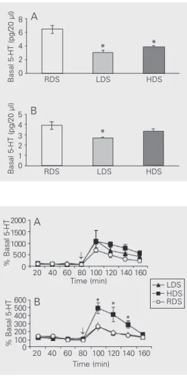
The means ± SEM of 5-HT basal concen-trations (pg/20 µl) for the rat lines are shown in Figure 1. One-way ANOVA showed that overall differences between lines for basal concentrations were highly significant for both prefrontal cortex [F(2,63) = 18.0, P<0.0001] and dorsal hippocampus [F(2,59) = 5.8, P<0.005]. Multiple comparison tests showed that basal levels for the prefrontal
Figure 1. Basal serotonin (5-HT) concentrations. Basal 5-HT con-centrations (mean ± SEM, pg/20 µl) of the high DPAT-sensitive (HDS), low DPAT-sensitive (LDS) and random DPAT-sensitive (RDS) lines in dialysates from the prefrontal cortex (A) and dor-sal hippocampus (B) obtained during the dark period for 5-6 animals/group. *P<0.05, com-pared with RDS (Newman-Keuls test).
Figure 2. The fenfluramine test. Effect of fenfluramine (10 mg/ kg, ip, arrow) on serotonin (5-HT) concentration in dialysates obtained during the dark period from the prefrontal cortex (A) and dorsal hippocampus (B) of rats selectively bred for high (HDS), low (LDS) and random (RDS) 8-OH-DPAT sensitivity. Data are reported as percentage (mean ± SEM) of basal concen-tration for 5-6 animals/group. *P<0.05, HDS compared with either LDS or RDS groups (Tukey test).
% Basal 5-HT
600 500 400 300 200
% Basal 5-HT
2000 1500 1000 500 0 100 0 LDS HDS RDS 20 40 60 80 100 120 140 160
Time (min)
20 40 60 80 100 120 140 160 Time (min)
* *
*
Basal 5-HT (pg/20 µl)
5 4 3 2 1 0
RDS LDS HDS
*
* *
RDS LDS HDS
Basal 5-HT (pg/20 µl)
cortex did not differ between HDS and LDS, whereas both lines had lower levels than RDS rats (Figure 1A). In contrast, basal con-centrations in the dorsal hippocampus only differed between the LDS and the RDS lines (Figure 1B).
Fenfluramine significantly increased ex-tracellular concentrations of 5-HT in both the prefrontal cortex [HDS, F(7,42) = 25.6, P<0.0001; LDS, F(7,35) = 4.7, P<0.01; RDS, F(7,35) = 33.9, P<0.0001] and dorsal hippo-campus [HDS, F(7,35) = 16.3, P<0.0001; LDS, F(7,35) = 4.8, P<0.01; RDS, F(7,35) = 9.8, P<0.0001]. The largest increase was observed in sample 5 (first sample after fen-fluramine injection) when compared with the four basal samples (post hoc test, P<0.05). In the dorsal hippocampus, the lines dif-fered in their response to fenfluramine [time x line factor, F(14,84) = 7.0, P<0.0001; line factor, F(2,12) = 20.9, P<0.0001] and the
post hoc test revealed that the HDS group had higher hippocampal 5-HT concentra-tions than either the LDS or RDS rats in three consecutive post-injection samples (see Fig-ure 2). In contrast, in the frontal cortex, the repeated measures test did not show signifi-cant line differences in response to fenflur-amine [time x line factor, F(14,91) = 1.0, NS; line factor, F(2,13) = 0.9, NS].
Discussion
The fact that basal 5-HT levels were lower in both selectively bred lines (HDS and LDS) compared with the RDS rats indicates that selection for either high or low 8-OH-DPAT sensitivity leads to decreased basal 5-HT levels in the hippocampus and frontal cor-tex. The basis for this lowered basal 5-HT release in both lines is not known, but might be related to the fact that both lines are more immobile than the RDS rats in the forced swim test (2). The lowered 5-HT levels can-not be related to 5-HT1A receptors or re-sponses to 8-OH-DPAT because HDS rats have greater cortical 5-HT1A binding and
greater hypothermic responses to 8-OH-DPAT than LDS rats (1,2).
The present finding of regional differ-ences in fenfluramine-induced release of 5-HT supports previous reports. It has been proposed that there are two anatomically and functionally distinct sets of serotonergic neurons projecting to the forebrain. These systems originate from separate nuclei in the brainstem, the dorsal (DRN) and median (MRN) raphe nuclei and project preferen-tially to the prefrontal cortex and dorsal hip-pocampus, respectively (10). There is evi-dence that these two systems differ in their pharmacological properties such as the vul-nerability to the toxic actions of amphet-amine derivatives (11). Furthermore, it has been suggested that fenfluramine has a greater acute effect on DRN than MRN terminals. Systemic administration of fenfluramine in-creased 5-HT concentrations in dialysates collected from the amygdala (a DRN termi-nal area) but not from the dorsal hippocam-pus (an MRN terminal area) (12). The in-crease in 5-HT observed in the three lines (HDS, LDS and RDS) following the fenflur-amine challenge was less in the dorsal hip-pocampus (about 350%) than in the prefron-tal cortex (about 900%). Thus, the present findings are consistent with the hypothesis that fenfluramine has a greater action on DRN than on MRN terminal areas.
than either LDS (248%) or RDS rats (243%). Therefore, it is possible that HDS rats have functional alterations of 5-HT reuptake sites in the hippocampus. The HDS rats have been reported to exhibit an abnormal re-sponse to 8-OH-DPAT following intrahip-pocampal administration. Unlike the LDS rats, which exhibit a typical anxiogenic
re-sponse in the social interaction test, the HDS rats do not (3). Thus, taken together, these results indicate that HDS rats exhibit abnor-mal responses following intrahippocampal administration of either 8-OH-DPAT or fen-fluramine due to functional alterations of 5-HT reuptake sites.
References
1. Knapp DJ, Overstreet DH & Crews FT (1998). Brain 5-HT1A receptor autoradiography and hypothermic responses in rats bred for differ-ences in 8-OH-DPAT sensitivity. Brain Research, 782: 1-10. 2. Overstreet DH, Rezvani AH, Knapp DJ, Crews FT & Janowsky DS
(1996). Further selection of rat lines differing in 5-HT-1A receptor sensitivity: behavioral and functional correlates. Psychiatric Genet-ics, 6: 107-117.
3. Gonzalez LE, File SE & Overstreet DH (1998). Selectively bred lines of rats differ in social interaction and hippocampal 5-HT1A receptor function: a link between anxiety and depression. Pharmacology, Biochemistry and Behavior, 59: 787-792.
4. File SE, Ouagazzal A-M, Gonzalez LE & Overstreet DH (1999). Chronic fluoxetine in tests of anxiety in rat lines selectively bred for differential 5-HT1A receptor function. Pharmacology, Biochemistry and Behavior, 62: 695-701.
5. Berger UV, Gu XF & Azmitia EC (1992). The substituted amphet-amines 3,4-methylenedioxymethamphetamine, methamphetamine, p-chloroamphetamine and fenfluramine induce 5-hydroxytryptamine release via a common mechanism blocked by fluoxetine and co-caine. European Journal of Pharmacology, 215: 153-160.
6. Bonanno G, Fassio A, Severi P, Ruelle A & Raiteri M (1994). Fenflur-amine releases serotonin from human brain nerve endings by a dual mechanism. Journal of Neurochemistry, 63: 1163-1166.
7. Poncet L, Denoroy L & Jouvet M (1993). Daily variations in in vivo
tryptophan hydroxylation and in the contents of serotonin and 5-hydroxyindoleacetic acid in discrete brain areas of the rat. Journal of Neural Transmission. General Section, 92: 137-150.
8. Hernandez L, Stanley BG & Hoebel BG (1986). A small, removable microdialysis probe. Life Sciences, 39: 2629-2637.
9. Paxinos G & Watson CH (1986). The Rat Brain in Stereotaxic Coordi-nates. Academic Press, New York, NY, USA.
10. Azmitia EC & Segal M (1978). An autoradiographic analysis of the differential ascending projections of the dorsal and median raphe nuclei in the rat. Journal of Comparative Neurology, 179: 641-667. 11. Mamounas LA, Mullen CA, O’Hearn E & Molliver ME (1991). Dual
serotoninergic projections to forebrain in the rat: morphologically distinct 5-HT axon terminals exhibit differential vulnerability to neu-rotoxic amphetamine derivatives. Journal of Comparative Neurol-ogy, 314: 558-586.
12. Viana MB, Silveira R & Graeff FG (1996). D-fenfluramine selectively releases 5-HT from dorsal raphe terminals. Brazilian Journal of Medi-cal and BiologiMedi-cal Research, 29: 639-642.