A Model of Post-Infection Fatigue Is
Associated with Increased TNF and 5-HT
2A
Receptor Expression in Mice
Yvonne Couch1*, Qin Xie1, Louise Lundberg1,2, Trevor Sharp1, Daniel C. Anthony1
1Department of Pharmacology, Mansfield Road, Oxford, OX1 3QT, United Kingdom,2Public Health England, Centre for Radiation, Chemical and Environmental Hazards, Chilton, Didcot, Oxford OX11 0RQ, United Kingdom
Abstract
It is well documented that serotonin (5-HT) plays an important role in psychiatric illness. For example, myalgic encephalomyelitis (ME/CFS), which is often provoked by infection, is a disabling illness with an unknown aetiology and diagnosis is based on symptom-specific criteria. However, 5-HT2Areceptor expression and peripheral cytokines are known to be
upregulated in ME. We sought to examine the relationship between the 5-HT system and cytokine expression following systemic bacterial endotoxin challenge (LPS, 0.5mg/kg i.p.), at a time when the acute sickness behaviours have largely resolved. At 24 hours post-injection mice exhibit no overt changes in locomotor behaviour, but do show increased immobility in a forced swim test, as well as decreased sucrose preference and reduced mar-ble burying activity, indicating a depressive-like state. While peripheral IDO activity was increased after LPS challenge, central activity levels remained stable and there was no change in total brain 5-HT levels or 5-HIAA/5-HT. However, within the brain, levels of TNF and 5-HT2Areceptor mRNA within various regions increased significantly. This increase in
receptor expression is reflected by an increase in the functional response of the 5-HT2A
receptor to agonist, DOI. These data suggest that regulation of fatigue and depressive-like moods after episodes of systemic inflammation may be regulated by changes in 5-HT receptor expression, rather than by levels of enzyme activity or cytokine expression in the CNS.
Introduction
Diseases such as myalgic encephalomyelitis/chronic fatigue syndrome (ME/CFS) are often pro-voked by infection and have been shown to be associated with altered expression of peripheral pro-inflammatory cytokines. Indeed, cytokine expression is higher in patients with ME/CFS than in individuals suffering from major depression, which is often argued to have a strong cytokine component [1]. The change in expression of 5-HT receptors, such as 5HT2A, is also a
feature of fatigue, which suggests that dysregulation of the serotonergic system may also play a
OPEN ACCESS
Citation:Couch Y, Xie Q, Lundberg L, Sharp T, Anthony DC (2015) A Model of Post-Infection Fatigue Is Associated with Increased TNF and 5-HT2A
Receptor Expression in Mice. PLoS ONE 10(7): e0130643. doi:10.1371/journal.pone.0130643
Editor:Yael Abreu-Villaça, Universidade do Estado do Rio de Janeiro, BRAZIL
Received:March 24, 2015
Accepted:May 21, 2015
Published:July 6, 2015
Copyright:© 2015 Couch et al. This is an open access article distributed under the terms of the
Creative Commons Attribution License, which permits unrestricted use, distribution, and reproduction in any medium, provided the original author and source are credited.
Data Availability Statement:All relevant data are within the paper and its Supporting Information files.
Funding:This work was supported by the Biology and Biotechnology Research Society (UK) in vivo additional studentship to YC/DA/TS.
role in post-infection-associated behaviours [2]. Post-infection ME/CFS-like behaviours can be modeled by the injection of bacterial endotoxin (LPS). The metabolic response and decreased motility that usually accompany LPS administration quickly resolve, but many depression-like behaviours persist beyond the acute phase. The molecular mechanisms that underlie these behavioural changes in both humans and animals are complex and poorly understood. A pop-ular theory argues that circulating cytokines such as tumor necrosis factor (TNF) and interleu-kin(IL)-1βincrease the activity of the enzyme indoleamine 2,3-dioxygenase (IDO) within the brain which reduces the availability of tryptophan, a precursor for 5-HT synthesis [3] and thus the overall levels of 5-HT in the CNS. However, the peripheral injection ofM.BovisBCG has
been shown to not alter the CNS kynurenine/tryptophan ratio [4], which is reported to be an indirect measure of IDO activity and therefore it is unclear whether this is solely a peripheral phenomenon, or whether IDO activity in the brain is important in regulating these behaviours.
An alternative hypothesis to the IDO theory is that the behaviours may be dependent on post-synaptic changes, rather than changes in 5-HT release or overall levels of 5-HT in the brain. We have previously shown that there is no significant decrease in brain 5-HT in a model of systemic inflammation [5], but that there is an increase in the expression certain 5-HT receptors in the CNS and that this change is related to a functional outcome. Animal models of depression also frequently show altered receptor expression levels, even when the genetic manipulation is in an unrelated gene. Pang and colleagues found altered levels of 5-HT1Band
5-HT2Areceptors in the hippocampus and cortex in a model of Huntington’s after observing
depression-like behaviour [6]. Both Maes [7] and Dantzer [8] suggest that sickness behaviour is an acute response, characterized by similar behavioural phenomenology as depression but with a pyretic component, and that behavioural responses persisting after 24 hours should be considered depression, rather than sickness.
Currently, the literature remains unclear as to whether the persistent behavioural changes that result from systemic inflammatory challenges are the result of systemic cytokines or CNS cytokines. Work studying the behavioural changes in response to inflammatory challenges often focuses on the role of cytokines in the CNS, usually TNF and IL-1β. There is significant evidence that both TNF and IL-1βplay important roles in modulating behaviour [9,10] but controversy still exists over the relative contributions of one cytokine vs the other [9]. We have demonstrated previously that there is significant up-regulation of TNF in the prefrontal cortex of animals defined as behaviourally‘depressed’in a model of chronic stress, and that this may be associated with changes in the 5-HT system [11].
With these facts in mind, we aimed to study LPS-induced fatigue symptoms in the mouse 24 hours after a systemic challenge, directly measuring tryptophan breakdown and 5-HT levels in the CNS, in addition to cytokine and 5-HT receptor expression, in order to establish whether there is a correlation between the 5-HT system and LPS-induced sickness behaviours. Our results suggest that some behavioural changes associated with CFS/ME-like depressive behav-iour may occur independently of CNS tryptophan levels but rather may be the result of changes in 5-HT receptor expression within the CNS.
Materials and Methods
Animals
Adult, outbred, male CD-1 mice (8 weeks) were obtained from Harlan (UK) and housed under a standard 12 hour light/dark cycle and provided with food/waterad libitum. Animals were
All procedures were carried out in accordance with the UK Animals (Scientific Procedures) Act, 1986. The protocols were carried out with approval of local ethical committees (University of Oxford, Clinical Medicine AWERB) and all efforts were made to minimise pain and suffer-ing. Animals were allowed to acclimatize to housing conditions for 1 week before any testing was carried out. Peripheral LPS challenge constituted one i.p. injection of LPS (026:B6; Sigma, UK) at 0.5mg/kg. All animals were healthy at the time of injection.
Behaviour
All testing was carried out between the hours of 10:00 and 16:00 of the light phase of the light/ dark cycle. Light levels within home cages were 5–8 Lux. In order to avoid differences in odour cues from one mouse to the other, a non-experimental mouse (of the same strain, sex and age) was allowed to explore the equipment for 10 min shortly before tests began. All behavioural testing, unless otherwise stated, was recorded on video and analysed post-hoc and blinded according to ARRIVE criteria (S1 ARRIVE Checklist) [12].
Elevated Plus Maze (EPM). 24 hours post injection mice were placed individually in the central square of an elevated plus-maze facing a closed arm, and behaviour was observed for 5 minutes. Time spent in the open arm, number of open arm entries, total arm entries and the latency to enter the first open arm were measured subsequently off-line from video recordings. An animal was considered to have entered an arm when all four legs were out of centre square. EPM testing was carried out during the light phase of the animals light:dark cycle at 350–400 Lux. However, it should be noted this test measures anxiety behaviours and is principally used to measure rodents’aversion to elevated and open spaces, the‘closed’arms have sides but no top and therefore are exposed to the same levels of light as the open arms when the light source is from above [13].
Open Field. Animals were subjected to open field testing as described previously [14,15] using an open-topped, black, rectangular box (50 × 30 × 30 cm) with the floor divided into 10 × 10 cm squares, under light levels of 350–400 Lux. Mice were placed individually in a cor-ner square, and the number of squares entered in a 3 min period was measured post-hoc on video recordings. The number of rears (both front paws off the ground, but not as part of grooming) were also counted. The latency for the mouse to leave (with all four feet) the corner square, and latency to the first rear, were also measured. As the open field is a large inescapable and novel environment, mice tend to show a preference towards the perimeter rather than the central area and thus measuring centre:perimeter ratio provides a relative metric of anxiety [16]. Sick animals also show reduced thigmotaxis during the acute phase of inflammation and it is therefore a good measure of general exploratory behaviour.
Forced Swim Test (FST). Mice were placed individually for 6 min into clear Perspex cyl-inders (height 25 cm, diameter 20 cm) containing 15 cm of water (23–25°C). The duration of immobility was recorded during the last 4 minutes of the 6 minute testing period. Water was changed after each test. A mouse was considered to be immobile when it floated in an upright position, and made only small movements to keep its head above the water. Climbing activity was considered when it used all its paws to reach the wall of the cylinder and tried to climb. The immobility, climbing and swimming time were scored blindly after the test. The FST shows both face and construct validity when measuring depression-like behaviours [17].
beginning of the test started with the onset of the dark phase of animals’cycle. To prevent the possible effects of side-preference in drinking behaviour, the position of the bottles in the cage was switched at 6 hours. No previous food or water deprivation was applied before the test. Percentage preference for sucrose is calculated using the following formula: Sucrose preference = Volume (Sucrose solution)/(Volume (Sucrose solution) + Volume (Water)) × 100. No naïve animals ever exhibited a preference for sucrose of<65% and, accordingly, mice
exhibiting a sucrose preference of<65% were defined as showing a depression-like phenotype.
This is in accordance with animal models of depression-like behaviour brought on by chronic stress [18,19].
Marble Burying. Mice were placed in clear plastic cages (25cm x 19cm) with a 5cm deep layer of sawdust on which was arranged 15 marbles in a 3 x 5 configuration [20]. Digging behaviour was filmed over 30 minutes noting latency to dig and number of marbles buried at intervals after starting recording. Marble burying shows stereotypical mouse digging behaviour and is often used to demonstrate anxiety, with an increase in marbles buried indicating an anx-ious state [20]. However, Thomas and colleagues [21] have suggested this is not the case, but rather that it reflects perseverative and repetitive behaviours.
(±)-1-(2,5-dimethoxy-4-iodophenyl)-2-aminopropane (DOI)-Induced Head-Twitch Behav-iour. Mice received a single dose of DOI (1mg/kg i.p.) and were observed for 20 minutes, during which time the number of head-twitches were counted as a measure of 5-HT2Aactivity [22].
5-HT and 5-HIAA HPLC
Animals were killed by overdose of anaesthetic (sodium pentobarbitone). Blood was removed via cardiac puncture for kynurenine analysis. This was followed by intracardial perfusion of ice-cold 0.9% heparinised-saline, and tissue was rapidly removed from specific CNS regions (cortex, hippocampus, striatum and cerebellum). Samples were analysed using HPLC with electrochemical detection and separated with an ACE column (C18, 3μm, 125 × 3mm + ACE
C18 guard, 10 × 3mm run at 35°C). Samples were carried in a mobile phase (12.5% methanol, 130mM NaH2PO4, 0.85mM Na2EDTA, 0.1mM 1-octanesulphonic acid, pH 3.55) pumped at
0.6ml/min (Waters 2695 HPLC Pump). Samples were detected using a glassy carbon electrode held at + 0.75 V (Dionex ED40). The sample content was determined with reference to daily-calibrated standard solutions in 0.06 M perchloric acid (5pmol 5-HT and 5-HIAA). Chromato-grams were displayed and analysed using Waters Empower 2 software.
IDO Activity
Animals were killed as above and tissue was rapidly removed from the same CNS regions as well as from the intestine, specifically a 1 inch portion of the proximal duodenum. Intestinal material was washed prior to homogenization with ice-cold saline. Tissue was weighed and homogenized in cold suspension buffer (250mM sucrose, 50mM Tris-HEPES (pH7.5), 0.2mM EDTA). Homogenate was centrifuged and supernatants analysed for IDO activity. Supernatant was combined 1:1 with incubation medium at 37°C (0.8mM TRP, 40mM ascorbin acid, 20μM
methylene blue, 200units/ml catalase in 100mM phosphate buffer) and agitated for 180 min-utes. Addition of 30% TCA stopped the reaction and incubation at 50°C for 30 minutes allowed N-formylkynurenine to convert to kynurenine. Samples were centrifuged at high speed and supernatants were analysed by HPLC for the presence of kynurenine.
Kynurenine HPLC
HPLC was performed as described by Zhanget al. [23]. Standard stocks of kynurenine and
or control plasma for calibration purposes. For CNS dialysates, samples (20μl) were analysed
without further treatment; plasma (50μl) was deproteinised with 10μl 26% perchloric acid,
centrifuged at 13,000 g for 10 min at 4°C, and 20μl used for analysis. Concentrations were
determined by HPLC with absorbance detection, using an ACE C18 column, 3μm, 150 x 3 mm
and eluent 5% acetonitrile, 15 mM potassium acetate, pH 4.0, flow rate 0.5 ml/min, using a diode array absorbance detector monitored at 360 nm (kynurenine). Time during the initial incubation phase and initial weight of tissue were taken into account and final data was pre-sented as the concentration of kynurenine produced (pmol) per mg of tissue per hour.
RNA Extraction and Quantitative RT-PCR
RNA extraction was performed as previously described from specifically microdissected snap-frozen brain regions [11,24]. Specific primers were designed by PrimerDesign as follows: TNF-F GCCTCCCTCTCATCAGTTCTAT; TNF-R TTTGCTACGACGTGGGCTA; IL-1β-F CAACCAACAAGTGATATTCTCCAT; IL-1β-R GGGTGTGCCGTCTTTCATTA; 5-HT2A-F
CAGGCAAGTCACAGGATAGC; 5-HT2A-R TTAAGCAGAAAGAAAATCCCACAG;
5-HTT-F TGCCTTTTATATCGCCTCCTAC; 5-HTT-R CAGTTGCCAGTGTTCCAAGA; IDO-F TGCTTACTCTCTTTTCCCTTCC; IDO-R CATCAGACCTGGTGCTTCA. Quantita-tive RT-PCR was run using SYBR green based technology (Primer Design Ltd.). Results are expressed as relative-fold expression compared to control animals and corrected to the house-keeping gene, glyceraldehyde 3-phosphate dehydrogenase (GAPDH).
Statistics
Statistical analysis were performed using GraphPad Prism 5.0 and InVivoStat software using two-way ANOVA and Student’s t-tests, and appropriate post-hoc analysis, as described in the text. Data are presented as mean ±SEM.
Results
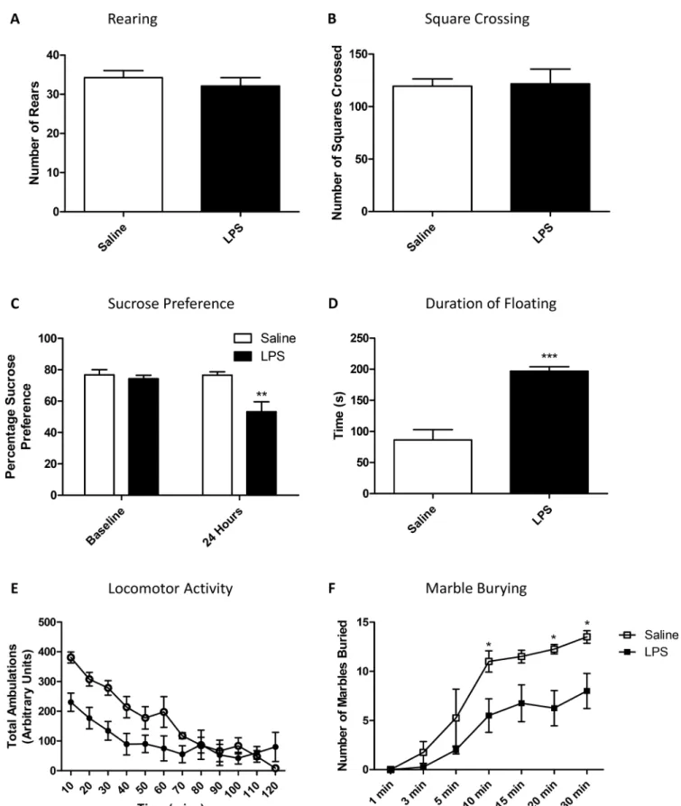
LPS causes depression-like behaviours at 24 hours
To determine whether basic exploratory and mood-related behaviours were altered 24 hours after an LPS challenge, animals were subjected to a standard 3-minute open field test, as well as a longer locomotor activity test, to examine both exploratory and anxiety-type behaviours. Open field behaviour was not different in terms of either rearing behaviour (Fig 1A) or distance travelled (Fig 1B) during a 3-minute window. During a longer 2 hour locomotor activity task, all animals slowed down over time, and there was an interaction between LPS-challenge and time but no main effect of LPS (RM-ANOVA time p<0.001 F11,110= 26.90; LPS p = 0.07
F1,110= 3.85; LPS:time p<0.001 F11,110= 5.09;Fig 1E). It should be noted that this dose of LPS
is capable of inducing sickness behaviours at an acute time pointS1andS2Figs) as well as a hepatic acute phase response (S2 Fig). It has been previously shown that peripheral inflamma-tion can cause depression like behaviour in mice [25]. Here, 24 hours after a single LPS chal-lenge, mice showed significant changes in sucrose preference, forced swim behaviours and marble burying. Specifically, after 24 hours LPS-treated animals showed a decrease in sucrose preference to<65% showing main effects of both time after testing and LPS, as well as an
inter-action (RM-ANOVA time p<0.05 F1,6= 6.51; LPS p<0.01 F1,6= 13.21; LPS:time p<0.05 F1,6=
6.30;Fig 1C). Bonferroni post-hoc tests showed a decrease in sucrose preference in LPS treated animals, compared to saline treated animals (p<0.01). In the forced swim test, floating
behav-iour reached almost twice saline treated levels in animals receiving LPS (p<0.001;Fig 1D).
Fig 1. Open field and locomotor activity in saline and LPS treated animals at 24 hours.Animals received a single dose of LPS (0.5 mg/kg) or vehicle 24 hours prior to testing, and were tested using a standard 3-minute open field measuring rearing (A) and square crossing (B). Animals were also tested using a two-bottle sucrose preference test (C) and a forced swim test studying duration of floating behaviour (D), as well as a longer 2 hour locomotor activity study (E) and marble burying test (F). Data are mean±SEM n = 6;*p<0.05,**p<0.01 and***p<0.001 compared to saline injected controls.
LPS treated animals (RM-ANOVA time p<0.001 F6,36= 33.69; LPS p<0.05 F1,36= 8.71; LPS:
time p = 0.053 F6,36= 2.32;Fig 1F) with post-hoc analysis showing significant differences
between groups after 10 minutes of testing (Bonferroni post-hoc p<0.05). It should be noted
that there was no impairment in the animals’overall levels of locomotor activity in this test (p = 0.93;S3 Fig).
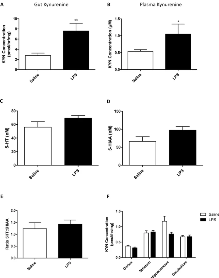
Systemic Inflammation does not cause changes in CNS 5-HT levels
Systemic inflammation is known to alter the activity of IDO, which is strongly expressed in the gut and the CNS. This enzyme is known to metabolize tryptophan and thus reduce its availabil-ity for 5-HT synthesis. In order to determine whether inflammation affected IDO activavailabil-ity, and as a consequence, 5-HT and kynurenine levels in the CNS, all of the above were measured in plasma, CNS and gut. At 24 hours after an LPS challenge kynurenine concentrations in the gut were significantly increased (Student’s t-test p<0.01;Fig 2A). Concomitantly, plasma levels of
kynurenine also increased in LPS-treated animals (p<0.05;Fig 3B). Total brain 5-HT, and its
metabolite 5-HIAA were assessed by HPLC 24 hours after a single LPS injection (0.5mg/kg) and a ratio of 5-HT:5-HIAA was used to assess turnover (5-HIAA/5-HT). There was no signifi-cant change in either 5-HT or 5-HIAA (Student’s t-test p = 0.16 and 0.10, respectively;Fig 2C and 2D), and also no change in the ratio of 5-HT to 5-HIAA (p = 0.5;Fig 2E). Furthermore, kynurenine levels within specific regions were investigated to determine whether the immune axis within the brain had any effects on tryptophan breakdown. LPS and the region studied both had significant effects on kynurenine accumulation (two-way ANOVA region p<0.001
F4,50= 25.71; LPS p<0.05 F1,50= 4.15; LPS:region p<0.05 F4,50= 3.82;Fig 4D), however,
post-hoc analysis revealed no significant effect of LPS in any brain region (Bonferroni p>0.05 in all
cases). The lack of change in IDO related activities was backed up by no change in IDO mRNA expression after an LPS challenge, even at acute time points where changes are likely to occur (S4 Fig).
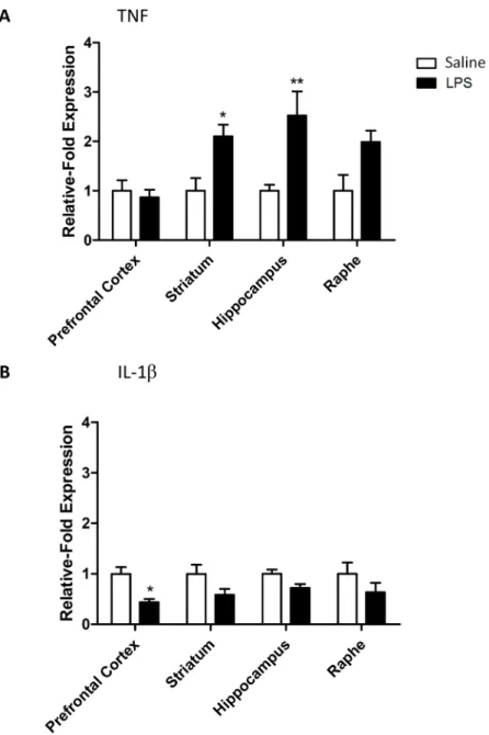
LPS increases TNF mRNA in the CNS at 24 hours
Bacterial LPS is known to cause a number of systemic effects, including changes in body tem-perature, which are largely resolved by 24 hours [26]. We wished to determine whether the same was the case for CNS inflammation caused by a systemic challenge. TNF mRNA expres-sion was up-regulated in the striatum, hippocampus and raphe (RM-ANOVA brain region p<0.05 F3,24= 3.34; LPS p<0.001 F1,24= 20.30; LPS:brain region p<0.05 F3,24= 3.34;Fig 3A)
with significant increases in the striatum and hippocampus (Bonferroni post-hoc p<0.05 and
p<0.01, respectively). Conversely, IL-1βexpression was not increased in any brain region but rather fell in the prefrontal cortex (RM-ANOVA brain region p = 0.7 F3,24= 0.36; LPS p<0.001
F1,24= 16.67; LPS:brain region p = 0.7 F3,24= 0.36;Fig 3B).
Systemic inflammation increases 5-HT
2Areceptor expression and
function
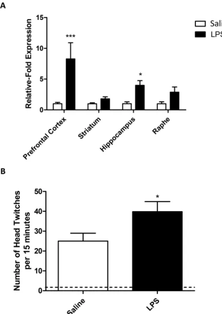
After determining that LPS induces behavioural effects and an increase in central TNF expres-sion, we decided to establish whether the expression of specific 5-HT receptors was altered by peripheral inflammation. 5-HT2Areceptor mRNA expression was increased after exposure to a
systemic inflammatory challenge and was affected in all brain regions (RM-ANOVA brain region p<0.05 F3,24= 3.77; LPS p<0.001 F1,24= 19.54; LPS:brain region p<0.05 F3,24= 3.77;
Fig 4A). 5-HT2Aexpression in the pre-frontal cortex and hippocampus was significantly
increased in LPS treated animals compared to controls (Bonferroni post-hoc p<0.001 and
systemic inflammatory challenge, we therefore decided to find out whether this had an effect on 5-HT2A-mediated behaviours. (±)-1-(2,5-dimethoxy-4-iodophenyl)-2-aminopropane
(DOI) induces a head-twitch response in mice after systemic exposure, and this occurs in a 5-HT2A-dependent manner [22]. DOI had a significant effect on head-twitch behaviours in
animals treated with both saline and LPS, however, this was elevated in animals receiving a sin-gle dose of LPS 24 hours before, compared to those receiving DOI alone (p<0.05;Fig 4B).
assessed for 5-HT (C); 5-HIAA (D); and the ratio of 5-HT:5-HIAA (E) as well as kynurenine production by IDO in different brain regions (F). Data are mean
±SEM n = 6;*p<0.05;**p<0.01 and***p<0.001 compared to saline injected controls.
doi:10.1371/journal.pone.0130643.g002
Fig 3. Cytokine mRNA expression in the CNS 24 hours after an LPS challenge.(A) TNF and (B) IL-1β
transporter mRNA levels expressed as relative-fold expression compared to saline controls within each region. mRNA expression is normalized to the housekeeping gene GAPDH prior to analysis. Data are mean
±SEM n = 6;*p<0.05 and**p<0.01 compared to saline injected controls.
Discussion
The acute phase of LPS-induced sickness behaviour is associated with a febrile response, decreased locomotor behaviour and a general reduction in activity. In the next phase of recov-ery from sickness, usually after 24 hours, animals continue to display depression-like behav-iours and fatigue. Here, we sought to characterize the changes in expression of 5-HT related genes associated with these behavioural changes. Together, the data suggest the novel idea that systemic inflammation may alter behaviour by changing the expression of 5-HT receptors,
Fig 4. 5-HT2Areceptor changes after LPS.mRNA expression in the CNS 24 hours after an LPS challenge
(A), mRNA levels are expressed as relative-fold expression compared to saline controls within each region. mRNA expression is normalized to the housekeeping gene GAPDH prior to analysis. LPS-induced changes in 5-HT2A-mediated head-twitch responses (B). Animals received a single dose of LPS (0.5mg/kg i.p.) and
were allowed to recover for 24 hours prior to receiving a single dose of DOI (1mg/kg i.p.). Head twitch behaviours were observed over a period of 15 minutes. Dotted line represents baseline (no DOI) data for both LPS and saline treated animals. Data are mean±SEM n = 6;*p<0.05,**p<0.01 and***p<0.001 compared to saline injected controls.
rather than by reducing the availability of 5-HT in the CNS, which is consistent with observa-tions in CFS/ME and depressed patient cohorts [2,27,28].
Depression-like behaviour is often evaluated using the sucrose preference test, with depres-sive anhedonia indicated by a low preference for sweet drinking solutions, as well as decreased activity in the forced swim test [29]. The forced swim test, however, can pose ethical problems when combined with sickness. Introducing rodents to a wet environment while they may be suffering from hypothermia might be considered unnecessary. Here, we have demonstrated that the marble burying test effectively demonstrates reduced activity, which is not associated with reduced locomotor behaviour, without the need to expose the animals to an aversive envi-ronment. Considering the issues that patients with mood problems have with initiation and perseverance tasks this may be significant [30].
It is well known that depression and depressive-like behaviours are associated with higher levels of circulating cytokines [31–35]. However, the causal nature of this relationship is cur-rently speculative. Here, we have introduced the circulating cytokines with a systemic immune challenge and therefore any changes in behaviour are likely to be due to the increased levels of inflammation, rather than the other way around. The IDO theory postulates that pro-inflam-matory cytokines up-regulate the activity of IDO and therefore deplete the levels of tryptophan available for making 5-HT [36]. Perhaps the most surprising result therefore, was that brain tis-sue levels of tryptophan breakdown to kynurenine and the levels of 5-HT and 5-HT metabo-lites were unaltered in the depressive-like late phase of sickness (>24 hours) in these animals.
The role of IDO in the breakdown of tryptophan suggests that any circulating factors altering the activity of this enzyme, should alter overall 5-HT metabolism [37–39]. It is clear from the work here that peripheral IDO activity is increased by systemic inflammation and this is reflected in the increased IDO activity in the gut and the increased levels of circulating kynure-nine. Others have demonstrated similarly large increases in the periphery [40], however, increases of this magnitude are rarely reflected by similarly large increases in the CNS. These data suggest that while the tryptophan/kynurenine pathway may be involved in inflammation-associated behavioural changes, this is likely to be an entirely peripheral phenomenon. The low n number used here may bias the HPLC results towards a type II error, however, the changes in behaviour and receptor expression were significant after LPS and therefore an increase in n to investigate the peripheral phenomenon theory was beyond the scope of this report. Indeed, a study by Hughes and colleagues [41] has suggested that changes in tryptophan occur indepen-dently of changes in IDO and inflammatory cytokines, so it is possible that the alteration in IDO activity has other functions that are unrelated to depression-like behaviours.
the IL-1 receptor antagonist mimic. With studies in depressed patients beginning to investigate the potential of inhibiting cytokines in treatment-resistant depression, there is a clear positive role for cytokines such as TNF in the regulation of mood [46]. However, the mechanisms by which TNF mediates changes in behaviour, and its potential role in both sickness and depres-sion, are yet to be explored.
Perhaps the most commanding results here are the changes we have observed in the 5-HT system. In this study, treatment with LPS caused a significant increase in 5-HT2Areceptor
expression in a number of discrete brain regions. We have also demonstrated that this results in a functional outcome, i.e. there is a significant alteration in 5-HT2A-mediated behaviours.
This is in accord with our previous studies on acute sickness, where increases in 5-HT2A
recep-tor expression were also associated with functional changes [24]. Our data also indicate that these changes are receptor specific, rather than a general change in 5-HT receptor expressing cells, as we do not see similar changes in other 5-HT receptors, such as 5-HT1A(S5 Fig). The
involvement of the 5-HT2Areceptor in mood is currently controversial. Studies from the late
80s indicate that receptor expression increases in patients with suicidal depression [47–49], with further data suggesting that this increase may be non-neuronal [50]. In terms of inflam-mation, and as a consequence sickness behaviour, increases in 5-HT2Areceptor expression
have been seen in models of inflammatory pain [51], as well as in other studies using LPS [5] indicating that systemic inflammation is capable of regulating this responsein vivo. The
mech-anisms by which changes in receptor expression occur were beyond the scope of this study; however, hypotheses can be generated based on existing research. Studies linking the 5-HT2A
receptor with inflammation have suggested that its activation may result in the down-regula-tion of inflammatory cytokines [52], specifically TNF expression [53]. Furthermore, studies in cultured glioma cells have demonstrated an inhibition of inducible nitric oxide synthase (iNOS) by 5-HT2Aagonists [54]. Work by Zhang and colleagues [51] demonstrated that the
up-regulation of 5-HT2Aafter inflammation was not co-localized with 5-HT, indicating that it
was not in serotonergic neurons. Furthermore, 5-HT2Areceptors have been shown to be
pres-ent on astrocytes and microglia [55]. Therefore, increased 5-HT2Areceptor expression after a
peripheral inflammatory stimulus could potentially occur in non-neuronal cells for the pur-poses of potentiating an anti-inflammatory response in the CNS.
Overall, this study has explored the association between late phase LPS-induced depression-like behaviour and the expression of 5-HT related genes. The data presented here demonstrate that inflammation does not appear to regulate CNS tryptophan levels, but rather may mediate depression-like behaviours by altering the expression and function of CNS 5-HT receptors. This could significantly affect the direction of future research into inflammation associated fatigue and mood disorders, and argues for the use of selective 5-HT receptor agonists and antagonists in their treatment.
Supporting Information
S1 ARRIVE Checklist. The ARRIVE (Animal Research: Reporting of In Vivo Experiments) guidelines were developed as part of an NC3Rs initiative to improve the design, analysis and reporting of research using animals–maximising information published and
minimis-ing unnecessary studies.This checklist confirms this study adhered to ARRIVE guidelines (S1 ARRIVE Checklist).
(PDF)
S1 Fig. Open field and locomotor activity in in saline and LPS treated animals at 24 hours.
as in a longer 2 hour locomotor activity test (C). Data are mean ±SEM n = 6. (PDF)
S2 Fig. TNF mRNA expression in the liver 6 hours after an LPS challenge, mRNA levels expressed as relative-fold expression compared to saline controls.mRNA expression is nor-malized to the housekeeping gene GAPDH prior to analysis. Data are mean ±SEM n = 6;
p<0.001 compared to saline injected controls.
(PDF)
S3 Fig. General locomotor activity in naïve, saline and LPS treated animals at 24 hours dur-ing marble burydur-ing.Animals received a single dose of LPS (0.5 mg/kg) or vehicle 24 hours prior to testing, and were tested in the marble burying field. Total locomotor activity within the field was assessed as number of arbitrary squares crossed during allotted time periods. A signif-icant increase in squares over time (p<0.001) was found but there was no difference between
groups (p = 0.93). Data are mean ±SEM n = 6. (PDF)
S4 Fig. IDO mRNA expression in the CNS at 6 and 24 hours after an LPS challenge, mRNA levels expressed as relative-fold expression compared to saline controls.mRNA expression is normalized to the housekeeping gene GAPDH prior to analysis. Data are mean ±SEM n = 6. (PDF)
S5 Fig. 5-HT1AmRNA expression in the CNS at 6 hours after an LPS challenge, mRNA
lev-els expressed as relative-fold expression compared to saline controls.mRNA expression is normalized to the housekeeping gene GAPDH prior to analysis. Data are mean ±SEM n = 6. (PDF)
Acknowledgments
The authors would like to thank Dr Michael Stratford (CRUK/MRC Oxford Institute for Radi-ation Oncology and Biology) for assistance with running HPLC and analysis.
Author Contributions
Conceived and designed the experiments: YC QX TS DA. Performed the experiments: YC QX LL. Analyzed the data: YC QX LL. Contributed reagents/materials/analysis tools: YC QX LL. Wrote the paper: YC LL DA.
References
1. Maes M, Twisk FN, Ringel K. Inflammatory and cell-mediated immune biomarkers in myalgic encepha-lomyelitis/chronic fatigue syndrome and depression: inflammatory markers are higher in myalgic encephalomyelitis/chronic fatigue syndrome than in depression. Psychotherapy and psychosomatics. 2012; 81(5):286–95. Epub 2012/07/27. doi:10.1159/000336803PMID:22832503.
2. Smith AK, Dimulescu I, Falkenberg VR, Narasimhan S, Heim C, Vernon SD, et al. Genetic evaluation of the serotonergic system in chronic fatigue syndrome. Psychoneuroendocrinology. 2008; 33(2):188–
97. Epub 2007/12/15. doi:10.1016/j.psyneuen.2007.11.001PMID:18079067.
3. Maes M, Mihaylova I, Ruyter MD, Kubera M, Bosmans E. The immune effects of TRYCATs (tryptophan catabolites along the IDO pathway): relevance for depression—and other conditions characterized by
tryptophan depletion induced by inflammation. Neuro Endocrinol Lett. 2007; 28(6):826–31. Epub 2007/
12/08. doi: NEL280607A11 [pii]. PMID:18063923.
4. O'Connor JC, Lawson MA, Andre C, Briley EM, Szegedi SS, Lestage J, et al. Induction of IDO by bacille Calmette-Guerin is responsible for development of murine depressive-like behavior. J Immunol. 2009; 182(5):3202–12. Epub 2009/02/24. doi: 182/5/3202 [pii] doi:10.4049/jimmunol.0802722PMID:
5. Couch Y, Martin CJ, Howarth C, Raley J, Khrapitchev AA, Stratford M, et al. Systemic inflammation alters central 5-HT function as determined by pharmacological MRI. Neuroimage. 2013; 75:177–86.
Epub 2013/03/12. doi:10.1016/j.neuroimage.2013.02.046PMID:23473937.
6. Pang TY, Du X, Zajac MS, Howard ML, Hannan AJ. Altered serotonin receptor expression is associated with depression-related behavior in the R6/1 transgenic mouse model of Huntington's disease. Hum Mol Genet. 2009; 18(4):753–66. Epub 2008/11/15. doi: ddn385 [pii] doi:10.1093/hmg/ddn385PMID:
19008301.
7. Maes M, Berk M, Goehler L, Song C, Anderson G, Galecki P, et al. Depression and sickness behavior are Janus-faced responses to shared inflammatory pathways. BMC Med. 2012; 10:66. Epub 2012/07/ 04. doi:10.1186/1741-7015-10-66PMID:22747645; PubMed Central PMCID: PMC3391987.
8. Dantzer R, Kelley KW. Twenty years of research on cytokine-induced sickness behavior. Brain Behav Immun. 2007; 21(2):153–60. Epub 2006/11/08. doi:10.1016/j.bbi.2006.09.006PMID:17088043;
PubMed Central PMCID: PMC1850954.
9. Bluthe RM, Dantzer R, Kelley KW. Interleukin-1 mediates behavioural but not metabolic effects of tumor necrosis factor alpha in mice. Eur J Pharmacol. 1991; 209(3):281–3. Epub 1991/12/17. PMID:
1839150.
10. Jiang Y, Deacon R, Anthony DC, Campbell SJ. Inhibition of peripheral TNF can block the malaise asso-ciated with CNS inflammatory diseases. Neurobiol Dis. 2008; 32(1):125–32. Epub 2008/08/02. doi:
S0969-9961(08)00140-X [pii] doi:10.1016/j.nbd.2008.06.017PMID:18672064.
11. Couch Y, Anthony DC, Dolgov O, Revischin A, Festoff B, Santos AI, et al. Microglial activation, increased TNF and SERT expression in the prefrontal cortex define stress-altered behaviour in mice susceptible to anhedonia. Brain Behav Immun. 2013; 29:136–46. Epub 2013/01/12. doi:10.1016/j.bbi.
2012.12.017PMID:23305936.
12. Kilkenny C, Browne WJ, Cuthill IC, Emerson M, Altman DG. Improving bioscience research reporting: the ARRIVE guidelines for reporting animal research. PLoS biology. 2010; 8(6):e1000412. Epub 2010/ 07/09. doi:10.1371/journal.pbio.1000412PMID:20613859; PubMed Central PMCID: PMC2893951.
13. Walf AA, Frye CA. The use of the elevated plus maze as an assay of anxiety-related behavior in rodents. Nature protocols. 2007; 2(2):322–8. Epub 2007/04/05. doi:10.1038/nprot.2007.44PMID:
17406592; PubMed Central PMCID: PMC3623971.
14. Hale MW, Hay-Schmidt A, Mikkelsen JD, Poulsen B, Bouwknecht JA, Evans AK, et al. Exposure to an open-field arena increases c-Fos expression in a subpopulation of neurons in the dorsal raphe nucleus, including neurons projecting to the basolateral amygdaloid complex. Neuroscience. 2008; 157(4):733–
48. PMID:18951955. doi:10.1016/j.neuroscience.2008.09.050
15. Jennings KA, Loder MK, Sheward WJ, Pei Q, Deacon RM, Benson MA, et al. Increased expression of the 5-HT transporter confers a low-anxiety phenotype linked to decreased 5-HT transmission. J Neu-rosci. 2006; 26(35):8955–64. Epub 2006/09/01. doi:10.1523/JNEUROSCI.5356-05.2006PMID:
16943551.
16. Prut L, Belzung C. The open field as a paradigm to measure the effects of drugs on anxiety-like behav-iors: a review. Eur J Pharmacol. 2003; 463(1–3):3–33. Epub 2003/02/26. PMID:12600700.
17. Cryan JF, Valentino RJ, Lucki I. Assessing substrates underlying the behavioral effects of antidepres-sants using the modified rat forced swimming test. Neurosci Biobehav Rev. 2005; 29(4–5):547–69.
Epub 2005/05/17. doi:10.1016/j.neubiorev.2005.03.008PMID:15893822.
18. Willner P, Towell A, Sampson D, Sophokleous S, Muscat R. Reduction of sucrose preference by chronic unpredictable mild stress, and its restoration by a tricyclic antidepressant. Psychopharmacol-ogy (Berl). 1987; 93(3):358–64. Epub 1987/01/01. PMID:3124165.
19. Strekalova T, Couch Y, Kholod N, Boyks M, Malin D, Leprince P, et al. Update in the methodology of the chronic stress paradigm: internal control matters. Behav Brain Funct. 2011; 7:9. Epub 2011/04/29. doi: 1744-9081-7-9 [pii] doi:10.1186/1744-9081-7-9PMID:21524310; PubMed Central PMCID: PMC3111355.
20. Deacon RM. Digging and marble burying in mice: simple methods for in vivo identification of biological impacts. Nature protocols. 2006; 1(1):122–4. Epub 2007/04/05. doi:10.1038/nprot.2006.20PMID:
17406223.
21. Thomas A, Burant A, Bui N, Graham D, Yuva-Paylor LA, Paylor R. Marble burying reflects a repetitive and perseverative behavior more than novelty-induced anxiety. Psychopharmacology (Berl). 2009; 204 (2):361–73. Epub 2009/02/04. doi:10.1007/s00213-009-1466-yPMID:19189082; PubMed Central
PMCID: PMC2899706.
22. Gonzalez-Maeso J, Weisstaub NV, Zhou M, Chan P, Ivic L, Ang R, et al. Hallucinogens recruit specific cortical 5-HT(2A) receptor-mediated signaling pathways to affect behavior. Neuron. 2007; 53(3):439–
23. Zhang X, He Y, Ding M. Simultaneous determination of tryptophan and kynurenine in plasma samples of children patients with Kawasaki disease by high-performance liquid chromatography with pro-grammed wavelength ultraviolet detection. Journal of chromatography B, Analytical technologies in the biomedical and life sciences. 2009; 877(16–17):1678–82. Epub 2009/04/28. doi:10.1016/j.jchromb.
2009.04.013PMID:19394282.
24. Couch Y, Martin CJ, Howarth C, Raley J, Khrapitchev AA, Stratford M, et al. Systemic inflammation alters central 5-HT function as determined by pharmacological MRI. Neuroimage. 2013; 75C:185–94.
Epub 2013/03/12. doi:10.1016/j.neuroimage.2013.02.046PMID:23473937.
25. Pitychoutis PM, Nakamura K, Tsonis PA, Papadopoulou-Daifoti Z. Neurochemical and behavioral alter-ations in an inflammatory model of depression: sex differences exposed. Neuroscience. 2009; 159 (4):1216–32. PMID:19409213. doi:10.1016/j.neuroscience.2009.01.072
26. Cunningham C, Campion S, Lunnon K, Murray CL, Woods JF, Deacon RM, et al. Systemic inflamma-tion induces acute behavioral and cognitive changes and accelerates neurodegenerative disease. Biol Psychiatry. 2009; 65(4):304–12. Epub 2008/09/20. doi: S0006-3223(08)00895-0 [pii] doi:10.1016/j.
biopsych.2008.07.024PMID:18801476; PubMed Central PMCID: PMC2633437.
27. Benedetti F, Barbini B, Bernasconi A, Fulgosi MC, Colombo C, Dallaspezia S, et al. Serotonin 5-HT2A receptor gene variants influence antidepressant response to repeated total sleep deprivation in bipolar depression. Prog Neuropsychopharmacol Biol Psychiatry. 2008; 32(8):1863–6. Epub 2008/09/20. doi:
10.1016/j.pnpbp.2008.08.017PMID:18801406.
28. Bhagwagar Z, Hinz R, Taylor M, Fancy S, Cowen P, Grasby P. Increased 5-HT(2A) receptor binding in euthymic, medication-free patients recovered from depression: a positron emission study with [(11)C] MDL 100,907. Am J Psychiatry. 2006; 163(9):1580–7. Epub 2006/09/02. doi:10.1176/appi.ajp.163.9.
1580PMID:16946184.
29. Slattery DA, Cryan JF. Using the rat forced swim test to assess antidepressant-like activity in rodents. Nature protocols. 2012; 7(6):1009–14. Epub 2012/05/05. doi:10.1038/nprot.2012.044PMID:
22555240.
30. Murphy CF, Alexopoulos GS. Longitudinal association of initiation/perseveration and severity of geriat-ric depression. Am J Geriatr Psychiatry. 2004; 12(1):50–6. Epub 2004/01/20. PMID:14729559. 31. Alesci S, Martinez PE, Kelkar S, Ilias I, Ronsaville DS, Listwak SJ, et al. Major depression is associated
with significant diurnal elevations in plasma interleukin-6 levels, a shift of its circadian rhythm, and loss of physiological complexity in its secretion: clinical implications. J Clin Endocrinol Metab. 2005; 90 (5):2522–30. PMID:15705924.
32. Capuron L, Gumnick JF, Musselman DL, Lawson DH, Reemsnyder A, Nemeroff CB, et al. Neurobeha-vioral effects of interferon-alpha in cancer patients: phenomenology and paroxetine responsiveness of symptom dimensions. Neuropsychopharmacology. 2002; 26(5):643–52. Epub 2002/04/03. doi:10.
1016/S0893-133X(01)00407-9PMID:11927189.
33. Levine J, Barak Y, Chengappa KN, Rapoport A, Rebey M, Barak V. Cerebrospinal cytokine levels in patients with acute depression. Neuropsychobiology. 1999; 40(4):171–6. PMID:10559698.
34. Maes M, Vandoolaeghe E, Ranjan R, Bosmans E, Bergmans R, Desnyder R. Increased serum interleu-kin-1-receptor-antagonist concentrations in major depression. J Affect Disord. 1995; 36(1–2):29–36.
Epub 1995/12/24. PMID:8988262.
35. Maes M, Bosmans E, De Jongh R, Kenis G, Vandoolaeghe E, Neels H. Increased serum IL-6 and IL-1 receptor antagonist concentrations in major depression and treatment resistant depression. Cytokine. 1997; 9(11):853–8. Epub 1998/02/07. doi:10.1006/cyto.1997.0238PMID:9367546.
36. O'Connor JC, Lawson MA, Andre C, Moreau M, Lestage J, Castanon N, et al. Lipopolysaccharide-induced depressive-like behavior is mediated by indoleamine 2,3-dioxygenase activation in mice. Mol Psychiatry. 2009; 14(5):511–22. PMID:18195714. doi:10.1038/sj.mp.4002148
37. Barton DA, Esler MD, Dawood T, Lambert EA, Haikerwal D, Brenchley C, et al. Elevated brain seroto-nin turnover in patients with depression: effect of genotype and therapy. Arch Gen Psychiatry. 2008; 65 (1):38–46. Epub 2008/01/09. doi: 65/1/38 [pii] doi:10.1001/archgenpsychiatry.2007.11PMID:
18180427.
38. Gjerris A, Sorensen AS, Rafaelsen OJ, Werdelin L, Alling C, Linnoila M. 5-HT and 5-HIAA in cerebrospi-nal fluid in depression. J Affect Disord. 1987; 12(1):13–22. Epub 1987/01/01. PMID:2437171. 39. Sullivan GM, Oquendo MA, Huang YY, Mann JJ. Elevated cerebrospinal fluid 5-hydroxyindoleacetic
acid levels in women with comorbid depression and panic disorder. Int J Neuropsychopharmacol. 2006; 9(5):547–56. Epub 2005/11/02. doi:10.1017/S1461145705006231PMID:16259647.
41. Hughes MM, Carballedo A, McLoughlin DM, Amico F, Harkin A, Frodl T, et al. Tryptophan depletion in depressed patients occurs independent of kynurenine pathway activation. Brain Behav Immun. 2012; 26(6):979–87. Epub 2012/06/12. doi:10.1016/j.bbi.2012.05.010PMID:22683764.
42. Bluthe RM, Laye S, Michaud B, Combe C, Dantzer R, Parnet P. Role of interleukin-1beta and tumour necrosis factor-alpha in lipopolysaccharide-induced sickness behaviour: a study with interleukin-1 type I receptor-deficient mice. Eur J Neurosci. 2000; 12(12):4447–56. Epub 2000/12/21. PMID:11122355. 43. Cadusseau J, Ragunathan-Thangarajah N, Surenaud M, Hue S, Authier FJ, Gherardi RK. Selective
elevation of circulating CCL2/MCP1 levels in patients with longstanding post-vaccinal macrophagic myofasciitis and ASIA. Curr Med Chem. 2014; 21(4):511–7. Epub 2013/10/03. PMID:24083602. 44. Simen BB, Duman CH, Simen AA, Duman RS. TNFalpha signaling in depression and anxiety:
behav-ioral consequences of individual receptor targeting. Biol Psychiatry. 2006; 59(9):775–85. Epub 2006/
02/07. doi:10.1016/j.biopsych.2005.10.013PMID:16458261.
45. Tyring S, Gottlieb A, Papp K, Gordon K, Leonardi C, Wang A, et al. Etanercept and clinical outcomes, fatigue, and depression in psoriasis: double-blind placebo-controlled randomised phase III trial. Lancet. 2006; 367(9504):29–35. Epub 2006/01/10. doi:10.1016/S0140-6736(05)67763-XPMID:16399150. 46. Miyaoka T, Wake R, Furuya M, Liaury K, Ieda M, Kawakami K, et al. Minocycline as adjunctive therapy
for patients with unipolar psychotic depression: an open-label study. Prog Neuropsychopharmacol Biol Psychiatry. 2012; 37(2):222–6. Epub 2012/02/22. doi:10.1016/j.pnpbp.2012.02.002PMID:22349578. 47. Stanley M, Mann JJ. Increased serotonin-2 binding sites in frontal cortex of suicide victims. Lancet.
1983; 1(8318):214–6. Epub 1983/01/29. doi: S0140-6736(83)92590-4 [pii]. PMID:6130248. 48. Mann JJ, Stanley M, McBride PA, McEwen BS. Increased serotonin2 and beta-adrenergic receptor
binding in the frontal cortices of suicide victims. Arch Gen Psychiatry. 1986; 43(10):954–9. Epub 1986/
10/01. PMID:3019268.
49. Stanley M, Mann JJ, Cohen LS. Serotonin and serotonergic receptors in suicide. Ann N Y Acad Sci. 1986; 487:122–7. Epub 1986/01/01. PMID:2436528.
50. Serres F, Azorin JM, Valli M, Jeanningros R. Evidence for an increase in functional platelet 5-HT2A receptors in depressed patients using the new ligand [125I]-DOI. Eur Psychiatry. 1999; 14(8):451–7.
Epub 2000/02/23. doi: S0924933899002229 [pii]. PMID:10683631.
51. Zhang YQ, Gao X, Ji GC, Huang YL, Wu GC. Expression of 5-HT2A receptor mRNA in rat nucleus raphe magnus neurons after peripheral inflammation. Acta pharmacologica Sinica. 2001; 22(10):923–
8. Epub 2001/12/26. PMID:11749776.
52. Yu B, Becnel J, Zerfaoui M, Rohatgi R, Boulares AH, Nichols CD. Serotonin 5-hydroxytryptamine(2A) receptor activation suppresses tumor necrosis factor-alpha-induced inflammation with extraordinary potency. J Pharmacol Exp Ther. 2008; 327(2):316–23. Epub 2008/08/19. doi:10.1124/jpet.108.143461
PMID:18708586.
53. Nau F Jr., Yu B, Martin D, Nichols CD. Serotonin 5-HT2A receptor activation blocks TNF-alpha medi-ated inflammation in vivo. PLoS One. 2013; 8(10):e75426. Epub 2013/10/08. doi:10.1371/journal. pone.0075426PMID:24098382; PubMed Central PMCID: PMC3788795.
54. Miller KJ, Gonzalez HA. Serotonin 5-HT2A receptor activation inhibits cytokine-stimulated inducible nitric oxide synthase in C6 glioma cells. Ann N Y Acad Sci. 1998; 861:169–73. Epub 1999/02/03.
PMID:9928254.
55. Wu C, Singh SK, Dias P, Kumar S, Mann DM. Activated astrocytes display increased 5-HT2a receptor expression in pathological states. Exp Neurol. 1999; 158(2):529–33. Epub 1999/07/23. doi:10.1006/