Rev Bras Anestesiol SCIENTIFIC ARTICLE 2010; 60: 3: 302-310
302 Revista Brasileira de Anestesiologia
Vol. 60, No 3, May-June, 2010
Influence of Naloxone and Methysergide on the Analgesic
Effects of Low-Level Laser in an Experimental Pain Model
André Peres e Serra
1, Hazem A Ashmawi
2Summary: Serra AP, Ashmawi HA – Influence of Naloxone and Methysergide on the Analgesic Effects of Low-Level Laser in an Experimental Pain Model.
Background and objectives: Although the mechanism of action of laser phototherapy (LPT) is not known, it is a promising analgesic method. The aim of this study was to evaluate whether the action of LPT depends on the activation of peripheral opioid or serotonergic receptors.
Method: Inflammatory pain was induced through the injection of carrageenin in the left posterior paw of male Wistar rats. The InGaAIP visible laser diode (660 nm) with fluency of 2.5 J.cm-2 was used. Von Frey filaments were used to analyze mechanical hyperalgesia. Animals were separated into five groups: Carrageenin; Laser (LPT); Non-coherent light; LPT + Naloxone; and LPT + Methysergide.
Results:Low-Level Laser phototherapy proved to be an effective analgesic method, while non-coherent light did not show a similar effect. The use of naloxone blocked the analgesic effect of LPT, while methysergide did not affect LPT-induced analgesia.
Conclusions:According to the parameter used in this study, LPT produced analgesia. Analgesia induced by laser phototherapy is mediated by peripheral opioid receptors. Laser phototherapy does not seem to interact with peripheral serotonergic receptors.
Keywords: ANALGESIA: low-level laser; ANIMALS: Wistar rats.
[Rev Bras Anestesiol 2010;60(3): 302-310] ©Elsevier Editora Ltda.
INTRODUCTION
According to the International Association for the Study of Pain (IASP), pain is an unpleasant sensory or emotional ex-perience associated with real or potential tissue damage or described in terms of such damage 1.
The search for ways to control and treat the different types of pain has been constant. Currently, several methods of analgesia are used. Low-Level Laser (LLL) has been used by surgeons to reduce healing time and comorbidities in the surgical wound, by ophthalmologists to reduce pain caused by glaucoma and treatment of retinopathy of prematurity, by dermatologists in the treatment of lichen planus, dermatitis, and keloids, and by dentists in the treatment of oral mucositis and orofacial pain 2-9.
Several mechanisms of action have been proposed to ex-plain the therapeutic and analgesic properties of LLL. Anal-gesia seems to be due to the temporal and spatially cohe-rent light emission of parallel waves of the same length, with synchrony of peaks and valleys 10. Effects like biomodulation,
Received from the Faculdade de Medicina da Universidade de São Paulo (FMUSP), SP
1. Anesthesiologist; Researcher of the Department of Anesthesiology of the FMUSP 2. Anesthesiologist; Researcher of the Department of Surgery of the FMUSP
Submitted on February 25, 2009 Approved on February 3, 2010
Correspondence to: Dr. André Peres e Serra
Faculdade de Medicina, Departamento de Cirurgia, Disciplina de Anestesiologia Av. Dr. Arnaldo, 455
Cerqueira César
01246-903 – São Paulo, SP, Brasil E-mail: [email protected]
ability to stimulate cellular division, vasodilation by the release of nitrous oxide, increase in cortisol levels and protein synthe-sis, and an increase in intracellular calcium and in the activity of superoxide dismutase have also been associated with LLL analgesia 11-16. Another possible mechanism of action would be through the activation of intracellular chromophores present in mitochondria 17. The hypothesis of three mechanisms of ac-tion of LLL has also been suggested: a) photodynamic acac-tion on cellular membranes along with an increase in intracellular calcium levels and cellular stimulation; b) photoreactivity of copper-zinc superoxide dismutase (SOD), and c) photolysis of the metallic complexes of nitrous oxide (NO) with the release of this vasodilator, and those characteristics of the laser are responsible for its regenerative and vasodilator effects 15.
It has also been suggested that singlet oxygen would sti-mulate RNA and DNA synthesis due to changes in photo ab-sorbing molecules, such as porphyrins and flavoproteins 10.
INFLUENCE OF NALOXONE AND METHYSERGIDE ON THE ANALGESIC EFFECTS OF LOW-LEVEL LASER IN AN EXPERIMENTAL PAIN MODEL
Revista Brasileira de Anestesiologia 303
Vol. 60, No 3, May-June, 2010 METHODS
Fifty Wistar male rats weighing 250 to 300 grams, provided by the Vivarium of the Faculdade de Medicina da Universida-de Universida-de São Paulo (FMUSP) where they were kept for at least 15 days before the study with three animals per compartment for adequate adaptation, being fed with commercial balanced meals and water ad libitum, 12-hour day-light cycle, and tem-perature ranging from 19° to 25°C.
Ethical standards for experiments in conscious animals of the International Association for the Study of Pain (IASP) 21 were followed. This study was approved by the Research Projects Ethics Committee (CAPPesq) of the Hospital das Clí-nicas of FMUSP (study registered under number 1513, CA-PPesq project number 0956/07). All experiments were under-taken in the Medical Investigation Laboratory 08, FMUSP.
The software Power and Sample Size Program, version 2.1.30® was used to calculate the sample size with the follo-wing parameters: study power of 80% to detect differences between means above 0.33 with level of significance of 0.05, and standard deviation of 0.45 units. Ten rats per group were necessary to fulfill those parameters.
Animals were randomly separated into five groups, with 10 rats per group. Each animal received an injection of 200
µg of carrageenin λ (Sigma-Aldrich, Saint Louis, MO, USA) diluted in saline for a total of 100 µL, in the plantar region of the left hind paw to induce pain 22. Forty-five minutes after the injection of carra geenin animals were restrained and 50 µL of saline was injected in the left hind paw (Control, White Light, and LLL Groups); 1 µg/paw of naloxone (Cristália Prod. Quim. Farm., Brazil) in 50 µL volume in the posterior hind paw (LLL + Naloxone Group), and 1 µg/paw of methysergide (Sigma-Aldrich, Saint Louis, MO, USA) in 50 µL volume in the left hind paw (LLL + Methysergide Group).
Fifteen minutes after the administration of saline, naloxone, or methysergide, according to the group, the animals received the first application of white light or LLL. The second applica-tion was done one hour after the first one, according to the information in Box I.
A semiconductor InGaAIP visible laser diode (Photon Lase III, DMC, São Carlos, Brazil) with wavelength of 660 nm, fluency of 2.5 J.cm-2 was used based on the following para-meters: potency of 15x10-3 W, tip with 0.028 cm2, 2 mm from the skin of the rat paw for 5 seconds 14,23.
Von Frey filaments were used to evaluate carrageenin-induced mechanical hyperalgesia. Animals were placed over a raised plastic mesh (21x27x15 cm) with 12x12 mm openin-gs, with a transparent plastic cover and, after the adaptation period, they were tested with Frey nylon filaments (Stoelting Co, Wood Dale, IL, USA). Filaments were applied, perpen-dicular to the paw of the animal, through the openings of the plastic mesh, with increasing force, between 10 mN and 250 mN. If an animal did not show any response to the 250 mN filament, the use of the 522 mN (the following filament) fila-ment was considered. Three measurefila-ments, with intervals of 5 and 10 minutes, were obtained and the smallest of the three measurements was considered the threshold. Measurements were performed in both hind paws before the administration of the antagonists and 1 hour, 2 hours, 3 hours, and 4 hours after the administration of carrageenin.
The generalized linear model (GLM), a type of statistical test used to compare the curves obtained as a whole, was used for the statistical analysis. Analysis of Variance (ANO-VA) was used to compare the results at each moment (0, 1, 2, 3, and 4 hours). The level of significance smaller than five per cent (p < 0.05) was adopted. The statistical analysis software SPSS 13.0 was used. Results were expressed as mean ± SEM.
Moment 0
Moment 45 minutes
Moment 1 hour
Moment 2 hours
200 µg Carrageenin
Control Group
White Light
Group LLL Group
LLL + Naloxone
Group
LLL + Methysergide
Group
NS (50 µL)
NS (50 µL)
NS (50 µL)
White Light for 5 seconds
White Light for 5 seconds
LLL for 5 seconds
LLL for 5 seconds
Methysergide 1 µg (50 µL) Naloxone
1 µg (50 µL)
SERRA, ASHMAWI
304 Revista Brasileira de Anestesiologia
Vol. 60, No 3, May-June, 2010 Carrageenin, saline, and antagonists were administered by the
same investigator (APS), who also performed the algesimetric test.
RESULTS
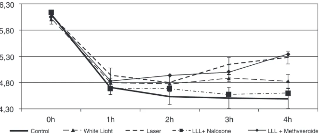
Table I shows the results obtained by the generalized line-ar model (GLM). The results presented line-are related with the effects of group interaction and time, i.e., comparing the cur-ves as a whole always between two groups.
Laser Group – it was different from the Control group in the generalized linear method analysis (p = 0.032). Comparing the results between different hours, a significant difference was ob-served at 3 and 4 hours when compared to the Control group (Chart 1).
White Light Group – it did not differ from the control group in the generalized linear model analysis, but it differed from the Laser group. Using the hour-to-hour comparison, the diffe-rence between both groups was seen at the fourth hour after the administration of carrageenin (Chart I).
Laser + Naloxone Group – the LLL + Naloxone group did not differ from the control group, but a significant difference was observed when it was compared to the Laser group, with an increase in hyperalgesia. Comparing the hour-to-hour re-sults, a difference was observed at moments 3 and 4 hours in relation to the Laser group (Chart 1).
Laser + Methysergide Group – the LLL + Methysergide group differed from the Control group in the generalized
line-ar model analysis, but differences were not observed when it was compared to the Laser group. Comparing the results hour-to-hour, the difference was observed at 2, 3, and 4 hours (Chart 1).
DISCUSSION
In the search for new analgesic and anti-inflammatory metho-ds Low-Level Laser (LLL) seems promising, since it is effec-tive in reducing inflammatory pain with few side effects 18. The effects of LLL are dose-dependent, and a response is not observed when sub- or super-dosage is used, but effecti-ve analgesia is obsereffecti-ved when used in adequate doses 14,23. As expected, the LLL in the parameters used here proved to produce analgesia, increasing significantly the mechanical hyperalgia threshold in animals in the Laser Group when com-pared to the Control Group. The White Light Group (source of common non-monochromatic, non-coherent light) used as a second control did not produce analgesia either.
The interaction between LLL-induced analgesia and opioid receptors is controversial. Low-Level Laser increases the peri-pheral release of opioids through migration of immune system cells, with local release of beta-endorphin, which is antagoni-zed by naloxone 18,19; on the other hand, another study using a carrageenin-induced pain model did not demonstrate that the effects of LLL were antagonized by naloxone 20. The models and laser doses used differed among the studies, as well as the route of naloxone administration, intraperitoneal or intraplantar 20. The present study demonstrated mediation of the analgesia by nalo-xone, which is similar to the results of previous studies 18,19.
Naloxone has an onset of action of two minutes, with du-ration of action dependent on the dose and route of adminis-tration. It has a half-life ranging from 43 to 90 minutes, and duration of action of approximately 1.5 hour 24,25. In the pre-sent study, naloxone was applied 15 minutes before the first dose of LLL and 1h15min before the second dose, and its concentrations were adequate during the applications of the LLL. Although naloxone crosses the blood-brain barrier, in the dose of 1 µg/paw it only antagonizes peripheral opioid effects without a systemic effect 26,27.
Table I – Descriptive analysis Obtained by the Generalized Linear Method (GLM)
Group 1 Group 2 DL p
Control LLL 4 0.032 *
Control White light 4 0.385
LLL + Methysergide Control 4 0.001 *
LLL + Methysergide LLL 4 0.563
LLL + Naloxone Control 4 0.979
LLL + Naloxone LLL 4 0.014 *
DL – degrees of liberty *p < 0.05.
6,30
5,80
5,30
4,30
0h
Control White Light Laser LLL+ Naloxone LLL + Methysergide
1h 2h 3h 4h
4,80
INFLUENCE OF NALOXONE AND METHYSERGIDE ON THE ANALGESIC EFFECTS OF LOW-LEVEL LASER IN AN EXPERIMENTAL PAIN MODEL
Revista Brasileira de Anestesiologia 305
Vol. 60, No 3, May-June, 2010
Naloxone is a non-specific opioid receptor antagonist, with higher affinity for µ and < receptors and lower affinity for δ re-ceptors. As a function of the higher affinity for µ and < recep-tors, it is possible that the analgesic effect of LLL is mediated by those two receptors. Since the affinity of naloxone for δ
receptors is smaller, it is not possible to determine the role of this receptor in LLL-induced analgesia, and the use of a specific antagonist would be interesting.
The role of serotonin (5-HT) on LLL analgesia had not been investigated. Serotonin has several different types of receptors (5-HT1, 5-HT2, 5-HT3, 5-HT4, 5-HT5, 5-HT6, 5-HT7) in different tissues with different functions. In the peripheral nervous system, serotonin has been associated with the pro-nociceptive effect 28,29. However, it has been indicated that the 5-HT1 receptor is pro-nociceptive and analgesic 29,30. Due to this uncertainty, we used methysergide, an antagonist of 5-HT1, 5-HT2, and 5-HT7 receptors, to determine whether LLL-induced analgesia is mediated by peripheral 5-HT1 receptors, which was not confirmed by the results observed, in which the analgesic pattern of LLL is maintained in the presence of
me-thysergide. Since the effects of the LLL were not potentiated either, the evidence indicates that its analgesic function is in-dependent of peripheral 5-HT1, 5-HT2, and 5-HT7 receptors.
The results of the present study support, at least partially, the hypothesis that LLL-mediated analgesia is mediated by peripheral opioid receptors, but not by peripheral serotonergic receptors.
ACKNOWLEDGEMENTS