Functio nal characte rizatio n and
lo calizatio n o f AQ P3 in the
hum an co lo n
Laboratorio de Fisiopatogenia, Departamento de Fisiología, Facultad de Medicina, Universidad Nacional de Buenos Aires, Buenos Aires, Argentina
C. Silberstein, A. Kierbel, G. Amodeo, E. Zotta, F. Bigi, D. Berkowski and C. Ibarra
Abstract
Water channels or aquaporins (AQPs) have been identified in a large variety of tissues. Nevertheless, their role in the human gastrointesti-nal tract, where their action is essential for the reabsorption and secretion of water and electrolytes, is still unclear. The purpose of the present study was to investigate the structure and function of water channels expressed in the human colon. A cDNA fragment of about 420 bp with a 98% identity to human AQP3 was amplified from human stomach, small intestine and colon by reverse transcription polymerase chain reaction (RT-PCR) and a transcript of 2.2 kb was expressed more abundantly in colon than in jejunum, ileum and stomach as indicated by Northern blots. Expression of mRNA from the colon of adults and children but not from other gastrointestinal regions in Xenopus oocytes enhanced the osmotic water permeability, and the urea and glycerol transport in a manner sensitive to an antisense AQP3 oligonucleotide, indicating the presence of functional AQP3. Immunocytochemistry and immunofluorescence studies in human colon revealed that the AQP3 protein is restricted to the villus epithelial cells. The immunostaining within these cells was more intense in the apical than in the basolateral membranes. The presence of AQP3 in villus epithelial cells suggests that AQP3 is implicated in water absorption across human colonic surface cells.
Co rre spo nde nce C. Ibarra
Departamento de Fisiología Facultad de Medicina, UBA Paraguay 2155, 7º piso 1121 Buenos Aires Argentina
Fax: + 54-1-963-6287 E-mail: ibarra@ fmed.uba.ar
Part of these data were presented at the International Symposium of the Molecular Physiology of Water Transport, Paris, France, April 27-30, 1997.
Research supported by the Universidad de Buenos Aires (TM23) and the Consejo Nacional de Investigaciones Científicas y Técnicas (PIP No. 867/98).
Received February 12, 1999 Accepted July 26, 1999
Ke y wo rds
·Absorptive epithelium
·Human colon
·Water channels
·Aquaporins
·AQ P3
Intro ductio n
Approximately 9 liters per day of water are involved in the digestion process and due to the great absorptive capacity of the intes-tine, only 0.2 liters are excreted with the stool (1). The opposite can also occur, i.e., the small and large intestine can secrete large amounts of water in the presence of toxins or endogenous secretagogues (2,3). It is gener-ally accepted that these important fluid
trans-fers are associated with salt reabsorption or secretion. Nevertheless, it is still a subject of controversy whether the absorptive processes are located in the surface (and villus) cells, if the secretory processes are located in the crypt cells (4,5) and if the transport-associ-ated water movements are transcellular or paracellular (6,7).
are found in those membranes which present high osmotic permeability and there is gen-eral agreement that the presence of func-tional AQPs in an epithelial barrier strongly indicates the existence of a transcellular route for the water transfers (8-10). In the gas-trointestinal tract, physiological and biophysi-cal studies have provided evidence against the expression of water channels in the epi-thelial cells from the stomach (11) and je-junum (12) or in Caco-2, a cell line derived from human colon carcinoma (13). In these cases, the paracellular route has been pro-posed as the main route for the transport-associated water transfer.
Alternatively, the presence of functional water channels has been postulated in rabbit rectal epithelium (7) and AQP expression in stomach, small intestine and colon has been reported in studies using Northern blots and immunolocalization (14-20).
In the present study we show that when mRNA from different sections of the
gas-trointestinal tract was injected into Xenopus
oocytes, only the portion corresponding to the colon expressed functional AQP3 which are more intensely localized in the apical mem-branes and less intensely localized in the basolateral membranes of villus epithelial cells.
Mate rial and Me tho ds
Patie nt spe cim e ns
The surgical specimens used in this study were obtained from 16 patients from the Hospital de Gastroenterología Dr. Bonorino Udaondo, Hospital de Niños Dr. Pedro de Elizalde and Hospital Israelita. The clinical conditions requiring removal of sections from stomach, small or large intestine in these patients were different carcinomas for adult specimens (N = 14) and Hirschsprungs dis-ease (N = 2, ages ranging between 3 and 7 years old) in the case of childrens speci-mens. Twenty-four hours prior to surgery,
25 to 40 ml kg body weight-1 h-1 of an
isos-motic lavage solution containing 25 mM
NaCl, 40 mM Na2SO4, 10 mM KCl, 20 mM
NaHCO3 and 60 g/l polyethylene glycol
(PEG) was orally administered to the pa-tients until the fecal discharge was clear and free of solid matter. This solution was re-ported to cause no net water or ion move-ment across the gut (21). Tissues used for these studies were taken from macroscopi-cally normal areas within the safety mar-gin from the pathological affected tissue. Segments were transported in ice-cold high
K+-Ringer solution to the laboratory and
pro-cessed within 2 h of excision. The high K+
-Ringer solution containing 120 mM KCl, 10
mM NaHCO3, 1.2 mM MgCl2, 1.2 mM CaCl2,
1.2 mM K2HPO4, 0.2 mM KH2PO4, and 25
mM glucose preserves the transport function across the colonic epithelial cells (6). This protocol was approved by the Ethics Com-mittee of the Gastroenterology, Children and Israelita Hospitals.
m RNA e xtractio n
The intestinal specimens were washed with 0.9% NaCl, opened along the mesenter-ic border and everted with a forceps on an iced Petri dish. The mucosa and submucosa were stripped from the muscular layers and underlying tissue and the mucosa was gently scraped with a glass microscope slide. The scrapings containing 1 g of epithelial cells were immediately suspended in 10 ml of 4 M guanidinium thiocyanate, 25 mM sodium citrate, pH 7, 0.5% sarcosyl and 0.1 mM ß-mercaptoethanol. The mixture was homog-enized for 30 s on ice using an Ultra-Turrax T25 blender at maximum speed. An equal volume of water-saturated phenol, 0.1 vol-ume of 2 M sodium acetate, pH 4, and 0.2 volume of chloroform were added and vortexed. The final suspension was cooled on ice for 15 min and then centrifuged at
10,000 g for 20 min at 4o
buf-fer, pH 7.4. RNA purity and concentration were assessed spectrophotometrically.
Ol-igo(dT)15
-cellulose columns (Sigma Chemi-cal Co., St. Louis, MO, USA) were used to isolate mRNA. The mean yield of mRNA was approximately 1 to 5 µg for each speci-men. RNA samples were stored at -80°C until the time for use.
RT-PCR assay
Reverse transcription was performed on 5 µg of total mRNA from human colon mucosa using Moloney murine leukemia vi-rus reverse transcriptase (Gibco, BRL,
Gaithersburg, MD, USA), oligo (dT)15
primer and 400 µM of each deoxyribonucleotide
triphosphate (dNTP) for 60 min at 37oC.
PCR (30 cycles at 94o
C 60 s, 50o
C 60 s and
72oC 60 s, followed by a final extension of
10 min at 72o
C) was carried out using 5 µM of two degenerate primers designed on the basis of highly conserved regions (NPA box) in the aquaporin family.
The sense [5'-CAC(C/T)T(G/C)AACCC (A/T)GC(G/T)GT(C/G)AC-3'] and antisense [5'-AAA(G/T)TC(C/A)CG(A/G)GC(T/ A)GGGTT-3'] oligonucleotide primers cor-responded to the nucleotides coding for amino acids 73-78 and 192-199, respectively, of the AQP1 sequence (X70257/S52660), and to amino acids 81-86 and 215-222 of the AQP3 sequence (D17695/D25280). Agarose gel electrophoresis of the RT-PCR products showed a 420-bp band that was cloned in the pGEM-T vector (Promega Corp., Madison, WI, USA) and the fragment was sequenced
using the ¦mol kit (Promega). RT-PCR was
also assayed with adult stomach, jejunum, ileum, and childrens distal colon mRNA using 2 µM of the specific sense [5'-CC TGAACCCTGCGGTGACC-3'] and anti-sense [5'-GGCATAGCCGGAGTTGAAGC-3'] oligonucleotide primers based on the RT-PCR product from colon. The protocol was
30 cycles at 94o
C 60 s, 55o
C 60 s, and 72o
C 60 s, followed by a final extension of 10 min
at 72o
C. PCR products were cloned and se-quenced as described before.
No rthe rn blo tting
About 10 µg of each total RNA was electrophoresed on a denaturing 1.4% aga-rose gel containing 2.2 M formaldehyde. One part of the gel was stained with ethi-dium bromide to evaluate the intensity and quality of 18S and 28S ribosomal bands for the different RNA samples. The other part was transferred to a reinforced nitrocellu-lose membrane (BAS85, Schleicher and Schuell, Dassek, Germany), fixed for 2 h at
80o
C and prehybridized for 4 h at 42o
C with a standard prehybridization buffer contain-ing 50% formamide, 5x SSC, 0.5% SDS and salmon sperm DNA (100 µg/ml) and
hybrid-ized overnight at 42oC with 10-20 µl 32
P-dCTP labeled probe diluted in a fresh 5-ml aliquot of warmed prehybridization buffer. Blots were then washed twice for 5 min in 2x SSC, 0.1% SDS at room temperature fol-lowed by 15 min in 0.1x SSC, 0.1% SDS at
37oC and 5 min in 0.1x SSC, 0.5% SDS at
55o
C. Next, blots were dried and exposed to X-ray films (X-Omat, Kodak) for 2-7 days.
Equivalent amounts of RNA were loaded as assessed on the basis of abundance of 18S and 28S RNAs and by Northern analysis using a chicken ß-actin probe (data not shown).
O o cyte inje ctio n
Oocytes were obtained from adult
Xeno-pus laevis by standard procedures (22) and stored immediately in Barths solution (88
mM NaCl, 1 mM KCl, 0.8 mM MgSO4, 0.3
mM Ca(NO3)2, 0.4 mM CaCl2, 2.4 mM
NaHCO3, 7.5 Tris, pH 7.6, 200 mOsm/kg)
containing penicillin (9.6 µg/ml) and strep-tomycin (10 µg/ml). The follicular cell layer was removed by incubation with 1 mg/ml collagenase (type 1A; Sigma) in sterile
Barths solution for 2 h at 25o
washed ten times with 10 ml of the same
solution and stored overnight at 4oC and then
used for mRNA microinjection. Selected defolliculated oocytes (~1.0 mm in diam-eter) were injected with 50 nl of water or mRNA (1 mg/ml) using an automatic micro-injection system. Oocytes were incubated
for 72 h at 18o
C in Barths solution with daily changes.
When the sense [5'-TTGCCATGTGCT TCCTGGCTCGTGAGCCCT-3'] or anti-sense [5'-AGGGCTCACGAGCCAGGA AGCACATGGCAA-3'] AQP3 oligonucle-otide derived from the 420-bp RT-PCR prod-uct was used, the mixture of each oligo-nucleotide (2.3 ng/oocyte) with mRNA (50
ng/oocyte) was warmed to 65oC for 2 min
and allowed to cool at room temperature just before co-injection.
O sm o tic wate r pe rm e ability
Experiments were performed in a tem-perature-jacketed chamber with a curved
bottom at 18o
C. An oocyte injected with water or with total mRNA was transferred from 200 mOsm Barths solution to a cham-ber containing ten-fold diluted Barths solu-tion (20 mOsm). The increase in osmotic volume was followed for the first 120 s by videomicroscopy by storing digitized im-ages at 10-s intervals in a computer. The time-dependent volume change of the oo-cyte was considered linear for this period of time and the Posm was calculated as previ-ously reported (23).
So lute uptake into o o cyte s
Solute uptake into Xenopus laevis oocytes
was studied as described (24). Briefly, indi-vidual oocytes previously microinjected with water or colon mRNA were incubated in 0.2 ml Barths solution containing 100 µCi/ml
[3H]-glycerol (200 mCi/mmol) and 2.7 µCi/ml
[14
C]-urea (8.6 mCi/mmol, New England Nuclear, Boston, MA, USA) and 1 mM urea
and glycerol with continuous shaking. The uptake was stopped after 10 min by adding 4 ml ice-cold Barths solution and each oocyte was placed on a filter (type GF-A, Whatman) and washed twice with 5 ml of the same solution under vacuum. Then, oocyte and fil-ter were placed in a glass vial with 0.1 ml formic acid for 5 min and mixed with 5 ml scintillation fluid to determine radioactivity.
Immuno blo tting studie s
Human colon was homogenized in 200 mM sucrose/10 mM Tris-HCl, pH 7.4, con-taining leupeptin (1 µg/ml) and 0.1 mg/ml phenylmethylsulfonyl fluoride. After homog-enization in a Potter apparatus and
centrifu-gation at 3,000 g for 10 min to remove nuclei
and incompletely homogenized membrane fragments, a high-speed pellet was prepared
by centrifugation at 100,000 g for 60 min.
Membranes were dissolved in SDS loading buffer, heated to 65°C for 5 min, and re-solved on 13% polyacrylamide gel. Proteins were electrotransferred to a nitrocellulose membrane by the semi-dry method. Mem-branes were blocked with 2% (w/v) bovine serum albumin in PBS for 30 min at room temperature and incubated overnight with the purified anti-AQP3 antibody (50 ng/ml). The secondary antibody was goat antirabbit IgG conjugated to alkaline phosphatase used at 1:1000 dilution. Sites of antibody-antigen reaction were visualized using 5-bromo-4-chloro-3-indolyl phosphate and nitroblue tet-razolium as color development reagents.
Im m uno cyto che m istry
paraformalde-hyde and cryoprotected overnight with PBS containing 30% (w/v) sucrose, embedded in ornithine carbamyl transferase (OCT) com-pound (Miles Laboratories Inc., Clifton, NJ,
USA), and frozen in liquid N2. Cryosections
(4-5 µm) of human colon and rat kidney (control) were placed on the same gelatin-coated glass slide.
Immunolabeling of sections. Rabbit af-finity-purified polyclonal antibody against a 26-amino acid synthetic peptide,
correspond-ing to the carboxyl terminus of AQP3 (NH2
- CHLEQPPPSTEAENVKLAHMKHKEQI-COOH) (25) was used.
For immunofluorescence, slides were pre-incubated with nonimmune goat serum and then incubated with the purified anti-AQP3
antibody (5 µg/ml) overnight at 4o
C. After rinsing, the slides were incubated with fluores-cein isothiocyanate-conjugated mouse anti-rabbit IgG (Sigma Clone RG-96, 1:40) for 4 h at room temperature. Omission of primary antibody was used as negative control.
For immunolocalization by light micros-copy, the sections were incubated with PBS containing 1% bovine serum albumin for 60 min at room temperature and then with puri-fied anti-AQP3 antibody (3-7 µg/ml)
over-night at 4oC in PBS containing 1% bovine
serum albumin. The slides were then rinsed
and incubated for 1 h at 25o
C with antirabbit IgG biotinylated species-specific whole an-tibody from donkey (Amersham RPN 1004, Amersham International, Amersham, Buck-ingham, UK) at a 1:100 dilution followed 1 h by streptavidin biotinylated horseradish per-oxidase complex (Amersham RPN 1051) at 1:100 dilution. The labeling was visualized by reaction with diaminobenzidine
contain-ing NiCl2 (26,27). Purified nonspecific
rab-bit IgG was used as negative control.
Re sults
AQ P3 m RNA e xpre ssio n
A 420-bp RT-PCR product was
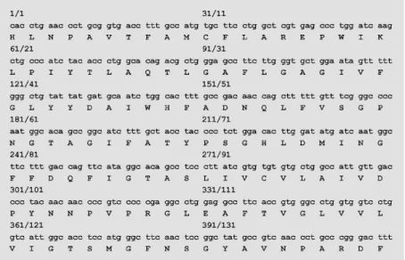
ampli-fied, cloned and sequenced from adult stom-ach, jejunum, ileum and colon and from childrens colon (Figure 1). This sequence showed 96% identity to rat kidney AQP3 (16,28), 98% identity to human kidney AQP3 (29) and 99% to human kidney glycerol intrinsic protein (GLIP) (30) in their respec-tive conserved regions. Furthermore, AQP3 mRNA expression in the different gastrointes-tinal sections was analyzed by Northern blot using the 420-bp RT-PCR product as a probe. A transcript of 2.2 kb was expressed more abundantly in colon than in jejunum, ileum or stomach (Figure 2).
O sm o tic wate r pe rm e ability
To determine the functional expression
of AQP3, Posm was measured in Xenopus
laevis oocytes injected with total mRNA from the different gastrointestinal sections. The results are summarized in Table 1. For each anatomic region, the Posm correspond-ing to oocytes injected with water or with mRNA were averaged from different speci-mens and their average differences from each other were calculated. Only in distal colon from adults and children did the average
differences significantly differ from zero (Table 1, columns 5 and 6). These differ-ences were inhibited by treatment of oocytes
with 0.3 mM HgCl2 for 10 min and restored
by incubation with ß-mercaptoethanol for 15 min (Figure 3A,B). An antisense but not a sense AQP3 primer co-injected with colon mRNA also inhibited AQP3 expression (Fig-ure 3A). In addition, oocytes expressing
AQP3 exhibited increases in [3
H]-glycerol
and [14
C]-urea uptake which were abolished when colon mRNA was co-injected with excess AQP3 antisense primer. In both cases, the AQP3-sense primer was used as control (Figure 4A,B).
Immuno lo calizatio n o f AQ P3 in human co lo n
Figure 5A shows the immunoblot of a
crude membrane preparation (100,000 g
membrane fraction) from human colon probed with the anti-AQP3 antibody. A pro-tein band of 27 kDa was seen and another faint band of higher apparent molecular mass (35 kDa) was also observed. This pattern is
similar to that reported for rat kidney me-dulla (14) where the upper band represents the glycosylated form of AQP3, and the lower band represents the nonglycosylated form. No bands were detected when nonim-mune goat serum was used or when the primary antibody was omitted (data not shown).
Immunofluorescence of human adult co-lon with AQP3 antibody showed strong stain-ing in villus but not in crypt epithelial cells (Figure 5C) when compared with a negative control (Figure 5B). At high power magnifi-cation, the labeling appeared to be localized predominantly in the apical membrane of surface cells while a considerably weaker signal was detected in the basolateral do-main of these cells (Figure 5D and E; for control, see Figure 5F). Strong staining in collecting ducts of rat kidney medulla (posi-tive control) is shown in Figure 5G.
Identification of labeling in villus epithe-lial cells was confirmed with immunoper-oxidase staining (Figure 6). At low power magnification intense staining was detected in some of the villus epithelial cells but not in globets cells, and no staining was ob-served in the crypt epithelial cells (B). Con-trol sections treated with nonimmune IgG revealed an absence of labeling (A). At high power magnification, strong staining was observed in the apical membrane and the subapical area just beneath the apical mem-brane of villus epithelial cells (C). Immuno-labeling of rat kidney sections revealed la-Figure 2 - Northern blot analysis
of AQP3 mRNA in the different human gastrointestinal sections. A transcript of 2.2 kb w as ex-pressed more abundantly in co-lon than in jejunum, ileum or stomach. Positions of RNA mark-ers are indicated by lines.
Table 1 - Osmotic w ater permeability (Posm: x 10-4 cm/s) of Xenopus laevis oocytes.
N indicates the number of patients. In each case 6 to 12 oocytes w ere injected w ith w ater (control) or w ith mRNA from each gastrointestinal section.
Adults Children
Stomach Jejunum Ileum Distal colon Distal colon
mRNA-injected 13.02 ± 2.9 16.51 ± 4.79 8.17 ± 0.77 18.27 ± 0.88 24.92 ± 1.69 Water-injected (control) 10.71 ± 1.66 15.04 ± 3.51 9.76 ± 0.97 10.96 ± 1.05 14.47 ± 0.35 M ean difference 2.31 ± 1.63 1.46 ± 1.28 -1.59 ± 1.72 7.31 ± 0.67 10.46 ± 1.34
t-test NS, N = 3 NS, N = 3 NS, N = 3 P<0.01, N = 5 P<0.05, N = 2
S
to
m
a
c
h
J
e
ju
n
u
m
2.4
1.35
Il
e
u
m
C
o
lo
n
beling in the basolateral membrane domain of collecting duct cells (D).
D iscussio n
The functional characteristics of water handling in the gastrointestinal tract has been
extensively studied both in vivo and in vitro
(1,4,7). Nevertheless, to our knowledge, this is the first study on the functional expression of water channels using total mRNA from different human gastrointestinal regions.
We demonstrate here the expression of functional water channels only when adult and children colon mRNA was injected into
Xenopus oocytes but not when mRNA from adult stomach, jejunum or ileum was used.
The HgCl2-sensitive expression of colon
mRNA was similar to that reported for the other tissues encoding AQPs (28,31,32). The ability of an AQP3 antisense sequence to block the mRNA expression suggests the presence of an AQP3. Increase of urea and
glycerol uptake by Xenopus oocytes injected
with colon mRNA and its inhibition by AQP3 antisense also support the functional expres-sion of AQP3.
We also reported here an amplification of a 420-bp DNA from stomach, jejunum, ileum and colon using RT-PCR. The amino acid sequence of all DNAs presented a high homology to AQP3 and GLIP, both cloned from a human kidney cDNA library (29,30). Expression of mRNA was demonstrated in all gastrointestinal sections by Northern blot and these results are coincident with those described by Ishibashi et al. (29) and Koyama et al. (33). However, we observed functional AQP3 only in human colon. A possible ex-planation could be a higher level of AQP3 mRNA in colon than in the other gastrointes-tinal sections as observed in the Northern blot studies. The lack of water-channel ex-pression in stomach and small intestine is consistent with functional data for vesicles derived from these tissues showing low plasma membrane water permeability (11,12).
Figure 3 - Functional expression of AQP3 present in human colon. Each bar indicated mean osmotic w ater permeability (Posm) measured in 6 to 10 oocytes, 3-4 days after injection w ith w ater (open bars) or w ith 50 ng of mRNA w ith the indicated treatment. A, The assay w as performed using mRNA from adult colon w ith or w ithout 0.3 mM HgCl2 (5-min
incubation), w ith HgCl2 treatment follow ed by incubation w ith ß-mercaptoethanol (BM E; 10
min) or preceded by incubation w ith AQP3 oligonucleotide sense or antisense primer (2.3 ng/oocyte). B, Injection of a child distal colon mRNA and its HgCl2 treatment. The Student t
-test w as applied and data w ere compared w ith those for colon mRNA-injected oocytes. * P<0.05, * * P<0.01, NS = nonsignificant.
Figure 4 - Selective AQP3 permeability to a small solute. Bars represent the mean ± SEM uptake of [3H]-glycerol (A) and [14C]-urea (B) measured in 6 to 10 oocytes, 3-4 days after
injection w ith w ater or w ith 50 ng of human colon mRNA. AQP3 oligonucleotide sense or antisense primers w ere co-injected w ith colon mRNA and data w ere compared by the Student t-test. * P<0.01 compared to colon mRNA-injected oocytes. NS = nonsignificant.
[ 3H ]-g ly c e ro l u p ta k e ( p m o l o o c y te
-1 1
0 m in -1) 120 100 80 60 40 20 0 A B * * * A n ti s e n s e S e n s e
Water Adult colon Water Adult colon
NS [ 1 4C ]-u re a u p ta k e ( p m o l o o c y te
-1 1
0 m in -1) 120 100 80 60 40 20 0 * A n ti s e n s e S e n s e NS P o s m ( c m /s x 1 0 -4) 30 25 20 15 10 5 0 P o s m ( c m /s x 1 0 -4) 30 25 20 15 10 5 0 A B * * * * * * * * * H g C l2 A n ti s e n s e S e n s e B M E H g C l2
Water Adult colon WaterChild colon
Colon
A
kDa
42
30
B C
G F
E D
Figure 5 - Immunoblot and immunofluorescence of AQP3 w ater channel protein in human colon using purified anti-AQP3 polyclonal antibody. Immunoblot of a colonic mucosa homogenate show ed tw o bands, 27 kDa and 35 kDa, corresponding to nonglycosylated and glycosylated forms of AQP3 protein, respectively (A). Immunofluorescence localization of AQP3 (C-E) and their control (B: X190, F: X450 magnification). Surface epithelial cells w ere strongly labeled w ith anti-AQP3, w hereas crypt epithelial cells w ere unlabeled (C: X190 magnification). Labeling is heavily localized in the apical membrane and w eakly localized in the basolateral membrane of villus epithelial cells (D: X375 magnification and E: X450 magnification). Extensive labeling in collecting duct of rat kidney medulla w as detected in the positive control (G: X190 magnification).
Recently, Yamamoto and Sasaki (34) dis-cussed the clear demonstration of AQP3 mRNA expression in stomach and small in-testine and the inability to detect AQP3 pro-tein in these epithelial cells by immunohis-tochemistry and functional studies. Further studies are needed to determine the
correla-tion between AQP3 mRNA and protein ex-pression.
cells. Previous immunolocalization studies have shown expression of GLIP and AQP3 in the basolateral domain of surface epitheli-al cells in rat colon but not in rat smepitheli-all intestine (14,33).
Thus, we have identified functional AQP3 in the human colon which may be implicated in fluid absorption across this particular
in-testinal section. The fact that the intercellu-lar junctions of colonic cells were reported to be tight (35) limits water transport across the paracellular route and suggests the transcellular route as the main pathway for the water movement driven by ion absorp-tion. If this is the case, regulation of AQP3 may contribute to maintaining the control of
A
B
C
D
water absorption and its alteration could lead to abnormal states such as constipation and diarrhea.
Ackno wle dgm e nts
We gratefully acknowledge the excellent assistance of Mr. Luis Masciotra and Mrs. Silvia Fariña in the immunoperoxidase and immunofluorescence studies. We thank Dr. Alberto Monserrat, Departamento de Pato-logía, Facultad de Medicina, UBA, for ad-vice in histology interpretation, and Dr. Miriam Echevarria, Departamento de
Fisio-logía y BioFisio-logía Animal, Facultad de Farma-cia, Universidad de Sevilla, España, for help-ful suggestions. We thank Dr. M.A. Knepper, National Institutes of Health, Bethesda, MD, USA, for kindly providing the antibody to aquaporin-3 and for the suggestions con-cerning the immunoblots. We also thank Dr. Fernando Galindo and Mrs. Estrella Alvarez, Hospital de Gastroenterología Dr. Bonorino Udaondo, and Drs. Luis Voyer and Graciela Bortolazzo, Hospital de Niños Dr. Pedro de Elizalde, for providing the gastrointestinal specimens.
Re fe re nce s
1. Binder HJ (1983). Absorption and secre-tion of w ater and electrolytes by small and large intestine. In: Sleisenger M H & Fortran JS (Editors), Gastrointestinal Dis-ease: Pathophysiology, Diagnosis, M an-agement. 3rd edn. WB Saunders, Phila-delphia, 811-840.
2. Bennish M L (1994). Cholera: pathophysi-ology, clinical features and treatments. In: Wachsmuth K, Blake P & Olsvik O (Edi-tors), Vibrio Choleraeand Cholera: M olec-ular to Global Perspectives. American So-ciety of M icrobiology, Washington, DC, 229-255.
3. Pow ell DW (1987). Intestinal w ater and electrolyte transport. In: Johnson LR (Edi-tor), Physiology of the Gastrointestinal Tract. 2nd edn. Raven Press, New York, 1267-1305.
4. Field M (1993). Intestinal electrolyte se-cretion: History of a paradigm. Archives of Surgery, 128: 273-278.
5. Singh SK, Binder HJ, Boron WF & Geibel JP (1995). Fluid absorption in isolated per-fused colonic crypts. Journal of Clinical Investigation, 96: 2373-2379.
6. Escobar E, Galindo F & Parisi M (1990). Water handling in the human distal colon in vitro: role of Na+, Cl- and HCO
3-.
Bio-chimica et Biophysica Acta, 1027: 257-263.
7. Parisi M & Ibarra C (1996). Aquaporins and w ater transfer across epithelial barri-ers. Brazilian Journal of M edical and Bio-logical Research, 29: 933-939.
8. Agre P, Brow n D & Nielsen S (1995). Aq-uaporin w at er channels: unansw ered
questions and unresolved controversies. Cell Biology, 7: 472-483.
9. Nielsen S & Agre P (1995). The aquaporin family of w ater channels in kidney. Kidney International, 48: 1057-1068.
10. Verkman AS, Shi LB, Frigeri A, Hasegaw a H, Farinas J, M itra A, Skach W, Brow n D, Van Hoek AN & M a T (1995). Structure and function of kidney w ater channels. Kidney International, 48: 1069-1081. 11. Priver NA, Rabon EC & Zeidel M L (1993).
Apical membrane of the gastric cell: w a-ter, proton and nonelectrolyte permeabili-ties. Biochemistry, 32: 2459-2468. 12. Van Heesw ijk M PE & Van Os PH (1986).
Osmotic w ater permeability of brush bor-der and basolateral membrane vesicles from rat cortex and small intestine. Jour-nal of M embrane Biology, 92: 183-193. 13. Parisi M , Pisam M , Calamita G, Gobin R,
Toriano R & Bourguet J (1995). Water pathw ays across a reconstituted epitheli-al barrier formed by Caco-2 cells: effects of medium hypertonicity. Journal of M em-brane Biology, 143: 237-245.
14. Frigeri A, Gropper M A, Turck CW & Verkman AS (1995). Immunolocalization of the mercurial insensitive w ater channel and glycerol intrinsic protein in epithelial cell plasma membrane. Proceedings of the National Academy of Sciences, USA, 92: 4328-4331.
15. Hasegaw a H, Zhang R, Dohrman A & Verkman AS (1993). Tissue-specific ex-pression of mRNA encoding rat kidney w ater channel CHIP28k by in situ hybrid-ization. American Journal of Physiology,
264: C237-C245.
16. Ishibashi K, Sasaki S, Fushimi K, Uchida S, Kuw ahara M , Sait o H, Furukaw a T, Nakajima K, Yamaguchi Y, Gojobori T & M arumo F (1994). M olecular cloning and expression of a member of the aquaporin family w ith permeability to glycerol and urea in addition to w ater expressed at the basolateral membrane of kidney collect-ing duct cells. Proceedings of the National Academy of Sciences, USA, 91: 6269-6273.
17. Koyama Y, Yamamoto T, Kondo D, Funaki H, Yaoit a E, Kaw asaki K, Sat o N, Hatakeyama K & Kihara I (1997). M olecu-lar cloning of a new aquaporin from rat pancreas and liver. Journal of Biological Chemistry, 272: 30329-30333.
18. M a T, Yang B & Verkman AS (1997). Clon-ing of a novel w ater and urea-permeable aquaporin from mouse expressed strong-ly in colon, placenta, liver, and heart. Bio-chemical and Biophysical Research Com-munications, 241: 53-58.
19. M isaka T, Abe K, Iw abuchi K, Kusakabe Y, Ichinose M , M iki K, Emori Y & Arai S (1996). A w ater channel closely related to rat brain aquaporin 4 is expressed in acid-and pepsinogen-secretory cells of human stomach. FEBS Letters, 381: 208-212. 20. Valent i G, Verbavat z JM , Sabolic I,
Fordtran JS (1980). Development of a lav-age solution associated w ith minimal w a-ter and electrolyte absorption or secre-tion. Gastroenterology, 78: 991-995. 22. Dascal N (1987). The use of the Xenopus
oocytes for the study of ion channels. CRC Critical Review s in Biochemistry, 22: 317-373.
23. Capurro C, Ford P, Ibarra C, Ripoche P & Parisi M (1994). Water permeability prop-erties of the ovarian oocytes from Bufo arenarum and Xenopus laevis. A compara-tive study. Journal of M embrane Biology, 138: 151-157.
24. M artial S, Ripoche P & Ibarra C (1991). Functional expression of urea channels in amphibian oocytes injected w ith frog uri-nary bladder mRNA. Biochimica et Bio-physica Acta, 1090: 86-90.
25. Ecelbarger CA, Terris J, Frindt G, Echevarria M , M arples D, Nielsen S & Knepper M (1995). Aquaporin-3 w ater channel localization and regulation in rat kidney. American Journal of Physiology, 269: F663-F672.
26. Hsu SM , Raine L & Fanger H (1981). Use of avidine-biot in-peroxidase com plex (ABC) in immunoperoxidase techniques:
A comparison betw een ABC and unla-beled antibody (PAP) procedures. Journal of Histochemistry and Cytochemistry, 29: 577-580.
27. Hsu SM & Soban E (1982). Color modifi-cation of diaminobenzidine (DAB) precipi-tation by metallic ions and its application for double immunohistochemistry. Jour-nal of Histochemistry and Cytochemistry, 30: 1079-1082.
28. Echevarria M , Frindt G, Prest on G, M ilovanovic S, Agre P, Fischbarg J & Windhager EE (1993). Expression of mul-tiple w ater channel activities in Xenopus oocytes injected w ith mRNA from rat kid-ney. Journal of General Physiology, 101: 827-841.
29. Ishibashi K, Sasaki S, Saito F, Ikeuchi T & M arumo F (1995). Structure and chromo-somal localization of a human w ater chan-nel (AQP3) gene. Genomics, 27: 352-355. 30. M a T, Frigeri A, Hasegaw a H & Verkman AS (1994). Cloning of a w ater channel homolog expressed in brain meningeal cells and kidney collecting duct that func-tions as a stilbene sensitive glycerol trans-porter. Journal of Biological Chemistry, 269: 21845-21849.
31. Sasaki S, Fushim i K, Ishibashi K & M arumo F (1995). Water channels in the kidney collecting duct. Kidney Interna-tional, 48: 1082-1087.
32. Zhang R, Skach W, Hasegaw a H, Van Hoek AN & Verkman AS (1993). Cloning, functional analysis and cell localization of a kidney proximal tubule w ater transporter homologous to CHIP28. Journal of Cell Biology, 120: 359-369.
33. Koyama Y, Yamamoto T, Tani T, Nihei K, Kondo D, Funaki H, Yaoita E, Kaw asaki K, Sato N, Hatakeyama K & Kihara I (1999). Expression and localization of aquaporins in rat gastrointestinal tract. American Jour-nal of Physiology, 276: C621-C627. 34. Yamamoto T & Sasaki S (1998).
Aquapor-ins in the kidney: Emerging new aspects. Kidney International, 54: 1041-1051. 35. Davis GR, Santa Ana CA, M oraw ski SG &

![Figure 4 - Selective AQP3 permeability to a small solute. Bars represent the mean ± SEM uptake of [ 3 H]-glycerol (A) and [ 14 C]-urea (B) measured in 6 to 10 oocytes, 3-4 days after injection w ith w ater or w ith 50 ng of human colon mRNA](https://thumb-eu.123doks.com/thumbv2/123dok_br/15807559.650319/7.918.384.835.646.1005/figure-selective-permeability-represent-glycerol-measured-oocytes-injection.webp)


