Abstract
The study prepared a nanoemulsion with a diterpenoid isoprenoid alcohol called phytol (PYT) and subsequently tested it for antioxidant capacity. For this, PYT-loaded nanoemulsion was prepared by phase inversion method and both PYT-containing nanoemulsion (PNE) and PYT-free nanoemulsion (PFNE) (2-16 µM) were tested for antiradical activity (DPPH•: 1,1-dipheny-picrylhydrazyl radical; ABTS•+: azino-bis-ethylbenzthiazoline-sulfonic acid; •OH: hydroxyl radical scavenging; NO•: nitrite oxide radical), lipid peroxidation (LP), reduction potential (RP), and inhibition of hemolysis (HL) in rat erythrocytes in compa-rison with an α-tocopherol analogue (Trolox - TRO - positive con-trol). In addition, an in vivo test was performed with wildtype and deficient Saccharomyces cerevisiae strains using hydrogen peroxide (H2O2) as a stressor. Results suggest that PNE exhibited higher anti-oxidant than the PFNE. Increasing doses reveled antianti-oxidant capacity in a dose-dependent manner. In the S. cerevisiae study, both PFNE- and PNE-treated groups exhibited decreased rates of survival with the highest doses, whichever in the presence of stressor increased the survival rates, which indicates antioxidative defense capacity of PYT. In this occasion, PNE exhibited prominent antioxidative defen-se in the predefen-sence of stressor rather than PFNE. In conclusion, PYT exhibited potential antioxidant activity but at high concentration it was toxic to the yeast cells. The production of PYT-nanoemulsions may be relevant to the pharmaceutical sciences.
Preparation of Phytol-Loaded Nanoemulsion and
Screening for Antioxidant Capacity
ORIGINALMd. Torequl Islam1,2,3, Leticia Streck4, Márcia Fernanda Correia Jardim Paz1,2,
João Marcelo de Castro e Sousa5, Marcus Vinícius Oliveira Barros de Alencar1,2, Ana Maria Oliveira Ferreira da Mata1,2, Ricardo Melo de Carvalho1, Jose Victor de Oliveira Santos1, Arnobio Antonio da Silva-Junior5, Paulo Michel Pinheiro Ferreira1,2,6,
Ana Amélia de Carvalho Melo-Cavalcante1,2
1 Northeast Biotechnology Network (RENORBIO), Post-graduation Program in Biotechnology, Federal University of Piauí, 64.049-550, Teresina, Brazil.
2 Post-graduation Program in Pharmaceutical Science, Federal University of Piauí, 64.049-550 Teresina, Brazil.
3 Department of Pharmacy, Southern University Bangladesh, 22-Shahid Mirza Lane (E), Academic building-II, 1st floor, 739/A, Mehedibag Road, Mehedibag-4000, Chittagong, Bangladesh.
4 Departament of Pharmacy, Federal Uuniversity of Rio Grande do Norte, 59012-570. Natal-RN, Brazil.
5 Department of Biological Sciences, Federal University of Piauí, 64.049-550, Picos, Brazil.
6 Department of Biophysics and Physiology, Federal University of Piauí, 64.049-550 Teresina, Brazil.
Contact information:
Md. Torequl Islam.
Address: Department of Biochemistry and Pharmacology, Post-graduation Program in Pharmaceutical Science, Federal University of Piauí, 64.049-550, Teresina, Brazil.
Tel: 0558681265804.
[email protected]Keywords
Antioxidant; Radicals; Nanoemulsion; Phytol;
Introduction
Oxidative stress results from the reactive oxygen/ nitrogen species (ROS/RNS) and to the damage of essential macromolecules, with apoptosis or necro-tic cell death. It is believed that the basic mechanism underlying toxicity may be extracellular reactive spe-cies ultimately reaching the nucleus, where it causes DNA damage. Furthermore, DNA damage followed by subsequent cellular proliferation propagates errors and may result in carcinogenesis [1]. Although oxidative DNA damage is repaired by a number of different enzymes, some believe that oxidants and repair enzymes must remain at steady state [2].
Phytol (3, 7, 11, 15-tetrametilhexadec-2-en-1-ol; PYT), along with antioxidant, is already evident to have a number of biological activities, including an-timicrobial, antispasmodic, antimutagenic, antibio-tic-chemotherapeutic, anticancer, anti-teratogenic, antidiabetic, lipid lowering, anticonvulsant, immu-noadjuvancy, antinociceptive, anti-inflammatory, anxiolytic, antidepressant, hair-fall defense and so on [3]. According to Andrade and Fasolo com-pounds with antioxidant and antimicrobial activities may promote overall health [4]. PYT is still used as a fragrance material in a number of cosmetics and non-cosmetics products [3]. Nowadays extensive research is focusing on the natural products, espe-cially derived from plants, despite of the synthesized medicaments. The use of natural antioxidant com-pounds as a therapy in diseases related to oxidative stress has gained massive interest for their abilities to quench free radicals and the protection of the body against oxidative stress-induced pathogenesis. It is noteworthy that we have a number of synthetic antioxidants. Having high adverse effects and rela-ted other unfavorable factors such as cost, availabi-lity, safety and efficacy are the stimulation to search new compounds in this context.
Otherwise, being an essential oil, PYT is a water non-soluble molecule, which limits its application and use in products for humans. So, we prepared PYT-loaded nanoemulsions (PNE), which along with
the nanoemulsion-free PYT (PFNE) undergone to in-vestigate the antioxidant capacity in a number of in vitro, ex vivo, and in vivo methods.
Materials and methods
Reagents and chemicals
Tween-80 (0.05%) dissolved in saline solution (0.9% NaCl) served as the vehicle. Trolox (6-Hydro-xy-2, 5, 7, 8-tetramethylchroman-2-carboxylic acid; TRO) was used as the standard for all in vitro and
ex vivo antioxidant assays. PYT, and all the neces-sary reagents and chemicals used in this study were obtained from Sigma-Aldrich (St. Louis, MO, USA).
Preparation of PNE
PNE was prepared by the emulsion phase inversion method. Briefly, an oil phase composed of medium chain triglycerides (1% w/w) and a surfactant (5% w/w) composed of soy phosphatidylcholine and so-dium oleate (1:7 w/w) was slowly titrated with an aqueous solution of 2-methylpyrrolidone (2% w/v) at 70 °C under constant magnetic stirring (1,500 rpm). This was emulsified using a basic Ultra-Turrax T18 equipped with the S 18 N-19 G dispersing tool at 11,000 rpm for one minute followed by four mi-nutes at 7,000 rpm. The surfactant mixture that produced transparent colloidal dispersions was se-lected in a previous study using a pseudo-ternary phase diagram [5].
Analysis of PYT-nanoparticles size distribution and zeta potential measurement
software. Zeta potential (ZP) measurements were performed using the same equipment applying the same field strength (approximately 5.9 V/cm). Five runs for each sample were used to determine the ZP value using the PALS ZP Analyzer software and the electrophoretic mobility according the Helmholtz-Smoluchowski equation. The samples were diluted 1:100 (v/v) with purified water.
Drug loading into the nanoemulsions
The ability of the nanoemulsions to load PYT was tested by adding an excess of the drug into the oil phase before the emulsification step described abo-ve. The samples were stored in a thermostatic bath at 25 °C and vortexed for one minute followed by 15 minutes in an ultrasonic bath every 12 h for 72 h. After centrifugation (1000 x g for 15 minutes), the samples were filtered through an acetate membra-ne (0.45 µm) and loaded PYT was analytically de-termined by UV-Vis spectrophotometry at 239 nm using the equation from the fitted standard curve plot constructed previously.
Experimental animals
The HL inhibition test was performed in eight week-old Wister albino rats (Rattus norvegicus; body weight: 180-220 g) from the Central Animal House of the Federal University of Piaui, Brazil. The animals were allowed free access to food (Purina, Brazil) and tap water ad libitum and were kept under contro-lled lighting (12 h dark/light cycles) at 24 ± 2 °C.
Test sample preparation
For the in vitro and ex vivo assays, PFNE and PNE were emulsified in 0.05% tween-80 dissolved in 0.9% NaCl at final concentrations ranging from 2-16 µM. Standard TRO was also dissolved in the same vehicle to give the same dilutions as PFNE and PNE. For the S. cerevisiae assay, PYT (PFNE and PNE) was adjusted to the same dose as above and in the same vehicle H2O2 (10 mM) was used as the stressor (STR) in this in vivo study.
In vitro antioxidant activity assays
DPPH• scavenging activity
The test for DPPH• scavenging activity was done
using a method slightly modified from that descri-bed by Manzocco et al. [6]. Briefly, 0.3 mL samples with concentrations from 2-16 µM were added to a 2.7 mL ethanolic solution of DPPH (0.5 mM). After 30 min, the absorbance was measured using a spectrophotometer at 517 nm. A similar con-centration of TRO served as the positive control, while only 0.3 mL vehicle was added to the DPPH solution for the negative control (NC). The blank contained no sample. The DPPH radical scaven-ging potential was calculated using the following equation:
% inhibition of DPPH• scavenging =
[(Abr – Aar)/Abr]×100
where Abr is the absorbance of DPPH free radicals before the reaction and Aar is the absorbance of DPPH free radicals after the reaction.
ABTS•+ decolorization activity
The ABTS•+ decolorization assay was done as des -cribed by Seeram et al. [7] with slight modification.
ABTS•+ radicals were produced by adding solid
manganese dioxide (80 mg) to a 5 mM aqueous stock solution of ABTS in 75 mM Na+/K+ buffer (pH 7.0). Then, 2.8 mL of the sample was added to
0.2 mL of ABTS•+ solution. The absorbance was
read at 750 nm after 5 minutes. The percentage of scavenging capacity was calculated as follows:
% ABTS•+ scavenged = [(Abr – Aar)/Abr]×100
where Abr is the absorbance of ABTS•+ free radi -cals before the reaction and Aar is the absorbance
of ABTS•+ free radicals after reaction with the test
•OH scavenging activity
The ability of test samples to scavenge hydrogen peroxide (H2O2) was determined according to the method described by Ruch et al. [8]. Briefly, a so-lution of 40 mM H2O2 was prepared in phosphate buffer (50 mM; pH 7.4). The concentration of H2O2 was measured spectrophotometrically by determi-ning the absorption at 230 nm. The test samples (2–16 µM) and standards were added to the H2O2 and the absorbance at 230 nm was determined after 10 minutes. A solution containing phosphate buffer without H2O2 served as the blank. The per-centage of H2O2 scavenging was calculated using the following equation:
% H2O2 scavenged = [(A0 – A)/A0]×100
where A0 is the absorbance of the control and A is the absorbance of the test sample.
Nitric oxide (NO•) scavenging activity
To test for NO• scavenging activity, 0.375 mL of the
test sample (PNE, FNE, and trolox), 1.5 mL of sodium nitroprusside (10 mM), and 0.375 mL phosphate bu-ffer saline (PBS; pH 7.4) was mixed, and then the absorbance (Abr) was read at 546 nm. After incu-bating the reaction mixture at 37 ˚C for 1 hour, 1 mL of the solution was mixed with 1 mL Griess reagent. The reaction mixture was then incubated at room temperature for 30 minutes and the final absorbance (Aar) was measured at 546 nm. The negative control contained only 0.375 mL of the
vehicle [9]. The amount of NO• inhibition was cal -culated by the following equation:
% inhibition of NO radical =
[(Abr – Aar)/Abr]×100
where Abr is the absorbance of NO• free radicals before the reaction and Aar is the absorbance of
NO• free radicals after the reaction with the Griess
reagent.
Lipid peroxidation inhibition activity
The TBARS (thiobarbituric acid radicals) assay was used to measure the quantity of lipid peroxidation activity. Briefly, 0.1 mL of sample (PFNE/PNE/TRO/ NC) was added to 1 mL homogenized egg yolk (1% w/v) in 20 mM phosphate buffer (pH 7.4). Lipid oxidation was induced by adding 0.1 mL of 2,2'-azobis (2-methylpropionamidine) dihydrochlori-de solution (AAPH; 0.12 M). The reaction mixture was incubated at 37 °C for 15 minutes. After coo-ling, 0.5 mL of the sample was centrifuged after the addition of 0.5 mL of trichloroacetic acid (15%) at 1,200 × g for 10 minutes. An aliquot of 0.5 mL from the supernatant was mixed with 0.5 mL TBA (0.67%) and heated at 95 °C for 30 minutes. Af-ter cooling, the absorbance was measured at 532 nm using a spectrophotometer. The levels of lipid peroxides were expressed as nM of TBARS/mg egg yolk using an extinction coefficient of 1.56x10⁵ mL/ cm. Finally, the results were expressed as the per-centage of inhibition of lipid peroxidation [10].
Reduction potential (RP) capacity
The RP test was conducted using a method descri-bed by Oyaizu [11] with the following modifications. A 0.2 mL sample was added to 0.5 mL of 0.2 M phosphate buffer (pH 6.6) and 0.5 mL of K3Fe(CN)6 (1% w/v) and the reaction mixture was heated at 50 ˚C for 20 minutes. Then, 0.5 mL of trichloroacetic acid (10% w/v) was added with constant shaking, followed by the addition of 1.175 mL distilled water and 0.125 mL of FeCl3 (0.1% w/v) after 5 minutes. The absorbance of the sample was taken at 700 nm relative to a blank (without sample) For the NC, only 0.2 mL of the vehicle was added and the samples were treated similarly. The reducing power capacity activity was calculated as follows:
Reducing potential (%) = [(Ats – Abs)/Ats]×100
Ex vivo antioxidant activity assay
HL inhibition capacity in rat erythrocytes
To test the inhibition of HL induced by H2O2, blood was collected from the retro-orbital plexus of anaesthetized (sodium pentobarbital 35 mg/kg; intraperitoneal) Wistar rats, and the erythrocytes were prepared for 10% suspension in PBS (pH 7.4). Next, 0.15 mL hydrogen peroxide (200 mM in PBS; pH 7.4) and 0.2 mL of the sample solution were added to 0.5 mL of the 10% erythrocytes suspension. The reaction mixture was incubated at 37 °C for 30 minutes and immediately centrifuged at 2,500 rpm for 3 minutes. Next, 0.2 mL of the supernatant was mixed with 2.8 mL PBS (pH 7.4) and the absorbance was measured at 475 nm [12]. For the NC, 0.2 mL of the vehicle was added. The percent rate of hemolysis was calculated using the following formula, taking into account that hemo-lysis 100% was induced by H2O2 (blank):
Inhibition of hemolysis (%) =
[(Absblank – Abssample) x100] / Absblank
where Abs (blank) is the control absorbance with 100% hemolysis induced by H2O2 and reactive Abs (sample) is the absorbance of aliquots containing various concentrations of the sample being studied.
In vivo antioxidant activity
S. cerevisiae assay
Previously sub-cultured yeast strains were linearly swabbed onto sterile YEPD media (0.5% yeast ex-tract, 2% peptone, 2% dextrose, and 2%
bacte-riological agar). Next, 0.01 mL of either a test sam-ple or control were applied to sterile paper disks and treated accordingly. For sole and co-treated groups, the test samples (PFNE and PNE) and the samples plus STR were added, respectively. NC and STR groups were treated with sterile vehicle (0.05% tween-80 dissolved in 0.9% NaCl) or H2O2, res-pectively. Treatments were done immediately after swabbing the organisms onto the petri dishes. The dishes were then inverted and kept in an incubator at 35 ± 1 °C for 72 h. The inhibition zones (in mm) were then measured. They ranged from 0 mm (full growth) to 40 mm (no growth), based on radius in the petri-dishes. All treatments were performed in duplicate [13].
Statistics
Results are presented as the mean ± standard devia-tion (SD). For in vitro and ex vivo antioxidant tests, data were analyzed by means of analysis of variance (one-way ANOVA) followed by t-Student-Newman-Keuls's post-hoc test. For S. cerevisiae, one-way
Bonferoni’s test and two Tukey post-tests were performed by using the GraphPadPrism software (version 6.0), considering p < 0.05.
Results and discussion
Preparation and characterization of PNE
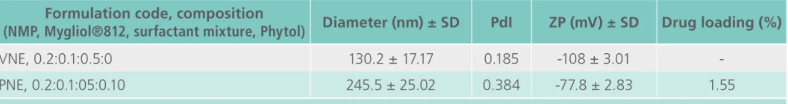
Table 1 summarizes the physicochemical charac-teristics of the transparent oil-in-water nanoemul-sions successfully produced in the absence (VNE) or containing PNE. The colloidal dispersions exhibited small and uniformly distributed droplet sizes, with
Table 1. Effect of PYT on droplet size (diameter), zeta potential (ZP) at VNE and drug loading at PNE.
Formulation code, composition
(NMP, Mygliol®812, surfactant mixture, Phytol) Diameter (nm) ± SD PdI ZP (mV) ± SD Drug loading (%)
VNE, 0.2:0.1:0.5:0 130.2 ± 17.17 0.185 -108 ± 3.01
-PNE, 0.2:0.1:05:0.10 245.5 ± 25.02 0.384 -77.8 ± 2.83 1.55
VNE showing a mean diameter of 130.20 nm and polydispersity index (PdI) of 0.185. Drug loading caused the PNE droplet size to increase to a mean
diameter of 245.50 nm (PdI = 0.384). Both VNE
and PNE nanoemulsions showed large negative ZP values. The drug loading was calculated using the
linear regression equation, A = 0.0045C + 0.005,
which was extracted from the standard curve. The measured absorbance was substituted for "C", ex-pressed as µg/mL (PYT). The correlation coefficient of the straight line was 0.9999. The maximum PYT loading on the oil-in-water (O/W) nanoemulsions
was 1.55 ± 0.012% (n = 3), which allowed us to
produce nanoemulsions containing 1% w/v (10 mg/ mL) PNE.
Nanoemulsions are commonly stabilized by rela-tively large amounts of surfactant in order to pro-duce transparent dispersions [14]. In this study, we have produced extraordinarily transparent nanoe-mulsions with droplet sizes in the range of 130 to 250 nm using low levels of surfactant (about 5% w/w). This was the result of using a suitable sur-factant pair, composed of soy phosphatidylcholine and sodium oleate (1:7 w/w) associated with the co-solvent 2-methylpyrrolidone (2% w/v). The effect of surfactant composition [5] and co-solvent ratio [15] on both droplet size and droplet size distribu-tion has been previously described. In addidistribu-tion, the composition of oil phase/surfactant/co-solvent used in this work produced stable nanoemulsions with high negative ZPs, guaranteeing suitable electros-tatic resilience against flocculation.
PYT loading contributed to the increased nanoe-mulsion droplet size, which probably occurred due to PYT solubilizing in the oil phase. In previous stu-dies with microemulsions containing the antitumor drug doxorubicin, we observed similar results, with drug loading increasing the droplet size of the mi-croemulsions [16]. The same effect was also ob-served for amphotericin B loaded oil-in-water mi-croemulsions [17]. Thus, PYT is a liquid immiscible with water and, as observed with amphotericin B, its solubilization into oil phase contributes to
increa-sed droplet size. The mechanism of the latter case is not clear, but the ZP reduction observed for drug-loaded nanoemulsions confirmed that PYT also dis-rupts the surfactant film at the oil-water interface. It is possible that the drug is partially located in this region, which is important for PYT’s antioxidant effects.
Antioxidant activity assays
Although living systems coexist with free radicals by developing diverse mechanisms for adapting them to advantageous physiological functions, excessive production of ROS and RNS are implicated in va-rious diseases [18].
In vitro antioxidant activity assays
DPPH• scavenging activity
The DPPH• test is the most commonly performed
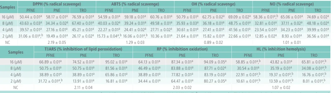
assay for determining antioxidant activity by moni-toring the decreased absorbance of the test sample. It assesses radical scavenging activity. Trolox, (+)-ca-techin, ethyl gallate, ascorbic acid, and a-tocopherol are generally used as standards for screening new compounds [19]. Table 2 shows that PNE produced greater inhibitory effect when compared to PFNE for all four doses tested, but the activity was lower than the standard (TRO) treated with the same do-ses. The highest inhibitions were observed for 16 µM of PFNE, PNE, and TRO and were 50.44 ± 0.01, 58.17 ± 0.01, and 76.59 ± 0.01%, respectively. The-re was a dose-The-response The-relationship between the tested doses and inhibition.
ABTS•+ decolorization activity
The ABTS assay, which monitors the scavenging of
ABTS•+ is another common method used to test for
Table 2. Antioxidant potential of PFNE/PNE in comparison to controls.
Samples DPPH (% radical scavenge) ABTS (% radical scavenge) OH (% radical scavenge) NO (% radical scavenge)
PFNE PNE TRO PFNE PNE TRO PFNE PNE TRO PFNE PNE TRO
16 (µM) 50.44 ± 0.01a 58.17 ± 0.01a 76.59 ± 0.01a 54.59 ± 0.01a 59.18 ± 0.01a 60.76 ± 0.01a 50.79 ± 0.01a 62.75 ± 0.02a 69.09 ± 0.02a 58.36 ± 0.01a,b 65.06 ± 0.01a 74.69 ± 0.02a
8 (µM) 43.63 ± 0.03a 34.34 ± 0.02a 67.40 ± 0.01a 40.03 ± 0.02a 39.24 ± 0.01a 49.58 ± 0.01a 35.93 ± 0.03a 36.18 ± 0.01a 48.75 ± 0.01a 32.81 ± 0.01a 37.11 ± 0.02a 48.18 ± 0.02a 4 (µM) 39.57 ± 0.01a 27.16 ± 0.01a 45.21 ± 0.01a 22.27 ± 0.01a 24.41 ± 0.02a 27.71 ± 0.02a 30.61 ± 0.01a 27.41 ± 0.01a 41.56 ± 0.01a 23.54 ± 0.01a 34.23 ± 0.01a 39.99 ± 0.01a 2 (µM) 31.06 ± 0.01a,b 19.49 ± 0.01a 26.17 ± 0.02a 15.73 ± 0.04a,b16.06 ± 0.01a,b 10.36 ± 0.01a 21.64 ± 0.01a 15.82 ± 0.01a 22.66 ± 0.01a 12.85 ± 0.02a 8.93 ± 0.01a 36.56 ± 0.01a
NC 2.19 ± 0.05 1.29 ± 0.03 0.89 ± 0.02 1.01 ± 0.01
Samples TBARS (% inhibition of lipid peroxidation) RP (% inhibition oxidation) HL (% inhibition hemolysis)
PFNE PNE TRO PFNE PNE TRO PFNE PNE TRO
16 (µM) 66.89 ± 0.01a 74.52 ± 0.01a 95.02 ± 0.01a 64.13 ± 0.01a 87.34 ± 0.01a 94.09 ± 0.05a 58.85 ± 0.01a,b 43.82 ± 0.01a 65.81 ± 0.01a,b 8 (µM) 50.75 ± 0.01a 50.75 ± 0.01a 81.56 ± 0.01a 46.49 ± 0.01a 83.88 ± 0.01a 87.71 ± 0.02a 30.54 ± 0.01a 35.19 ± 0.01a 34.08 ± 0.01a,b 4 (µM) 38.89 ± 0.01a 38.89 ± 0.01a 65.86 ± 0.01a 38.89 ± 0.01a 77.82 ± 0.01a 83.19 ± 0.03a 22.91 ± 0.01a,b 19.37 ± 0.01a,b 16.76 ± 0.01a,b 2 (µM) 31.72 ± 0.01a,b 13.91 ± 0.01a 16.81 ± 0.01a 34.44 ± 0.01a 64.47 ± 0.01a 80.27 ± 0.05a 10.61 ± 0.01a,b 13.59 ± 0.01a,b 8.01 ± 0.01a,b
NC 2.11 ± 0.04 2.03 ± 0.02 1.07 ± 0.02
ap < 0.05 compared to NC (0.05% tween 80 in 0.9% NaCl); bp < 0.05, compared to TRO (ANOVA and t-Student-Newman-Kewls as post hoc test);
PFNE: phytol free with nano-emulsion; PNE: phytol in nano-emulsion; TRO: trolox (standard); values are the mean ± SD (n = 5).
Table 3. IC50 of antioxidant potential of PYT and standard (trolox).
Parameters DPPH test ABTS test OH test NO test
PFNE PNE TRO PFNE PNE TRO PFNE PNE TRO PFNE PNE TRO
IC50 (mM/L) 1.39 ± 0.25 4.14 ± 0.53 3.14 ± 0.27 4.23 ± 0.47 4.50 ± 0.47 4.20 ± 0.24 2.76 ± 0.39 4.77 ± 0.55 3.26 ± 0.39 5.17 ± 0.58 4.76 ± 0.70 2.72 ± 0.51 CI 0.63-3.06 1.34-12.82 2.19-4.14 2.18-8.21 2.19-9.26 3.44-5.13 1.07-7.15 1.87-12.16 1.61-6.62 2.12-12.60 1.73-13.09 0.49-15.14
r2 0.96 0.85 0.98 0.94 0.93 0.99 0.89 0.89 0.93 0.90 0.88 0.75
Parameters TBARS test RP test HL test
PFNE PNE TRO PFNE PNE TRO PFNE PNE TRO
IC50 (mM/L) 2.52 ± 0.38 4.22 ± 0.45 3.21 ± 0.58 2.08 ± 0.43 1.05 ± 0.12 0.41 ± 0.38 5.63 ± 0.66 3.81 ± 0.46 6.72 ± 0.76 CI 0.95-6.71 2.35-7.59 2.30-4.47 0.42-10.27 0.78-1.41 0.01-16.81 2.19-14.49 2.05-7.08 3.52-12.83
r2 0.89 0.95 0.98 0.82 0.99 0.84 0.89 0.94 0.94
60.76 ± 0.01%, respectively (Table 2). There was also a dose-response relationship among the test samples.
•OH scavenging activity
The free hydroxyl radical (•OH) generally reacts with
polyunsaturated fatty acid moieties of cell mem-brane phospholipids and causes cellular destruction
[20]. Measuring the hydroxyl anionic radical (-•OH)
is done similarly to the DPPH and ABTS assays,
whe-re •OH produced from the H2O2 is scavenged by the sample tested. We found that PNE, rather than PFNE, showed higher inhibition. The greatest inhi-bition was observed for the highest dose (16 µM), resulting in 50.79 ± 0.01%, 62.75 ± 0.02%, and 69.09 ± 0.02% by PFNE, PNE, and TRO,
respec-tively. The CI revealed that both PFNE (CI =
1.07-7.15 µM; r2 = 0.89) and PNE (CI = 1.87-12.16 µM; r2 = 0.89) had improved activity in comparison to
TRO (CI = 1.61-6.62 µM; r2 = 0.93; Table 3). There
was also a dose-response relationship for the tested sample doses.
Nitric oxide (NO•) scavenging activity
The sodium nitroprusside in the NO• scavenging
test was used as the source of nitric oxide (NO) radical. Under aerobic conditions, NO reacts with oxygen to produce a stable nitrate (NO3•−) and ni -trite (NO2•−) radicals, the quantities of which can be determined using the Griess reagent. In this test,
PNE, rather than PFNE, had strong NO• scavenging
activity, with an inhibition of 65.06 ± 0.01% by the 16 µM dose, while TRO by 74.69 ± 0.02%. However, increasing doses exhibited greater inhibi-tion, thus indicating a dose-response relationship. The CIs for PFNE, PNE, and TRO were 2.12-12.60 µM (r2 = 0.90), 1.73-13.09 µM (r2 = 0.88), and 0.49-15.14 µM (r2 = 0.75), respectively (Table 3). Since
PNE showed strong inhibition of NO• production, it
could have physiologically important roles and could potentially prevent nitrosative stress [21].
Lipid per-oxidation inhibition activity
In the TBARS assay, lipid peroxidation was detec-ted by measuring the amount of malonyl aldehyde (MDA) formed from unsaturated fatty acids. The more MDA that reacted with thiobarbituric acid (TBA), the stronger the pink coloration of the sam-ple and the greater the absorbance [19]. Lipid pe-roxidation is usually completed by AAPH (in vitro), but it was strongly inhibited by TRO, followed by PNE and PFNE. The CI parameter demonstrates the prominence activity of PNE with a coefficient of 0.95 (r2) (Table 3). However, in this study, 16 µM of PNE and PFNE exhibited inhibition of LP by 74.52% and 66.89% respectively. The oxidation/peroxidation of lipids by foreign chemicals is a major cause of liver injury [22]. In this in vitro test, the dose-response relationship and the significant (p < 0.05) inhibition of LP suggests PYT has potent antioxidant capacity.
Reduction potential (RP) capacity
The RP assay is a rapid and sensitive antioxidant test, in which absorbance increases upon formation of a color complex with potassium ferricyanide, trichloro acetic acid, and ferric chloride. An increase in the absorbance indicates antioxidant activity of the test sample [23]. Antioxidants are the reducing agents. According to the Table 2, PNE improved the reduc-tion activity more than PFNE for all of the doses tested. PFNE (16 µM) showed a similar reduction capability as the lowest dose of PNE (2 µM). The CI calculated for PFNE, PNE, and TRO were 0.42-10.27 µM (r2 = 0.82), 0.78-1.41 µM (r2 = 0. 99), and 0.01-16.81 µM (r2 = 0.84), respectively (Table 3). Even though the activity was lower than the TRO, there was a dose-response relationship in all cases. A possible simplified mechanism for the antioxidant activity of PYT is shown in Figure 1.
Ex vivo antioxidant activity assay
HL inhibition capacity in rat erythrocytes
in-hibition was assessed by measuring the hemoglo-bin concentration in the reaction [12]. In this study, PFNE proficiently up-regulated erythrocyte mem-brane stability and, thus, reduced HL. Erythrocyte lysis was decreased by 58.85 ± 0.01%, 43.82 ± 0.01%, and 65.81 ± 0.01% after treatment with 16 µM of PFNE, PNE, and TRO, respectively. Oxidative
radicals, especially NO•, can bind to Fe2+-bound
heme, which alters its physiological functions [24], since free hemoglobin (Hb) is crucial for dissolved oxygen (O2) and carbon-di-oxide (CO2) respiration during normal metabolic processes. Since PYT
exhi-bited strong NO• scavenging activity in this study,
it may alter the detrimental in vivo effects of this radical. However, the lower inhibition of HL with PNE may be due to its greater hydrophobicity at high concentration and antioxidant-induced pro-oxidative effects [25].
In vivo antioxidant activity
S. cerevisiae assay
Currently, there is evidence that the body prevents free radical damage [26] using some enzymes and substrates [27]. For example, the dismutation en-zyme, superoxide dismutase (SOD), can convert
O2• into ordinary oxygen (O2) or H2O2. On the other hand, the catalase (CAT) enzyme, which is ubiquitous in most living organisms, is responsible for converting H2O2 to water (H2O) and oxygen (O2) [28]. These two enzymes are essential for the neutralization of ROS and keeping cells free from oxidative stress-mediated damage [29]. S. cerevi-siae have antioxidant compounds in the cytoplasm, mitochondria, and on the entire surface of the cell [30]. In addition, one of its cell wall structural po-lymers (1-3), (1-6)-b-D-glucan) has antioxidant [31] and immunomodulatory activities [32]. Therefore, this organism can be used to measure the anti-oxidant action of a wide variety of organic and inorganic substances. In addition, this eukaryotic model allows for the investigation of the effects of antioxidants at the cellular level and is, thus, an at-tractive alternative to mammalian cell lines. Based on colony formation tests performed by Bakkali et al. [25], certain EOs are cytotoxic to S. cerevisiae, mainly in the stationary growth phase. However, it depends on the state of cell growth as the cells in vegetative stage are much more sensitive due to high penetration power of EOs more efficiently at the budding sites. EOs containing phenolic, al-dehyde, and alcoholic OH are cytotoxic in nature [33] and they may act against fungi [34].
Study-based conclusive talks
The use of PYT in aqueous dispersions requires a powerful solubilizing strategy, such as O/W nanoe-mulsion. Previous studies have reported the efficacy of loading insoluble terpenes into nanoemulsions and the increased stability of these mixtures [35]. In addition, an antioxidant synergy formulation (ASF) containing delta, alpha, and gamma toco-pherol loaded into nanoemulsions decreased in-flammation in CD-1 mice, which was attributed to the increased bioavailability of gamma (2.2 folds) and delta (2.4 folds) tocopherol when compared to pure compounds [36]. The antioxidant activity of the anticancer compound quercetin was also increa-sed when loaded into nanoemulsions, as measured
as a function of prophylactic antitumor efficacy against DMBA-induced breast tumors [37]. It was also shown to combat doxorubicin- and cyclosporin A-induced cardiotoxicity and nephrotoxicity [38].
EOs are known for their antioxidant and cytotoxic, rather than mutagenic, activities. At low concentra-tions, antioxidants cannot penetrate the mitochon-drial membrane and, thus, may retain antioxidant, rather than cytotoxic or prooxidant, activities. Low concentrations of retinol and tocopherol exhibit an-tioxidant and antimutagenic activities, whereas, at high concentration, they are genotoxic [39]. The-refore, both the concentration of the antioxidant and the integrity of the mitochondrial membrane are keys for the antioxidant and cytotoxic activities of the EOs.
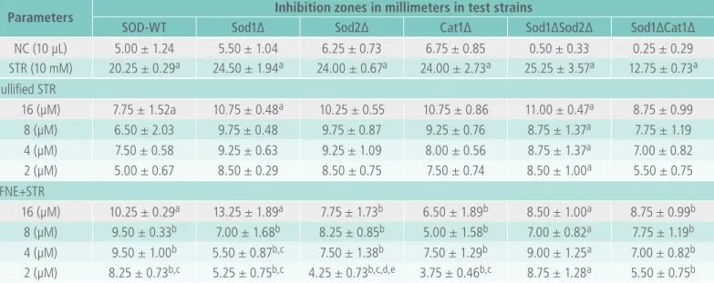
Based on the results shown in Table 4, it is clear that PFNE had oxidative activities in Sod1∆Sod2∆ at all tested doses, but the inhibition zones (7.00-11.00 mm) were lower than those seen for the STR (25.25 mm). The PFNE also exhibited an oxidative effect in the SOD-WT and Sod1∆ strains at 16 µM. Survival of the rest of the strains was significantly (p < 0.05) increased with the treated doses, where PFNE produced a statistical difference (p < 0.05) of 2 µM with 16 µM in SOD-WT, 2 µM and 4 µM with 16 µM in Sod1∆, 2 µM with 4 µM, 8 µM and 16 µM in Sod2∆ and 2 µM with 16 µM in Cat1∆ strains, respectively.
< 0.05), but PNE exhibited oxidative effects at all of the doses tested, except 2 µM. There may be an effect of the vehicle in which the PYT nanoparticles were emulsified. However, the data presented in both Tables 4 and 5 suggest lower zones of inhibi-tion for both formulainhibi-tions, which were lower than the STR-treated group. The data shown in Tables 3 and 4 reveal that both PYT formulations effectively
protected all S. cerevisiae strains tested, thus de-monstrating the antioxidant defense capacities of PFNE and PNE.
However, in the without-STR (nullified) treated groups and groups co-treated with 16 µM of PFNE/ PNE, the zones of inhibitions were slightly larger than for the other co-treated doses. There may be a hyperoxy effect; which was eventually minimized
Table 4. IC50 of antioxidant potential of PYT and standard (trolox).
Parameters Inhibition zones in millimeters in test strains
SOD-WT Sod1∆ Sod2∆ Cat1∆ Sod1∆Sod2∆ Sod1∆Cat1∆
NC (10 µL) 5.00 ± 1.24 5.50 ± 1.04 6.25 ± 0.73 6.75 ± 0.85 0.50 ± 0.33 0.25 ± 0.29 STR (10 mM) 20.25 ± 0.29a 24.50 ± 1.94a 24.00 ± 0.67a 24.00 ± 2.73a 25.25 ± 3.57a 12.75 ± 0.73a Nullified STR
16 (µM) 7.75 ± 1.52a 10.75 ± 0.48a 10.25 ± 0.55 10.75 ± 0.86 11.00 ± 0.47a 8.75 ± 0.99 8 (µM) 6.50 ± 2.03 9.75 ± 0.48 9.75 ± 0.87 9.25 ± 0.76 8.75 ± 1.37a 7.75 ± 1.19
4 (µM) 7.50 ± 0.58 9.25 ± 0.63 9.25 ± 1.09 8.00 ± 0.56 8.75 ± 1.37a 7.00 ± 0.82 2 (µM) 5.00 ± 0.67 8.50 ± 0.29 8.50 ± 0.75 7.50 ± 0.74 8.50 ± 1.00a 5.50 ± 0.75 PFNE+STR
16 (µM) 10.25 ± 0.29a 13.25 ± 1.89a 7.75 ± 1.73b 6.50 ± 1.89b 8.50 ± 1.00a 8.75 ± 0.99b 8 (µM) 9.50 ± 0.33b 7.00 ± 1.68b 8.25 ± 0.85b 5.00 ± 1.58b 7.00 ± 0.82a 7.75 ± 1.19b 4 (µM) 9.50 ± 1.00b 5.50 ± 0.87b,c 7.50 ± 1.38b 7.50 ± 1.29b 9.00 ± 1.25a 7.00 ± 0.82b
2 (µM) 8.25 ± 0.73b,c 5.25 ± 0.75b,c 4.25 ± 0.73b,c,d,e 3.75 ± 0.46b,c 8.75 ± 1.28a 5.50 ± 0.75b
ap < 0.05 compared to NC (0.05% tween 80 in 0.9% NaCl); bp < 0.05 compared to STR (H2O2); cp < 0.05 compared to PFNE 14.16 µM; dp < 0.05 compared to PFNE 7.08 µM; ep < 0.05 compared to PFNE 4.72 µM (one-way ANOVA followed by Bonferoni’s test within 95%
confidence interval); values are the mean ± SD (n = 4).
Table 5. Inhibition zones of PNE, NC and STR in proficient and mutant S. cerevisiae strains.
Parameters Inhibition zones in millimeters in test strains
SOD-WT Sod1∆ Sod2∆ Cat1∆ Sod1∆Sod2∆ Sod1∆Cat1∆
NC (10 µL) 5.00 ± 1.24 5.50 ± 1.04 6.25 ± 0.73 6.75 ± 0.85 0.50 ± 0.33 0.25 ± 0.29 STR (10 mM) 20.25 ± 0.29a 24.50 ± 1.94a 24.00 ± 0.67a 24.00 ± 2.73a 25.25 ± 3.57a 12.75 ± 0.73a Nullified STR
16 (µM) 9.25 ± 0.87a 10.00 ± 0.82 8.75 ± 1.09 8.25 ± 1.19a 10.25 ± 0.67a 9.50 ± 1.11a 8 (µM) 8.75 ± 0.87 8.50 ± 1.11 9.25 ± 0.55 8.75 ± 0.55a 9.50 ± 0.75a 9.25 ± 0.87a 4 (µM) 8.25 ± 0.55 7.00 ± 0.82 7.50 ± 1.00 8.25 ± 0.55a 8.00 ± 0.47a 8.25 ± 0.87a 2 (µM) 6.50 ± 0.75 6.25 ± 0.55 6.25 ± 0.55 6.50 ± 0.58a 6.25 ± 0.55a 7.00 ± 0.82a PNE+STR
16 (µM) 9.75 ± 0.85a 6.25 ± 0.55b 7.75 ± 1.52b 6.75 ± 1.79a 8.25 ± 0.99a 7.75 ± 1.78a 8 (µM) 7.00 ± 1.70b 8.25 ± 1.28b 8.00 ± 1.25b 6.00 ± 1.83a 9.00 ± 0.94a 8.50 ± 1.38a 4 (µM) 8.00 ± 0.47b 7.50 ± 1.53b 8.25 ± 1.59b 8.25 ± 0.98a 6.25 ± 0.29a 7.75 ± 0.73a 2 (µM) 5.50 ± 0.33b 6.00 ± 1.06b 5.50 ± 0.75b 5.00 ± 0.82b 6.00 ± 0.67a 6.00 ± 1.06a
ap < 0.05 compared to NC (0.05% tween 80 in 0.9% NaCl); bp < 0.05 compared to STR (H
2O2) (one-way ANOVA followed by Bonferoni’s test
after the application of STR encountered activity of both the PFNE and PNE. In the in vitro •OH scaven -ging test (Table 2), PYT significantly (p < 0.05)
aug-mented •OH scavenging potential, thus the STR in
this case produces OH radicals that may encounter PYT (PFNE/PNE). While the defense increased with decreasing doses of PFNE and PNE, the activity was significant (p < 0.05) when compared to the NC and STR-treated groups.
It is clear that, like other aerobes, S. cerevisiae
has a number of antioxidant defenses, such as: (i) a cytosolic copper-zinc superoxide dismutase (CuZn-SOD; Sod1) encoded by the SOD1 gene; (ii) a mitochondrial manganese superoxide dismutase (Mn-SOD; Sod2) encoded by the SOD2 gene; (iii) a cytosolic catalase encoded by the CTT1 gene; and (iv) a peroxisomal catalase encoded by the CTA1
gene. However, S. cerevisiae can perform both ae-robic and anaeae-robic metabolism depending on the nature of the carbon source. Under aerobic condi-tions, S. cerevisiae produces energy mainly within its mitochondria using a process similar to that of human cells. According to Costa et al. [40], when wildtype S. cerevisiae is exposed to ethanol-ge-nerating superoxide (O2•-) and H2O2 during both diauxic and post-diauxic growth and the mito-chondrial Sod mutant (Sod2∆) produces essential protection against oxidative stress. A similar study was suggested while working on L-ascorbic acid by Saffi et al. [41]. In this study, both formulations of PYT protected the cytosolic Sod1∆, mitochon-drial Sod2∆, and liver Cat1∆ mutants, which may confirm the potential protection against oxidative damage by PYT.
Previous in vivo studies showed that 6, 12, 24, and 48 µM PYT had antioxidant activity in mice. It
was also postulated to have signiicant •OH, O2•,
methoxy radical (root CH2O•-), carbon-dioxide anion radical (CO2•), NO•, and DPPH• scavenging activities. There is also ex vivo evidence for the an-tioxidant effects of PYT at doses of 2, 6, 12, and 18 µM in Swiss mice [3].
There are compounds [42] (Nunes et al., 2014), including diterpene and carnosic acid (C20H28O4), that have antioxidant activity due to the presence of active -OH groups [43]. There are a number of commercially used natural antioxidants, including some diterpenes- kahweol, cafestol [44], carnosic acid, and carnosol [19], and oxidation products of carnosic acid (rosmanol and isorosmanol) [45]. On the other hand, there are also numerous natural antioxidants with reactive hydroxyl (–OH) groups, including carvacrol, thymol, euginol, isoeuginol, gereniol, caffeic acid, p-cumaric acid, terpein-4-ol, alpha-terpenol, limonen, and p-cymene [46].
PYT is a phenolic EO containing an OH group at its C1 position, which is of interest for its antioxi-dant activity. In addition, an increased number of OH groups may increase the polarity of diterpenes. Recently, aldehyde, oxime, and ethyl bromocrotonyl derivatives of PYT have been shown to have 2, 4, and 6.5 folds more potent antitubercular activities, respectively [47], thus featuring a better pharmaco-logical profile with synthetic products of PYT.
Plant-based antioxidants (natural) have recently gained popularity due to their role as dietary supple-ments with minimal side effects. There is no doubt that earlier researches done on PYT are demons-trating its potential antioxidant capacity. Our study is also connecting the earlier reports on this diter-pene. Both pure phytol (PFNE) and phytol-loaded nanoemulsions (PNE) in this study exhibited potent antioxidant activities in in vitro, ex vivo, and in vivo
(PNE) exhibited better antioxidant activities than its free form (PFNE), which may suggest that emulsion preparation is possible by taking into pharmaceuti-cals consideration.
Abbreviations
AAPH: 2,2'-azobis(2-methylpropionamidine) dihy-drochloride; ABTS: azino-bis-ethylbenzthiazoline-sulfonic acid; CAT/Cat: catalase; DPPH: 1,1-diphenyl-2-picryl-hydrazyl; HL: hemolysis inhibition; LP: lipid peroxidation; MDA: malonyl aldehyde; NO: nitric oxide; RNS: reactive nitrogen species; OH: hydroxyl; OS: organoselenium; PBS: phosphate buffer saline; PFNE: phytol free with nano-emulsion; PNE: phytol in nano-emulsion; PYT: phytol; RBC: red blood cor-puscle; ROS: reactive oxygen species; RP: reduction potential; SOD/Sod: superoxide dismutase; SOD-WT: superoxide dismutase-wild type; STR: stressor; TBA: thiobarbituric acid; TBARS: thiobarbituric acid; TRO: 6-hydroxy-2, 5, 7, 8-tetramethylchroman-2-carbo-xylic acid; YEPD: yeast extract-peptone-dextrose media.
Acknowledgement
We are really very much thankful to Northest Bio-technology Network RENORBIO for funding this project. We also owe to the Nucleus Pharmaceuti-cal Technology (NTF), at Federal University of Piaui, (Teresina, Brazil) to provide laboratory facilities to conduct this study.
Ethical statement
This study is conducted under a doctoral project approved by the Ethics Committee of Federal Uni-versity of Piaui (UFPI), Brazil (#109/14)..
Conflict of interest
We do declare that we have no conflict of interest.
References
1. S. Jose, P. Jayesh, A. Mohandas, R. Philip, I. S. Bright Singh. Application of primary haemocyte culture of Penaeus monodon in the assessment of cytotoxicity and genotoxicity of heavy metals and pesticides. Marine Environmental Research 2011; 71: 169-177.
2. L. Risom, P. Møller, S. Loft. Oxidative stress-induced DNA damage by particulate air pollution. Mutation Research 2005; 592: 119-137.
3. M. T. Islam, M. V. O. B. Alencar, K. C. Machado, K. C. Machado, A. A. C. Melo-Cavalcante, D. P. Sousa, R. M. Freitas. Phytol in a pharma-medico-stance. Chemico-Biological Interactions 2015; 240: 60-73.
4. J. M. M. Andrade, D. Fasolo. Polyphenol Antioxidants from Natural Sources and Contribution to Health Promotion. Chapter 20, Polyphenols in Human Health Disease 2014; 1: 253-265.
5. F. Ostertag, J. Weiss, D. J. McClements. Low-energy formation of edible nanoemulsions: Factors influencing droplet size produced by emulsion phase inversion. Journal of Colloid Interface Science 2012; 388: 95-102.
6. L. Manzocco, M. Anese, M. C. Nicoli. Antioxidant properties of tea extracts as affected by processing. Lebens-mittel-Wissenschaft Und-Technology 1998; 31: 694-698.
7. N. P. Seeram, S. M. Henning, R. Lee, Y. Niu, H. S. Scheuller, D. Heber. Catechin and caffeine contents of green tea dietary supplements and correlation with antioxidant activity. Journal of Agricultural and Food Chemistry 2006; 54: 1599-1603.
8. R. J. Ruch, S. J. Cheng, J. E. Klaunig. Prevention of cytotoxicity and inhibition of intercellular communication by antioxidant catechins isolated from Chinese green tea. Carcinogens 1989; 10: 1003-1008.
9. I. Marcocci, J. J. Marguire, M. T. Droy-lefaiz, L. Packer. The nitric oxide scavenging properties of Ginkgo biloba extract. Biochemical and Biophysical Research Communications 1994; 201: 748-755.
10. H. Esterbauer, K. H. Cheeseman. Determination of aldehydic lipid peroxidation products: Malonaldehyde and 4-hydroxynonenal. Methods in Enzymology 1990; 186: 407-421.
11. 11 M. Oyaizu. Studies on products of browning reactions: antioxidant activities of products of browning reaction prepared from glucosamine. Journal of Nutrition 1986; 44: 307-315.
12. T. K. Girish, P. Vasudevaraju, U. J. S. Prasada Rao. Protection of DNA and erythrocytes from free radical induced oxidative damage by black gram (Vigna mungo L.) husk extract. Food and Chemical Toxicology 2012; 50: 1690-1696.
13. V. Fragoso, N. C. Nascimento, D. J. Moura, A. C. R. Silva, M. F. Richter, J. Saffi. Antioxidant and antimutagenic properties of the monoterpene indole alkaloid psychollatine and the crude foliar extract of Psychotria umbellata Vell. Toxicology in Vitro 2008; 22: 559-566.
15. A. H. Saberi, Y. Fang, D. J. McClements. Effect of glycerol on formation, stability, and properties of vitamin-E enriched nanoemulsions produced using spontaneous emulsification. Journal of Colloid Interface Science 2013; 411: 105–113
16. T. P. Formariz, L. A. Chiavacci, M. V. Scarpa, A. A. Silva-Júnior, E. S. T. Egito, C. H. B. Terrugi, C. M. Franzini, V. H. V. Sarmento, A. G. Oliveira. Structure and viscoelastic behavior of pharmaceutical biocompatible anionic microemulsions containing the antitumoral drug compound doxorubicin. Colloids Surfaces B: Biointerfaces 2010; 77: 47-53.
17. K. C. Pestana, T. P. Formariz, C. M. Franzini, V. H. V. Sarmento, L. A. Chiavacci, M. V. Scarpa, E. S. T. Egito, A. G. Oliveira. Oil-in-water lecithin-based microemulsions as a potential delivery system for amphotericin B. Colloids Surfaces B: Biointerfaces 2008; 66: 253-259.
18. B. H. Neo, S. Kandhi, M. Ahmad, M. S. Wolin. Redox regulation of guanylate cyclase and protein kinase G in vascular responses to hypoxia. Respiratory Physiology & Neurobiology 2010; 174: 259-264.
19. M. Antolovich, P. D. Prenzler, E. Patsalides, S. McDonald, K. Robards. Methods for testing antioxidant activity. Analyst 2002; 127: 183-198.
20. M. N. Alam, N. J. Bristi, M. Rafiquzzaman. Review on in vivo
and in vitro methods evaluation of antioxidant activity. Saudi Pharmaceutical Journal 2003; 21: 143-152.
21. A. Carr, M. R. McCall, B. Frei. Oxidation of LDL by myeloperoxidase and reactive nitrogen species-reaction pathways and antioxidant protection. Arteriosclerosis, Thrombosis, and Vascular Biology 2000; 20: 1716-1723.
22. M. Dirlik, A. Karahan, H. Canbaz, M. Caglikulekci, A. Polat, L. Tamer, S. Aydin. Effects of sulfasalazine on lipid peroxidation and histologic liver damage in a rat model of obstructive jaundice and obstructive jaundice with lipopolysaccharide-induced sepsis. Current Therapeutic Research (CTR) 2009; 70: 299-315.
23. G. K. Jayaprakash, R. P. Singh, K. K. Sakariah. Antioxidant activity of grape seed extracts on peroxidation models in-vitro. Journal of Agricultural and Food Chemistry 2001; 55: 1018-1022.
24. S. Archer. Measurement of nitric-oxide in biological models. FASEB Journal 1993; 7: 349-360.
25. F. Bakkali, S. Averbeck, D. Averbeck, M. Idaomar. Biological effects of essential oils – A review. Food and Chemical Toxicology 2008; 46: 446-475.
26. M. Valko, D. Leibfritz, J. Moncol, M. T. Cronin, M. Mazur, J. Telser. Free radicals and antioxidants in normal physiological functions and human disease. The International Journal of Biochemistry and Cell Biology 2007; 39: 44-84.
27. S. Munné-Bosch, M. Mueller, K. Schwarz, L. Alegre. Diterpenes and antioxidative protection in drought-stressed Salvia officinalis plants. Journal of Plant Physiology 2001; 158: 1431-1437.
28. P. Chelikani, I. Fita, P. C. Loewen. Diversity of structures and properties among catalases. Cellulular and Molecular Life Science 2004; 61: 192-208.
29. T. J. A. S. Andrade, B. Q. Araújo, A. M. G. L. Citó, da Silva, J. Saffi, M. F. Richter, A. B. F. Ferraz. Antioxidant properties and chemical composition of technical Cashew Nut Shell Liquid (Tcnsl). Food Chemistry 2011; 126: 1044-1048
30. R. Pinheiro, I. Belo, M. Mota. Oxidative stress response of Kluyveromyces marxianus to hydrogen peroxide, paraquat and pressure. Applied Microbiology and Biotechnology 2002; 58: 842-847.
31. G. Kogan, A. Stasko, K. Bauerova, M. Polovka, L. Soltes, V. Brezova, et al. Antioxidant properties of yeast (1-3)-b-D-glucan studied by electron paramagnetic resonance spectrometry and its activity in the adjuvant arthritis. Carbohydrate Polymers 2005; 61: 18-28.
32. J. Li, D. F. Li, J. J. Xing, Z. B. Cheng, C. H. Lai. Effects of bglucan extracted from Saccharomyces cerevisiae on growth performance, and immunological and somatotropic responses of pigs challenged with Escherichia coli lipopolysaccharide. Journal of Animal Science 2006; 84: 2374-2381.
33. G. Sacchetti, S. Maietti, M. Muzzoli, M. Scaglianti, S. Manfredini, M. Radice, R. Bruni. Comparative evaluation of 11 essential oils of different origin as functional antioxidants, antiradicals and antimicrobials in food. Food Chemistry 2005; 91: 621-632.
34. E. M. Soylu, S. Soylu, S. Kurt. Antimicrobial activity of the essential oils of various plants against tomato late blight disease agent Phytophthora infestans. Mycopathology 2006; 161: 119-128.
35. M. Maswal, A. A. Dar. Formulation challenges in encapsulation and delivery of citral for improved food quality. Food Hydrocolloids 2014; 37: 182-195.
36. F. Kuo, B. Subramanian, T. Kotyla, et al. Nanoemulsions of an anti-oxidant synergy formulation containing gamma tocopherol have enhanced bioavailability and anti-inflammatory properties. International Journal of Pharmaceutics 2008; 363: 206-213.
37. A. K. Jain, K. Thanki, S. Jain. Self-nanoemulsifying formulation of quercetin: Implications of pro-oxidant activity on the anticancer efficacy. Nanomedicine: Nanotechnlogy Biology and Medicine 2014; 10: 959-969.
38. S. Jain, A. K. Jain, M. Pohekar, et al. Novel self-emulsifying formulation of quercetin for improved in vivo antioxidant potential: Implications for drug-induced cardiotoxicity and nephrotoxicity. Free Radical Biology and Medicine 2013; 65: 117-130.
39. G. Bronzetti, M. Cini, E. Andreoli, L. Caltavuturo, M. Panunzio, C. Della Croce. Protective effects of vitamins and selenium compounds in yeast. Mutation Research 2001; 496: 105-115.
41. J. Saffi, L. Sonego, Q. D. Varela, M. Salvador. Antioxidant activity of L-ascorbic acid in wild-type and superoxide dismutase deficient strains of Saccharomyces cerevisiae. Redox Report 2006; 11: 179-184.
42. G. B. L. Nunes, P. R. Policarpo, L. M. Costa, T. G. da Silva, G. C. G. Militão, C. A. Câmara, J. M. B. Filho, S. J. C. Gutierrez, M. T. Islam, R. M. de Freitas. In Vitro Antioxidant and Cytotoxic Activity of Some Synthetic Riparin-Derived Compounds. Molecules 2014; 19: 4595-4607.
43. O. I. Aruoma, B. Halliwell, R. Aeschbach, J. Löliger. Antioxidant and prooxidant properties of active rosemary constituents: carnosol and carnosic acid. Xenobiotic 1992; 22: 257-268.
44. K. J. Lee, J. H. Choi, H. G. Jeong. Hepatoprotective and antioxidant effects of the coffee diterpenes kahweol and cafestol on carbon tetrachloride-induced liver damage in mice. Food and Chemical Toxicology 2007; 45: 2118-2125.
45. S. Munné-Bosch, L. Alegre. Changes in carotenoids, tocopherols and diterpenes during drought and recovery, and the biological significance of chlorophyll loss in Rosmarinus officinalis plants. Planta 2000; 210: 925-931.
46. M. A. Shah, S. J. D. Bosco, S. A. Mir. Plant extracts as natural antioxidants in meat and meat products. Meat Science 2014; 98: 21-33.
47. D. Saikia, S. Parihar, D. Chanda, S. Ojha, J. K. Kumar, C. S. Chanotiya, K. Shanker, A. S. Negi. Antitubercular potential of some semisynthetic analogues of phytol. Bioorganic & Medicinal Chemistry Letters 2010; 20: 508-512.
International Archives of Medicine is an open access journal publishing articles encompassing all aspects of medical scien-ce and clinical practiscien-ce. IAM is considered a megajournal with independent sections on all areas of medicine. IAM is a really international journal with authors and board members from all around the world. The journal is widely indexed and classified Q1 in category Medicine.