Abstract
Background: Mucosal cyst of the maxillary sinus or antral pseudo-cyst is one of great importance injury, being the pathology that affects more the maxillary sinus. Their discovery, in most cases, it is for the interpretation of the images in routine panoramic radiography.
Aims: The research aimed to evaluate the prevalence of mucous cyst in maxillary sinus in radiology clinic at Ceara Federal University.
Material and Methods: To this study conduction, were analyzed 1996 panoramic radiographs from a digital file obtained between April 2011 to April 2013.
Results: Aspects as gender, affected side and teeth absence next to the cyst in the respective quadrant were evaluated.It was observed in the sample the occurrence of 45 patients with suggested images of mucous cysts in maxillary sinus,making a prevalence of 2.25%. From them, 26 (57.8%) were female and 19 (42.2%) were male. 48 maxillary sinuswere affected with the wound, from which28 (58.3%) it was in the left side and 20 (41.7%) in the right site. Three patients presented the wound in both sides, what represents 6.7% of the affected patients. From those 48 Mucous retention cyst, 40 (83.3%) were not related to an edentulous area in ipsilateral quadrant and 8 (16.7%) were shown next to an edentulous area.
Prevalence of Maxillary Sinus Jaw Mucuous Cysts
in University Dental Radiology Service
ORIGINAL
Joaquim Lira Saraiva Neto1, Lidia Audrey Rocha Valadas Marques1, Edilson Martins Rodrigues Neto2, Barbara Betty de Lima1, Francineudo Oliveira Chagas1, Ana Cristina de Mello Fiallos3, Henrique Clasen Scarparo1, João Hildo de Carvalho Furtado Junior1, Lúcio Mitsuo Kurita1,Patricia Leal Dantas Lobo4,
Allana Bezerra Capistrano1, Erika Saboia Guerra Diogenes1, Rávilla Alves da Silva1, Paulo Arthur Mendonça Bruno5, Sebastião da Silva Santos1, Márcio Glauber Lopes de Aguiar1, Carlos Ricardo de Queiroz Martiniano1
1 Department of Clinical Dentistry, Federal University of Ceará. 2 Department of Physiology and
Pharmacology, Federal University of Ceará/Pharmacy School, Catholic University Center of Quixadá. 3 Department of Restorative Dentistry,
Federal University of Ceará. 4 Dentistry School (Sobral)/Federal
University of Ceará.
5 Medical School, Federal University of Paraiba.
Contact information:
Edilson Martins Rodrigues Neto.
Introduction
Mucosal cyst of the maxillary sinus or antral pseu-docyst is one of great importance injury, being the pathology that affects more the maxillary sinus. Their discovery, in most cases, it is for the interpre-tation of the images in routine panoramic radiogra-phy [1-5].
The process of formation of this cyst usually consists of an inflammatory exudates that has ac-cumulated under the mucosa of the maxillary si-nus and caused a rise sessile. It is a condition that affects more the maxillary sinus and the aetiology is unknown, but may be attributed to allergic and inflammatory processes of the nasal sinus mucosa, trauma, and periapical periodontal infections, rela-tive humidity and temperature decrease [1, 2, 3, 5]. Radiographically appears as a slightly lesion, well-defined, homogeneous, oval-shaped or dome, the insertion base can be wide or narrow. The pa-noramic radiograph allows the maxillary sinus floor display, especially in its lower wall, which allows to clearly identify the presence of mucous cyst. This image has been proven very effective in the diag-nosis of this injury [6-8].
According to Rodrigues et al., 2013, the radiogra-phic image analysis gives opportunities for dentists to detect changes in the maxillary sinus. The maxi-llary sinus mucous cyst has unique characteristics that make it a classic lesion is diagnosed, but the differential diagnosis, one can cite the Dentigerous cysts, malignant and hyperplasia [1].
Works analyzed, show that in panoramic radio-graphs, this lesion presents between 2% and 10%
of samples, especially in Caucasian patients were male. It is also seen that the age group 20-30 years is the most prevalent, with no preference for the side of the affected breast, with the characteristic, and in most unilateral cases [3].
Usually, this lesion is asymptomatic requiring only the radiographic monitoring of the same, since in many cases it spontaneously regresses [7, 9]. A few clinicians prefer confirm your radiographic impres-sions and delete a tumor by puncture and drainage of the inflammatory exudates. In symptomatic ca-ses (headache, fatigue, facial neuralgia and vertigo extreme cases) surgical intervention is necessary by Caldwell-Luc surgery, allowing easy access and viewing of foreign bodies [3, 6]. Its growth is slow and does not affect the integrity of the cortical bone of the sinus floor [1, 6]. Their size varies from the size of a grain of corn to that of a walnut, and occasionally can grow so much to completely fill the antral cavity [7].
The Caldwell-Luc access is made in the anterior wall of the maxillary sinus, for it is made a linear incision 0,5 cm above the inserted mucosa in the region between canine and premolars, posterior total thickness of detachment to the infraorbital region, access and removal of foreign body or al-tered mucosa.Postoperatively it is recommended the use of antibiotics and nasal wash with saline [9].
The panoramic radiograph to be an easy exam and low cost,It has been indicated as a routine in the dental practice, thanks to its viewing range with low amount of radiation in a single examination.
Conclusion: The conclusion was that the cyst of retention mucous in the maxillary sinus had prevalence in males and in the left side of the maxillary sinus. It was not found a relation between the cyst and the edentulous area.
Keywords
Even the panoramic radiograph showing no maxi-llary sinus with details,continues to have a signifi-cant use by the dental profession to diagnose some diseases as well as some facial abnormalities [3, 7]. According to (Rodrigues et al., 2013), detecting ima-ges of mucous retention cysts of the maxillary sinus can help define their characteristics and behaviors, as well as establishing a therapeutic protocol. They also claim that clinical and radiographic examina-tions are essential to define alternative treatments and rule out other conditions such as mucocele, polyps and sinusitis [1].
Since the importance of epidemiological study and the performance of various dental specialties on the maxillary sinus. It aimed to evaluate the pre-valence of mucous cysts of the maxillary sinus in the Clinic of Radiology at the Federal University of Ceara Dental Science Course, Fortaleza-CE- Brazil, through the evaluation of panoramic radiographs.
Materials and Methods
Retrospective and radiological clinical study with a survey of 1,996 digital panoramic radiographs were performed in the radiology clinic at the Fe-deral University of Ceara Dental Science Course, Fortaleza-CE, between April 2011 and April 2013. Were excluded all panoramic radiographs did not provide adequate visualization of the maxillary si-nuses, regardless of patient age. The study of two dentist researchers where both reviewed the pa-noramic radiographs to mucous retention cyst de-tection, and to enter into agreement, the overview was included in the study. The criterion for judging the existence of the mucous cyst was the presence of a radiopaque image, oval-shaped or dome, on the floor or walls of the maxillary sinus. In dou-bts about similar anomalies the differentiation was made by computed tomography The data were analyzed according to gender, affected side and absence of teeth next to suggestive of mucinous cyst of the maxillary sinus. Furthermore, a
litera-ture review was conducted to compare the sam-ple submitted to the previously published studies. This study as required by Resolution nº466/12 was approved by the Ethics Committee of the Ceara Federal University an opinion 1.287.565.
Results
It was found that of 1996 patients evaluated radio-graphically, 38% were male and 62% female, get-ting this sample depending on the influx of patients to the service of Clinical Radiology of the Ceara Fe-deral University. It was found that of 1996 patients evaluated radiographically, getting this sample de-pending on the influx of patients to the service of Clinical Radiology at Ceará Federal University.
From those 45 patients, 26 (57.8%) were female and 19 (42.2%) were male. From those patients,48 maxillary sinuses were suggestive image of mucous cysts of the maxillary sinus, that is, three patients (6.7%) had bilateral lesions, 28 (58.3%) on the left and 20 (41.7%) on the right.
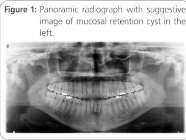
Of the 48 mucosal lesions suggesting cyst of the maxillary sinus 40 (83.3%) were not associated with an edentulous area in the ipsilateral quadrant and 8 (16.7%) presented near the edentulous area with one or more missing teeth. (Figure 1).
Discussion
According to Farman and Nortjé (2002) extensive lesions occupying the maxillary sinus can produce surprisingly few clinical signs [9]. For this reason, the first panoramic radiograph may indicate that other tests the presence of disease in the maxillary sinus, thus fitting the dentist understand panoramic radiological features of the disease and the nor-mal variations of the paranasal sinuses. [10] In cases where the panoramic radiograph occur overlapping images of adjacent structures, indicates the compu-ted tomography [7, 11].
The prevalence of mucous retention cyst of the maxillary sinus found in 45 of 1996 panoramic ra-diographs analyzed was 2.25%. Convergent result previous studies that found Mucous cysts in 1,5% to 14% of the sample [2, 3]. According to Rodri-gues et al., (2009), when a total of 6293 panoramic radiographs were evaluated, they realized that 201 were suggestive of mucosal retention cysts, resul-ting in a prevalence of 3.19%[4]. Gonçalves and Silveira (1993) analyzed 3.180 reports of panoramic radiographs in which the event occurred in 139 patients with mucous cysts of the maxillary sinus, giving a prevalence of 4.4% [6]. Rodrigues et al., (2013), reviewed 6.000 panoramic radiographs of a digital file, where 185 radiographs were suggestive of mucosal retention cyst of the maxillary sinus or 3.08% of the total [1].
Mucous retention cysts were found in 26 female patients (57.8%) and 19 males (42.2%). Showing a slight predilection for females when dealing with the number of cases, but referring to the total sam-ple, it has a predilection for males, which is in line with other studies that reported a greater predilec-tion for males cyst [2, 3].
Bósio et al., (2009) [5], in a study of 173 or-thodontic patients, found a prevalence of 5.8% of cases of maxillary sinus retention cyst, which was no significant difference between men and women. The prevalence of mucous retention cysts in the maxillary sinus is around an average of 5%, but
va-ries from study to study, perhaps due to the popu-lation, location and period.The prevalence is about two times higher in men than in women[7].
Of the 45 patients, 48 maxillary sinuses were suggestive image of mucous cysts of the maxillary sinus, that is, three patients (6.7%) had bilateral lesions, 28 (58.3%) on the left side and 20 (41.7%) on the right. Studies show a predilection of the cyst the left. Manhães Jr. (2002) claimed to be the mucous retention cyst, in most cases, unilateral, which draws more attention of the dentist in the analysis of panoramic radiographs by noting the di-fference between healthy sinus and the other with the injury [3].
Of the 48 lesions suggestive of mucous cyst of the maxillary sinus, 40 (83.3%) were not associa-ted with an edentulous area in the ipsilateral and 8 quadrant (16.7%) presented next to an edentulous area (one or more missing teeth). Those findings testify the studies of Soikkonen et al (1994) cited by Manhães Jr. (2012)they encounter in their work, cysts in dentate and edentulous patients. However in this study, most of the mucosal retention cysts (83.3%) were found in patients without dental ab-sences near the cyst, which makes us think that the etiology of it has no association with edentulous areas [3].
It can be seen that the mucous cysts of the maxi-llary sinus, in most cases is a radiographic finding, which means that the dentists have to pay attention more precisely to the region of the maxillary sinus lesions in search not only look eyes to the area of the teeth [1].
Conclusion
go against those described in the literature, and males the most affected proportionally, occurring predominantly unilateral, the left side more affected and showing no direct relationship with edentulous areas.
References
1. Rodrigues CD, Silveira MF, Alencar AHG, Silva MAGS, Mendonça EF, Estrela C. Three-dimensional images contribute to the diagnosis of mucous retention cyst in maxillary sinus. Med Oral Patol Cir Bucal, 18(1): e151-e157, 2013.
2. Andrade VC, Bacchi A, Salim MAA, Bourguignon Filho AM.Cisto Mucoso De Retenção – Relato De Caso Clínico. Saber Científico Odontológico, Porto Velho, 2 (1):66-72, 2012.
3. Manhães Júnior LRC. Cisto mucoso do seio maxilar: importância no diagnóstico radiográfico. [Monografia] Piracicaba/UNICAMP (SP), 2012.
4. Rodrigues CD, Freire GF, Silva LB, et al. Prevalence and risk factors of mucous retention cysts in a Brazilian population. Dentomaxillofacial Radiology, 38(1):480-3, 2009.
5. Bósio JA, Orlando T, Rovigatti E, Gruner SK. The incidence of maxillary sinus retention cysts in orthodontic patients. World Journal of Orthodontics, 10(1): e7-e8, 2009.
6. Gonçalves RCC, Silveira MMF. Cisto mucoso do seio maxilar: prevalência em radiografias panorâmicas. Revista odontológica do Brasil central, 3(8):19-22, 1993.
7. Batista OS, Rosário Júnior AF, Wishinieski C. Contribuição para o estudo do seio maxilar. Rev Port Estomatol Med Dent Cir Maxilofac, 52(4): 235-39, 2011.
8. Matheny KE, Dunacavage JA. Contemporaryindications for the Caldwell-Luc procedure. Curr Opin Otolaryngol Head Neck Surg, 11 (1), 23-6, 2003
9. Farman AG, Nortjé CJ. Pathologic conditions of the maxillary sinus. Panoramic Imaging News, 2(3):1-7, 2002.
10. Cury SEV, Cury DPN, Silva BSF, Molina OTF, Araújo JO, Manhães Jr, Oliveira LB. Pseudocisto Antral: Prevalência na cidade de Volta Redonda, Rio de Janeiro, Brasil. Antral pseudocyst: prevalence in the city of Volta Redonda, Rio de Janeiro, Brazil. Rev Cadernos UniFOA, 05 (1):69-84, 2013.
11. Castro AJR, Sassone LM, Amaral G. Maxillary sinus changes and the relationship with dentistry problems sources. Revista do Hospital Universitário Pedro Ernesto, UERJ, 12 (1):30-5, 2013.
International Archives of Medicine is an open access journal publishing articles encompassing all aspects of medical scien-ce and clinical practiscien-ce. IAM is considered a megajournal with independent sections on all areas of medicine. IAM is a really international journal with authors and board members from all around the world. The journal is widely indexed and classified Q2 in category Medicine.